Technical field
This invention relates to deep brain stimulation to treat neurological condition or disorders, more particularly, deafferentation disorder (e.g., tinnitus and phantom pain), and affective disorders.
Background of the invention
A. Auditory Dysfunction
Auditory dysfunctions are common. For example, in the United States, the prevalence of tinnitus when the whole population is considered is approximately 3%. This prevalence is only 1% under the age of 45 but increases significantly with age, rising to 9% in the population over 65 years (Adams et al., 1999). This roughly translates to 36 million Americans with tinnitus (Heller 2003). Tinnitus is a noise in the ears, often described as ringing, buzzing, roaring, or clicking. Subjective and objective forms of tinnitus exist, with objective tinnitus often caused by muscle contractions or other internal noise sources in the area proximal to auditory structures. In certain cases, external observers can hear the sound generated by the internal source of objective tinnitus. In subjective forms, tinnitus is audible only to the subject. Tinnitus varies in perceived amplitude, with some subjects reporting barely audible forms and others essentially deaf to external sounds and/or incapacitated by the intensity of the perceived noise.
Because auditory dysfunction often occurs secondary to a pathological state, initial treatment may focus on finding an underlying cause. A subject presenting with, for example, tinnitus may be asked for information regarding medications, recent or chronic noise exposure, and home and work environment. Common medications such as aspirin are known to cause tinnitus in some patients or in elevated dosages. Stress can be a direct cause of tinnitus and can aggravate existing cases. A thorough physical exam is typically made of a subject with complaints of tinnitus to eliminate pathologies such as hypertension, tumors, and infections. Objective tinnitus may be diagnosed using a stethoscope if the source of the noise can be localized. For example, hypertension or arterial disorders may produce objective tinnitus, as the carotid arteries pass close to the auditory organs in humans, and excessive pressure or arterial blockage may cause detectable noise to both the subject and to an outside observer.
If a treatable underlying cause to the auditory dysfunction is identified, treatment may focus on elimination of the cause. For example, hypertensive patients may see a reduction or elimination of tinnitus once anti-hypertensive therapy begins. However, a significant number of patients have untreatable underlying pathologies or have auditory dysfunction in the absence of any identifiable cause. For these patients, treatments for directly reducing or eliminating the auditory dysfunction are desirable.
Tinnitus research is actively pursued in the hope of finding efficacious treatments. Recently published work has utilized drug delivery systems such as the system described in U.S. Pat. No. 5,713,847, which includes a catheter inserted into a patient's auditory cortex or thalamus for microinfusing drugs.
Another example of published drug delivery techniques is U.S. Pat. No. 6,656,172, which describes a tinnitus treatment that includes inserting intrathecally a catheter for infusing a drug. Other treatment methods may try to mask the perceived tinnitus noise by generating an audible signal of appropriate frequency. WO 01/08617 describes a system with a vibrating probe placed in proximity to the inner ear.
Nerve stimulation has been shown to be helpful in treating patients with chronic intractable pain. For those patients who prove unresponsive to conservative pain management techniques, peripheral nerve stimulation may be a successful therapy for pain management when the pain is known to result from a specific nerve. Peripheral nerve stimulation is based in part on the Melzack-Wall gate control theory of pain. Sweet and Wespic first used electrical stimulation of peripheral nerves in the 1960s to mask the sensation of pain with a tingling sensation (paresthesia) caused by the electrical stimulation. Subsequent refinements in the technology, surgical technique and patient selection have led to improved long term results.
B. Depression
Depression is an important public health problem affecting about 15% of the general population (Rauch 2003). The lifetime rates for major depression vary widely across countries, ranging from 1.5 cases per 100 adults in the sample in Taiwan to 19.0 cases per 100 adults in Beirut. The annual rates range from 0.8 cases per 100 adults in Taiwan to 5.8 cases per 100 adults in New Zealand. The mean age at onset shows less variation (range, 24.8-34.8 years). In every country, the rates of major depression are higher for women than men. Insomnia and loss of energy occur in most persons with major depression at each site. Persons with major depression are also at increased risk for co-morbidity with substance abuse and anxiety disorders at all sites. Persons who were separated or divorced have significantly higher rates of major depression than married persons in most of the countries, and the risk is somewhat greater for divorced or separated men than women in most countries (Weissman, Bland et al. 1996).
Efforts have been made to treat psychiatric disorders with peripheral/cranial nerve stimulation. Recently, partial benefits with vagus nerve stimulation in patients with depression have been described in U.S. Pat. No. 5,299,569. Another example of electrical stimulation to treat depression is described in U.S. Pat. No. 5,470,846, which discloses the use of transcranial pulsed magnetic fields to treat depression. Yet further, U.S. Pat. No. 5,263,480 describes that stimulation of the vagus nerve may control depression and compulsive eating disorders and U.S. Pat. No. 5,540,734 teaches stimulation of the trigeminal or glossopharyngeal nerves for psychiatric illness, such as depression.
Significant advances in the treatment of depression have been made in the past decade. Since the introduction of selective serotonin reuptake inhibitors (SSRIs), i.e., Prozac.RTM., many patients have been effectively treated with anti-depressant medication. New medications to treat depression are introduced almost every year, and research in this area is ongoing. However, an estimated 10 to 30 percent of depressed patients taking an anti-depressant are partially or totally resistant to the treatment. Those who suffer from treatment-resistant depression have almost no alternatives. Thus, there is a need to develop alternative treatments for these patients.
C. Deep Brain Stimulation to Treat Neurological Conditions
Deep brain stimulation (DBS) has been applied to the treatment of central pain syndromes and movement disorders, and it is currently being explored as a therapy for epilepsy. For instance, U.S. Pat. No. 6,016,449 and U.S. Pat. No. 6,176,242 disclose a system for the electrical stimulation of areas in the brain for the treatment of certain neurological diseases such as epilepsy (See Vonck et al., 2005; Vonck et al., 2002 and Velasco et al., 2001), cluster headaches (Benabid, Wallace et al. 2005) and Parkinson's disease (Benabid, Wallace et al. 2005).
The use of stimulating electrodes to treat tinnitus has been published. U.S. Pat. Nos. 5,735,885 and 5,496,369 describe the placement of an electrode in the primary auditory cortex of a patient. U.S. Pat. Nos. 6,456,886 and 5,697,975 also use an electrode placed in the auditory cortex, and further describe placement of an electrode in the medial geniculate body of the thalamus.
Thus, various electrical stimulation and/or drug infusion devices have been proposed for treating neurological disorders. Some devices stimulate through the skin, such as electrodes placed on the scalp. Other devices require significant surgical procedures for placement of electrodes, catheters, leads, and/or processing units. These devices may also require an external apparatus that needs to be strapped or otherwise affixed to the skin.
However, despite the aforesaid available treatments, there are patients with major depression, auditory dysfunction and other neurological conditions that remain disabled. For these severely ill and disabled patients, novel therapies are required. Thus, the present invention is the first to utilize deep brain stimulation to treat a variety of neurological conditions, for example depression and auditory dysfunction.
Brief summary of the invention
Embodiments of the present invention comprise a therapeutic system for treating a neurological condition or disorder having a surgically implanted device in communication with a predetermined brain region, for example the amygdala, hippocampus, parahippocampus, perirhinal cortex, and entorhinal cortex. The device can include a distal probe, such as, for example, an electrode assembly or electrical stimulation lead. The proximal end of the probe is coupled to an electrical signal source, which, in turn, is operated to stimulate the predetermined brain region.
In certain embodiments of the present invention, the neurological disorder and/or condition is an auditory dysfunction, for example, but not limited to tinnitus, hyperacousis, phonophobia, misophonia, auditory agnosia, auditory spatial dysfunction or auditory hallucinations. Yet further, the neurological disorder and/or condition can be a mood and/or anxiety disorder, for example, but not limited to depression, biopolar dysthymic disorder, panic disorder, posttraumatic stress disorder, obsessive-compulsive disorder, phobic disorder. In further embodiments, the neurological disorder and/or condition is phantom pain.
Other stimulation devices used in certain embodiments are drug pumps which provide chemical stimulation of a predetermined brain region. Chemical stimulation can be provided by delivery of pharmaceuticals or neuroactive substances that, for example, disrupt or block pathological activity.
Magnetic stimulation of certain brain regions for the treatment of neurological conditions and/or disorders is used in certain embodiments of the present invention. Magnetic stimulation can be provided by internally implanted probes or by externally applied directed magnetic fields.
Yet further, thermal stimulation can be provided via implanted probes that are regulated to heat and/or cold temperatures. In other embodiments, ultrasound stimulation is used as a stimulation source, either by itself or in combination with another stimulation source. For example, in certain embodiments of the invention, ultrasound is used to stimulate active tissue by propagating ultrasound in the presence of a magnetic field as described by Norton (2003), herein incorporated by reference in its entirety. Combinations of stimulation sources are used in some embodiments of the invention.
Devices used with the invention can operate with various stimulation parameters. One example of stimulation parameters used with an electrical stimulation device to treat neurological conditions and/or disorders uses an amplitude in the range of about 2 mA to about 100 mA, a frequency in the range of about 3 Hz to about 50 Hz, and a pulse width in the range of about 5 microseconds to about 100 microseconds. However, other parameters are used in other embodiments of the invention, such as, for example, higher and lower frequencies, various current amplitudes, and/or pulse width durations. In another embodiment of the invention, a frequency stimulation parameter of about 80 Hz is used. Burst mode stimulation is used in preferred embodiments of the invention. The burst stimulus comprises a frequency in the range of about 1 Hz to about 300 Hz, more particular, in the range of about 1 Hz to about 12 Hz, and more particularly, in the range of about 1 Hz to about 4 Hz, 4 Hz to about 7 Hz or about 8 Hz to about 12 Hz, 18 Hz to 20 Hz, and 40 Hz. The burst stimulus comprises at least two spikes, for example, each burst stimulus can comprise about 2 to about 100 spikes, more particularly, about 2 to about 10 spikes. Each spike can comprise a frequency in the range of about 50 Hz to about 1000 Hz, more particularly, in the range of about 200 Hz to about 500 Hz. The interval between spikes can be about 0.5 milliseconds to about 100 milliseconds. More particularly, the maximum inter-spike interval may be about 5 milliseconds. Those of skill in the art realize that this can vary depending upon the patient and the treatment. The frequency of the spikes within the burst does not need to be constant or regular, in fact, typically, the frequency of the spikes is random or variable. In further embodiments, the burst stimulus is followed by an inter-burst interval. The inter-burst interval has a duration in the range of about 5 milliseconds to about 5 seconds, more preferably, in the range of about 10 milliseconds to about 300 milliseconds, or any range therebetween, for example, the minimum inter-burst interval may be about 20 milliseconds. It is envisioned that the burst stimulus has a duration in the range of about 10 milliseconds to about 5 seconds, more particularly in the range of about 250 milliseconds to 1 second. The burst stimulus and the inter-burst interval can have a regular pattern or an irregular pattern (e.g., random or irregular harmonics).
The invention also comprises a method for treating neurological conditions and/or disorders. The method comprises surgically implanting an electrical stimulation lead such as a multiple electrode lead. Following implantation, the proximal end of the lead is attached to a signal generator. The signal generator then generates a signal that stimulates a predetermined brain region.
In some embodiments of the invention, electrical stimulation parameters are varied after implantation to optimize treatment of a neurological disorder or condition. The parameters varied may include modification of the predetermined implantation site, or modification of, for example, signal amplitude, frequency, pulse width or pulse shape of the stimulation signal.
The foregoing has outlined rather broadly the features and technical advantages of the present invention in order that the detailed description of the invention that follows may be better understood. Additional features and advantages of the invention will be described hereinafter which form the subject of the claims of the invention. It should be appreciated that the conception and specific embodiment disclosed may be readily utilized as a basis for modifying or designing other structures for carrying out the same purposes of the present invention. It should also be realized that such equivalent constructions do not depart from the invention as set forth in the appended claims. The novel features which are believed to be characteristic of the invention, both as to its organization and method of operation, together with further objects and advantages will be better understood from the following description when considered in connection with the accompanying figures. It is to be expressly understood, however, that each of the figures is provided for the purpose of illustration and description only and is not intended as a definition of the limits of the present invention.
Brief description of the drawings
For a more complete understanding of the present invention, reference is now made to the following descriptions taken in conjunction with the accompanying drawing, in which:
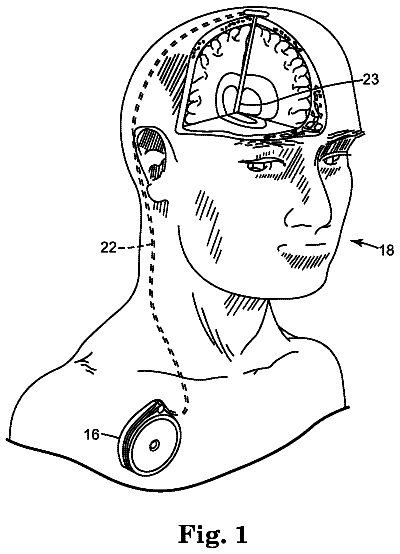
FIGS. 1A and 1B illustrate example electrical stimulation systems.
FIGS. 2A-2I illustrate example electrical stimulation leads that may be used to electrically stimulate brain regions;
FIG. 3 illustrates example placement of a single stimulation lead and stimulation electrode in communication with the amygdalohippocampal complex;
FIG. 4 is a block diagram of processes according to a method for treating auditory dysfunction using a stimulation system; and
FIG. 5 is a table showing the results of an amytal test.
Detailed description of the invention
I. Definitions
Unless defined otherwise, technical and scientific terms used herein have the same meaning as commonly understood by one of ordinary skill in the art to which this invention belongs. For purposes of the present invention, the following terms are defined below.
As used herein, the use of the word "a" or "an" when used in conjunction with the term "comprising" in the claims and/or the specification may mean "one," but it is also consistent with the meaning of "one or more," "at least one," and "one or more than one." Still further, the terms "having", "including", "containing" and "comprising" are interchangeable and one of skill in the art is cognizant that these terms are open ended terms.
As used herein the term "affective disorders" refers to a group of disorders that are commonly associated with co-morbidity of depression and anxiety symptoms.
As used herein the term "anxiety" refers to an uncomfortable and unjustified sense of apprehension that may be diffuse and unfocused and is often accompanied by physiological symptoms.
As used herein the term "anxiety disorder" refers to or connotes significant distress and dysfunction due to feelings of apprehension, guilt, fear, etc. Anxiety disorders include, but are not limited to panic disorders, posttraumatic stress disorder, obsessive-compulsive disorder and phobic disorders.
As used herein, the term "depression" refers to a morbid sadness, dejection, or melancholy.
As used herein, the terms "amygdala" and "amygdalohippocampal complex" or "amygdaloid complex" or "amygdaloid nucleus" refer to the gray and white matter associated with the amygdala, as well as the projections associated with, or projecting to and/or from the amygdala, for example, projections associated with the hippocampus. The amygdala is an almond-shaped brain region located in the medial temporal lobes of the brain. It is a subcortical structure located at the dorsomedial tip of the temporal lobe and continuous with the uncus of the parahippocampal gyrus. The amygdala comprises several separately-functioning nuclei that have been grouped together by their anatomical proximity, including the basal and lateral nuclei, the central nucleus, the cortical nuclei, and medial nuclei. Those of skill in the art are aware that the nuclei that compose the amygdala are reciprocally connected to the hypothalamus, hippocampal formation, neocortex, and thalamus. As used herein, the term amygdala includes the defined area of the amygdala as known by one of skill in the art, as well as the surrounding or adjacent gray matter or white matter tracts leading to and from amygdala and/or gray matter or white matter tracts that are contiguous with amygdala. The surrounding or adjacent gray matter or white matter can include up to approximately a 1 cm radius of amygdala.
As used herein, the term "auditory dysfunction" refers to conditions or dysfunctions associated with the auditory pathway. Such auditory dysfunctions can include, but are not limited to tinnitus, hyperacousis, phonophobia, misophonia, auditory agnosia in all its forms, auditory spatial dysfunction (localizing sound) and auditory hallucinations, inclusive of musical hallucinosis. Auditory hallucinations can occur in schizophrenia or use of certain drugs (e.g., antimuscarinic agents, antiparkinsonian drugs, antidepressants, beta adrenoceptor antagonists and opiates). Auditory dysfunction can also include hearing loss. Hearing loss can be conductive hearing loss (mechanical transmission of sound into the sensory receptors in the cochlea is impaired), sensorineural hearing loss (a loss of function in the sensory receptors in either the cochlea or the auditory nerve), or central hearing loss (a lesion in the brain stem or auditory cortex).
As used herein, the terms "auditory nerve" and "cochlear nerve" are interchangeable and refer to the nerve fibers along which the sensory cells of the ear communicate information to the brain. The auditory or cochlear nerve are part of the vestibulocochlear nerve which carries two kinds of sensation, vestibular (balance) and audition (hearing) from sensory receptors in the inner ear. The auditory nerve consists of the vestibular nerve and the cochlear nerve. The vestibulocochlear nerve is also known as the eighth cranial nerve.
As used herein, the term "brain region" refers to any tissue comprising that part of the central nervous system contained within the cranium. The brain stem tissue is also encompassed by the term brain region, including the diencephalon.
As used herein, the term "in communication" refers to the stimulation lead being adjacent, in the general vicinity, in close proximity, or directly next to or directly on the predetermined stimulation site. Thus, one of skill in the art understands that the lead is "in communication" with the predetermined site if the stimulation results in a modulation of neuronal activity. In certain embodiments, the predetermined site is the amygdalohippocampal complex or amygdaloid complex.
As used herein the term "limbic system" encompasses the amygdala, hippocampus, septum, cingulate gyrus, cingulate cortex, hypothalamus, epithalamus, anterior thalamus, mammillary bodies, nucleus accumbens, formix, parahippocampus, perirhinal cortex, and entorhinal cortex. The limbic system has connections throughout the brain, more particularly with the primary sensory cortices, including the rhinencephalon for smell, the autonomic nervous system via the hypothalamus, and memory areas. Yet further, the limbic system is involved in mood, emotion and thought. Two limbic or paralimbic divisions have been described, one associated with the archencephalic hippocampus, one associated with the paleocephalic amygdala. The hippocampal division connects predominantly to the following structures: hippocampus, posterior parahippocampal, retrosplenium, posterior cingulated cortex, and the supracallosal cingulated cortex. The hippocampal division is involved in explicit processing, memory encoding, visual spatial analysis, skeletomotor effector, attentional and motivational functions. The amygdala division connects predominantly to the anterior parahipocampal area, the insula, temporal pole, infracallosal cingulated cortex and the orbitofrontal cortex. The amygdala division is involved in implicit processing, visceral integration, visual feature analysis, appetite drives, social awareness and mood (Cummings and Mega 2003).
As used herein the term "modulate" refers to the ability to regulate positively or negatively neuronal activity. Thus, the term modulate can be used to refer to an increase, decrease, masking, altering, overriding or restoring of neuronal activity.
As used herein, the term "burst firing" or "burst mode" or "burst mode stimulation" refers to an action potential that is a burst of high frequency spikes (300-1000 Hz) (Beurrier et al., 1999). Burst firing acts in a non-linear fashion with a summation effect of each spike. One skilled in the art is also aware that burst firing can also be referred to as phasic firing, rhythmic firing (Lee 2001), pulse train firing, oscillatory firing and spike train firing, all of these terms used herein are interchangeable.
As used herein, the term "tonic firing" or "tonic mode" refers to an action potential that occurs in a linear fashion.
As used herein, the term "burst" refers to a period in a spike train that has a much higher discharge rate than surrounding periods in the spike train (N. Urbain et al., 2002). Thus, burst can refer to a plurality of groups of spike pulses. A burst is a train of action potentials that occurs during a `plateau` or `active phase`, followed by a period of relative quiescence called the `silent phase` (Nunemaker, Cellscience Reviews Vol 2 No. 1, 2005.) Thus, a burst comprises spikes having an inter-spike interval in which the spikes are separated by 0.5 milliseconds to about 100 milliseconds. Those of skill in the art realize that the inter-spike interval can be longer or shorter. Yet further, those of skill in the art also realize that the spike rate within the burst does not necessarily occur at a fixed rate; this rate can be variable.
As used herein, the term "spike" refers to an action potential. Yet further, a "burst spike" refers to a spike that is preceded or followed by another spike within a short time interval (Matveev, 2000), in other words, there is an inter-spike interval, in which this interval is generally about 10 ms but can be shorter or longer, for example 5 milliseconds or 0.5 milliseconds.
As used herein, the term "neuronal" refers to a neuron which is a morphologic and functional unit of the brain, spinal column, and peripheral nerves.
As used herein, the term "neurology" or "neurological" refers to conditions, disorders, and/or diseases that are associated with the nervous system. The nervous system comprises two components, the central nervous system, which is composed of the brain and the spinal cord, and the peripheral nervous system, which is composed of ganglia and the peripheral nerves that lie outside the brain and the spinal cord. One of skill in the art realizes that the nervous system may be separated anatomically, but functionally they are interconnected and interactive. Yet further, the peripheral nervous system is divided into the autonomic system (parasympathetic and sympathetic), the somatic system and the enteric system. Thus, any condition, disorder and/or disease that effects any component or aspect of the nervous system (either central or peripheral) is referred to as a neurological condition, disorder and/or disease. As used herein, the term "neurological" or "neurology" encompasses the terms "neuropsychiatric" or "neuropsychiatry" and "neuropsychological" or "neuropsychological". Thus, a neurological disease, condition, or disorder includes, but is not limited to cognitive disorders, auditory disorders, affective disorders, movement disorders, mental disorders, pain disorders, sleep disorders, etc.
As used herein, the term "neuropsychiatry" or "neuropsychiatric" refers to conditions, disorders and/or diseases that relate to both organic and psychic disorders of the nervous system.
As used herein, the term "neuropsychological" or "neuropsychologic" refers to conditions, disorders and/or disease that relate to the functioning of the brain and the cognitive processors or behavior.
As used herein, the term "somatosensory system" refers to the peripheral nervous system division comprising primarily afferent somatic sensory neurons and afferent visceral sensory neurons that receive sensory information from skin and deep tissue, including the 12 cranial and 21 spinal nerves.
As used herein, the term "stimulate" or "stimulation" refers to electrical, chemical, thermal stimulation and/or magnetic stimulation and/or ultrasound stimulation that modulates neuronal tissue of brain regions. Ultrasound stimulation is used as a stimulation source, either by itself or in combination with another stimulation source. For example, in certain embodiments of the invention, ultrasound is used to stimulate active tissue by propagating ultrasound in the presence of a magnetic field as described by Norton (2003), herein incorporated by reference in its entirety.
As used herein, the term "treating" and "treatment" refers to modulating certain areas of the brain so that the subject has an improvement in the disease, for example, beneficial or desired clinical results. For purposes of this invention, beneficial or desired clinical results include, but are not limited to, alleviation of symptoms, diminishment of extent of disease, stabilized (i.e., not worsening) state of disease, delay or slowing of disease progression, amelioration or palliation of the disease state, and remission (whether partial or total), whether detectable or undetectable. One of skill in the art realizes that a treatment may improve the disease condition, but may not be a complete cure for the disease.
II. Electrical Stimulation Sources
FIGS. 1A-1B illustrate example neurological stimulation systems 10 for electrically stimulating a predetermined brain region to treat auditory dysfunctions such as, for example, tinnitus, as well as treat other possible neurological conditions and/or diseases. In general terms, stimulation system 10 includes an implantable electrical stimulation source 12 and one or more implantable electrical stimulation leads 14 for applying electrical stimulation pulses to a predetermined site. In operation, one or both of these primary components are implanted in or on a subject's body, as discussed below. In certain embodiments, stimulation source 12 is coupled directly to a connecting portion 16 of stimulation lead 14. In certain other embodiments, stimulation source 12 is incorporated into the stimulation lead 14 and stimulation source 12 instead is embedded within stimulation lead 14. For example, such a stimulation system 10 may be a Bion.RTM. stimulation system manufactured by Advanced Bionics Corporation. Whether stimulation source 12 is coupled directly to or embedded within the stimulation lead 14, stimulation source 12 controls the stimulation pulses transmitted to one or more stimulation electrodes 18 located on a stimulating portion 20 of stimulation lead 14, positioned in communication with a predetermined site, according to suitable stimulation parameters (e.g., duration, amplitude or intensity, frequency, pulse width, etc.). One example of stimulation parameters used may use an amplitude in the range of about 2 mA to about 100 mA, a frequency in the range of about 3 Hz to about 50 Hz, and a pulse width in the range of about 5 microseconds to about 100 microseconds. However, other parameters are used in other embodiments of the invention, such as, for example, higher and lower frequencies, various current amplitudes, and/or pulse width durations. In another embodiment of the invention, a frequency stimulation parameter of about 80 Hz is used. The predetermined site in communication with the stimulation lead 14 is a brain region such as the amygdala or amygdaloahippocampal complex in a preferred embodiment. A doctor, the patient, or another user of stimulation source 12 may directly or indirectly input stimulation parameters to specify or modify the nature of the stimulation provided.
Preferred embodiments employ a burst stimulus. Examples of burst stimulus are found in U.S. application entitled "New Stimulation Design for Neuromodulation", filed Oct. 20, 2005, which is incorporated by reference. The burst stimulus comprises a frequency in the range of about 1 Hz to about 300 Hz, more particular, in the range of about 1 Hz to about 12 Hz, and more particularly, in the range of about 1 Hz to about 4 Hz, 4 Hz to about 7 Hz or about 8 Hz to about 12 Hz, 18 Hz to 20 Hz, and 40 Hz. The burst stimulus comprises at least two spikes, for example, each burst stimulus can comprise about 12 to about 100 spikes, more particularly, about 2 to about 10 spikes. Each spike can comprise a frequency in the range of about 50 Hz to about 1000 Hz, more particularly, in the range of about 200 Hz to about 500 Hz. The interval between spikes can be about 0.5 milliseconds to about 100 milliseconds. The frequency of the spikes within the burst does not need to be constant or regular, in fact, typically, the frequency of the spikes is random or variable. In further embodiments, the burst stimulus is followed by an inter-burst interval. The inter-burst interval has a duration in the range of about 5 milliseconds to about 5 seconds, more preferably, about 10 milliseconds to about 300 milliseconds. It is envisioned that the burst stimulus has a duration in the range of about 10 milliseconds to about 5 seconds, more particularly in the range of about 250 milliseconds to 1 second. The burst stimulus and the inter-burst interval can have a regular pattern or an irregular pattern (e.g., random or irregular harmonics).
In one embodiment, as shown in FIG. 1A, stimulation source 12 includes an implantable pulse generator (IPG). One of skill in the art is aware that any commercially available implantable pulse generator can be used in the present invention, as well as a modified version of any commercially available pulse generator. Thus, one of skill in the art would be able to modify an IPG to achieve the desired results. An exemplary IPG is one that is manufactured by Advanced Neuromodulation Systems, Inc., such as the Genesis.RTM. System, part numbers 3604, 3608, 3609, and 3644. Another example of an IPG is shown in FIG. 1B, which shows stimulation source 12 including an implantable wireless receiver. An example of a wireless receiver may be one manufactured by Advanced Neuromodulation Systems, Inc., such as the Renew.RTM. System, part numbers 3408 and 3416. In another embodiment, the IPG can be optimized for high frequency operation as described in U.S. Provisional Application Ser. No. 60/685,036, filed May 26, 2005, entitled "SYSTEMS AND METHODS FOR USE IN PULSE GENERATION," which is incorporated herein by reference. The wireless receiver is capable of receiving wireless signals from a wireless transmitter 22 located external to the person's body. The wireless signals are represented in FIG. 1B by wireless link symbol 24. A doctor, the patient, or another user of stimulation source 12 may use a controller 26 located external to the person's body to provide control signals for operation of stimulation source 12. Controller 26 provides the control signals to wireless transmitter 22, wireless transmitter 22 transmits the control signals and power to the wireless receiver of stimulation source 12, and stimulation source 12 uses the control signals to vary the signal parameters of electrical signals transmitted through electrical stimulation lead 14 to the stimulation site. Thus, the external controller 26 can be for example, a handheld programmer, to provide a means for programming the IPG. An example wireless transmitter 122 may be one manufactured by Advanced Neuromodulation Systems, Inc., such as the Renew.RTM. System, part numbers 3508 and 3516.
Conventional neuromodulation devices can be modified to apply burst stimulation to nerve tissue of a patient by modifying the software instructions stored in the devices. Specifically, conventional neuromodulation devices typically include a microprocessor and a pulse generation module. The pulse generation module generates the electrical pulses according to a defined pulse width and pulse amplitude and applies the electrical pulses to defined electrodes. The microprocessor controls the operations of the pulse generation module according to software instructions stored in the device.
These conventional neuromodulation devices can be adapted by programming the microprocessor to deliver a number of spikes (relatively short pulse width pulses) that are separated by an appropriate inter-spike interval. Thereafter, the programming of the microprocessor causes the pulse generation module to cease pulse generation operations for an inter-burst interval. The programming of the microprocessor also causes a repetition of the spike generation and cessation of operations for a predetermined number of times. After the predetermined number of repetitions have been completed, the microprocessor can cause burst stimulation to cease for an amount of time (and resume thereafter). Also, in some embodiments, the microprocessor could be programmed to cause the pulse generation module to deliver a hyperpolarizing pulse before the first spike of each group of multiple spikes.
The microprocessor can be programmed to allow the various characteristics of the burst stimulus to be set by a physician to allow the burst stimulus to be optimized for a particular pathology of a patient. For example, the spike amplitude, the inter-spike interval, the inter-burst interval, the number of bursts to be repeated in succession, the amplitude of the hyperpolarizing pulse, and other such characteristics could be controlled using respective parameters accessed by the microprocessor during burst stimulus operations. These parameters could be set to desired values by an external programming device via wireless communication with the implantable neuromodulation device.
In another embodiment, a neuromodulation device can be implemented to apply burst stimulation using a digital signal processor and one or several digital-to-analog converters. The burst stimulus waveform could be defined in memory and applied to the digital-to-analog converter(s) for application through electrodes of the medical lead. The digital signal processor could scale the various portions of the waveform in amplitude and within the time domain (e.g., for the various intervals) according to the various burst parameters.
FIGS. 2A-2I illustrate example stimulation leads 14 that may be used for electrically stimulating a predetermined brain region for treating neurological conditions and/or disorders. As described above, each of the one or more stimulation leads 14 incorporated in stimulation system 10 includes one or more stimulation electrodes 18 adapted to be positioned in communication with the predetermined brain region and used to deliver the stimulation pulses received from stimulation source 12. A percutaneous stimulation lead 14, such as example stimulation leads 14a-d, includes one or more circumferential electrodes 18 spaced apart from one another along the length of stimulating portion 20 of stimulation lead 14. Circumferential electrodes 18 emit electrical stimulation energy generally radially (i.e., generally perpendicular to the axis of stimulation lead 14) in all directions. Directional stimulation electrodes 18 emit electrical stimulation energy in a direction generally perpendicular to the surface of stimulation lead 14 on which they are located. Although various types of stimulation leads 14 are shown as examples, the present invention contemplates stimulation system 10 including any suitable type of stimulation lead 14 in any suitable number. In addition, stimulation leads 14 may be used alone or in combination. For example, unilateral stimulation of an ipsilateral or contralateral side of a brain region may be accomplished using a single electrical stimulation lead 14 implanted in communication with the region in one hemisphere of the subject's brain, while bilateral electrical stimulation of the brain region may be accomplished using two stimulation leads 14 implanted in communication with the region in both brain hemispheres. Multi-region implantation of stimulation leads can be used.
In one embodiment, the stimulation source is transcutaneously in communication with the electrical stimulation lead. In "transcutaneous", electrical nerve stimulation (TENS) the stimulation source is external to the patient's body, and may be worn in an appropriate fanny pack or belt, and the electrical stimulation lead is in communication with the stimulation source, either remotely or directly.
In addition to electrical stimulation, other forms of stimulation can be used, for example magnetic. Magnetic stimulation can be provided by internally implanted probes or by externally applied directed magnetic fields, for example, U.S. Pat. Nos. 6,592,509; 6,132,361; 5,752,911; and 6,425,852, each of which is incorporated herein in its entirety. Quick pulses of magnetic stimulation can be applied externally or transcranially, for example repetitive transcranially magnetic stimulation (rTMS).
Whether using percutaneous leads, laminotomy leads, or some combination of both, the leads are coupled to one or more conventional neurostimulation devices, or signal generators. The devices can be totally implanted systems and/or radio frequency (RF) systems. An example of an RF system is a MNT/MNR-916CC system manufactured by Advanced Neuromodulation Systems, Inc.
The description continues in the full USPTO document.
 Lapsed, fee not paid11 drawings