 Lapsed, fee not paid9 drawings
Biotech & Lab · US 9,804,156 B2
Biomarkers for systemic lupus erythematosus
The disclosure provides SLE biomarkers.
US 9,804,168 B2 · Assignee: OPKO Pharmaceuticals, LLC · Inventors: Moola; Muralidhar Reddy et al.
Sheet 1 of 65 from the published document. All sheets in the USPTO PDF
The present invention is useful in screening for biomarkers associated with any other disease or condition. Such diseases and conditions range from the neurological diseases, autoimmune diseases and cancers identified above as well as any other disease or condition that has a biomarker such as an antibody or other characterizing protein or biomolecule associated with the disease or progression of the disease. The large ligand libraries of the invention can be used directly in biological fluid, under the appropriate experimental conditions and according to the processes recited herein, to screen for such markers and without the need to use fewer support members (e.g. about 100,000 or less) or without the need to transfer such peptoids or ligands to a microarray before screening the biological fluid. In addition, the ligand libraries may also be used to screen for cell based receptors that specifically relate to a particular cell surface marker.
U.S. patent publication 2007/0003954 discloses protein and antibody profiling using small molecule microarrays. The application discloses ligands, which bind to ligand binding moieties wherein the ligands are arranged in arrays of synthetic molecules, which are used to screen for biomarkers and molecular fingerprints. The specific arrays described therein include, for example, a peptoid microarray having 7680 different compounds bound to the array. In that disclosure, bead based libraries were utilized as the initial means to make peptoids which were then transferred to microarrays with addressable locations on the microarray to screen biological fluids. The screening results in a unique pattern or molecular fingerprint on the array for any particular protein in a complex biological mixture. U.S. patent application 2010/0303805, hereby incorporated by reference, discloses certain peptoid
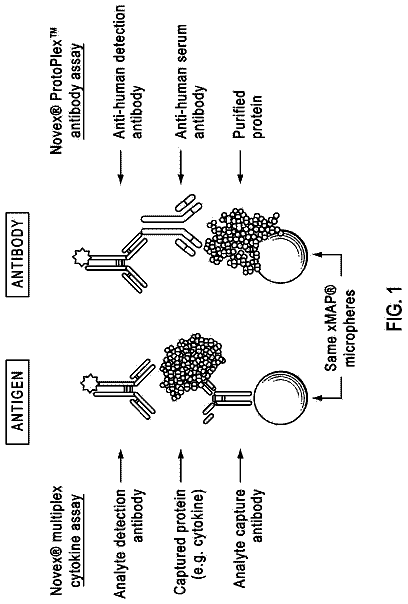
1 of 65 drawing sheets so far from the published document, cropped to the drawing. Every sheet is in the USPTO PDF.
What the patent claimed, word for word. All of it is now free to use.
The present invention relates to a new screening methodology and diagnostic and therapeutic products derived therefrom. In particular, new large bead-based libraries containing a rich assortment of small molecules, peptides, peptoids and/or other oligomers are used to screen biological samples for disease related biomarkers. The invention allows for both rapid and direct screening of plasma, serum or other biological fluid to find disease associated antibodies in a host of diseases and further finds antibody-specific molecules, which can serve as diagnostic tools or as therapeutics for said diseases. Diagnostic kits containing such antibody specific molecules can be prepared for virtually any disease state that has an antibody or immunogenic component such as autoimmune diseases, central nervous system disorders and cancer. Such kits can be made from virtually any known support system provided said system can support or bind the antibody specific molecule such as a peptoid or other ligand binding moiety. Similarly, any known detection method including ELISA or other known detection means can be used to detect the antibody subsequent to either the initial screening to find putative hits and/or after a diagnostic screen using such putative hits in the diagnostic assay. Such methods may also be used to screen for other biomarkers including proteins and/or other biomolecules on the surface of cells to distinguish between cells expressing disease related markers versus healthy cells not expressing such markers.
U.S. patent publication 2007/0003954 discloses protein and antibody profiling using small molecule microarrays. The application discloses ligands, which bind to ligand binding moieties wherein the ligands are arranged in arrays of synthetic molecules, which are used to screen for biomarkers and molecular fingerprints. The specific arrays described therein include, for example, a peptoid microarray having 7680 different compounds bound to the array. In that disclosure, bead based libraries were utilized as the initial means to make peptoids which were then transferred to microarrays with addressable locations on the microarray to screen biological fluids. The screening results in a unique pattern or molecular fingerprint on the array for any particular protein in a complex biological mixture. U.S. patent application 2010/0303805, hereby incorporated by reference, discloses certain peptoids and diagnostic arrays useful in screening biological fluids for biomarkers associated with central nervous system disorders. The specific monomers disclosed therein utilized to form the arrays therein may also be utilized in the new screening methodology of the present invention provided the libraries are enlarged to a much greater number of beads/peptoids or beads/ligands—e.g., between greater than 100K to 150 MM.
The present inventors have found that significantly larger bead based libraries (relative to microarray based screens for antibody biomarkers or bead based screens for cells) can, under the right conditions, be used to directly screen complex biological samples to find disease associated biomarkers as well as a significantly larger pool of ligands which bind to such ligand-binding moieties. This significantly larger pool includes a significantly improved number of high affinity ligands that serve as diagnostic tools as well as potential therapeutics. This approach also permits a significantly improved screening rate for any particular complex biological fluid because the need to make microarrays or similar addressable support systems is obviated in the first instance. Once the screening is performed, microarrays or other support systems including diagnostic arrays comprising the hits found in the screen may be manufactured and are included within the scope of this invention.
The present invention relates to a composition or a plurality of “compositions” comprising a large, random bead-based library of ligands. The library or plurality of libraries that form any particular “composition” is selected to screen for a target disease or condition and each library may be used to screen against a different disease or condition or the same libraries may be used to screen multiple disease states or conditions. The term “random” includes those libraries that have a rich assortment of side chains on the mono-substituted amines that form any particular monomer in an oligomeric chain. This assortment of R groups on the amine starting material is “random” even if some of the chemical and/or physical features on any particular monomer such as functionality/solubility are part of a desired feature or characteristic of the target oligomers. For plasma based screens or serum screens, for example, it is desired that any particular ligand bound to a bead has solubility characteristics which facilitate interaction, in solution, with a ligand binding moiety such as an antibody. In addition, the size of the oligomer is also a feature that is considered when forming a library of ligands that can bind to a ligand-binding moiety when the target moiety is, for example, an antibody or protein. The bead-based library comprises beads or similar support structures (i.e., polymeric resins) having bonded thereto (or to a linker on such resin) a ligand selected from the group consisting of small molecules, peptides, peptoids, polysaccharides or any oligomer based compound including nucleic acids or modified nucleic acid moieties. In a preferred embodiment, the bead-based library comprises peptoids. The peptoids are oligomers having monomeric units of between 5 to 15 monomers linked covalently to form the oligomer. The oligomer may have additional moieties linked to a terminal end of the oligomer to bond to a support or to a linker which links the oligomer to the support. The oligomeric peptoids are generated using, for example, a hybrid combination of a typical solid state peptide synthesis merged with a sub-monomer synthetic approach and comprise glycine or carbon substituted glycine-like moieties having a mono-substituted amide wherein the substituent on the amide nitrogen or α-carbon is selected from a wide range of moieties depending upon the monosubstituted amine or glycine α carbon substituent utilized in the synthesis. The peptoid libraries may generally be prepared as described in, for example, Kodadek and Reddy, Proceedings of the National Academy of Sciences, Sep. 6, 2005, volume 102, No. 36 or as described herein. As referenced above, the mono-substituted amine pool is generally selected from a wide range of monomers. The size of the library can range from about 200,000 to 150 MM beads having said number of distinct ligands per bead. Alternatively, and depending upon the size of the bead or support, each support or bead may have more than one ligand per bead/support and the ligand(s) may be the same ligand or distinct ligands.
The beads/supports having the ligand(s) which comprise the library are then taken further in the process of the invention. The invention comprises a process for screening a biological fluid for disease associated biomarkers comprising the steps of screening a biological control sample and a biological diseased sample with at least one bead-based ligand library and finding disease associated biomarkers using such a screen. The invention comprises a process for screening a complex biological sample for the presence of a disease-associated biomarker, which comprises exposing said sample to a plurality of ligand-bearing supports wherein at least one ligand detectably binds to the disease associated biomarker. The invention further comprises a method of screening a complex biological sample for disease associated biomarkers comprising the steps of
exposing a random ligand library to a control sample to identify and remove any non-specific ligand hits and
exposing the remaining ligand library to a diseased sample to identify any ligands which bind to a disease associated biomarker in the diseased sample. In particular, the invention comprises a process for screening a biological sample for a disease associated biomarker, comprising
pre-treating a ligand-bearing bead (LBB) library with a suitable solvent to form treated LLBs;
exposing the treated LBBs to a normal control (NC) biological sample having control sample ligand binding moieties;
exposing the treated LBBs from the control sample to a Dynabead screen (iron tagged anti-IgG antibody) and removing the hits;
washing the remaining LBB library and exposing said library to an NC biological sample having any remaining control sample ligand binding moieties using quantam dot labeled secondary anti IgG antibodies and removing the hits;
washing the remaining LBB library and exposing said library to a biological sample from a patient having a disease;
exposing the treated LBBs from the diseased sample to a Dynabead screen and removing the hits;
washing the remaining LBBs and exposing said library to the biological sample from a patient having a disease;
adding quantam dot labeled secondary anti IgG antibodies to the washed LBBs and identifying the disease-associated ligand binding moieties bound to a ligand on the LBB and, optionally, after washing the Dynabeads from step (6), repeating step
using the Dynabead hits from step
and identifying the Dynabead Qdot hits. In a preferred embodiment, Tentagel beads (having embedded PEG linkers) are utilized in the preparation of the LBB. Alternative beads and/or particles and having different and/or optional linkers may also be utilized along with alternative detecting means. Beads may also be selected from, for example, Luminex beads. In a preferred process, the Dynabead steps are not utilized except as initial validation steps to confirm the Qdot hits.
Any or all of the separated hits from the step or steps identified above may be further characterized, chemically identified and synthesized as the same moiety or as a modified version thereof. In particular, the preferred characterization relates to taking the ligands on the LBB from step
and sequencing the particular oligomer or ligand bound to the biomarker or disease associated ligand-binding moiety. In a preferred embodiment, the ligand is a peptoid and the peptoid is sequenced to identify and/or confirm or reconfirm the identity of the putative diagnostic probe, which can be further utilized in a diagnostic kit or as the basis for a therapeutic drug or vaccine candidate depending upon the particular disease or condition. In a preferred embodiment, the ligand hit is sequenced, indentified and then resynthesized or synthesized in a larger scale using a bead or support based synthetic method to produce the identified/sequenced ligand. In this instance, the preferred synthetic ligand includes, for example, a cysteine amino acid as a functional ligand that is linked to a resin/bead or support (or to a linker on said support) and this amino acid is further treated with, for example, submonomers comprising bromoacetic acid and substituted amines having the selected R groups for the particular sequenced ligand (peptoid or α-substituted peptoid). The cysteine residue or other analogous amino acid residue provides a sulfhydryl, which can react with electrophilic groups on glass slides or supports. In a different manner, in the initial library formation and on tentagel beads or beads or resins without linkers, methionine is used as the first monomer to permit cleavage from the bead or resin following synthesis of the peptoid oligomer. The methionine forms part of the oligomer when cleaved from the resin or bead. Rink resins, on the other hand, have cleavage linkers or linkers that facilitate, under the right conditions, cleavage of the molecule from the linker and resin without also cleaving the other amide bonds in the oligomer.
The invention also comprises diagnostic kits using the ligands (or modified versions thereof) identified in the biological sample screening methodology. The power of the particular screen results in the rapid identification of a significant number of actual hits that are further utilized in such diagnostic kits. The term “rapid” in this instance means that the present process avoids the complicated and unnecessary step of building a microarray before having to analyze complex biological fluid, which thus results in a significant savings in time. In addition, the present method permits a much larger number of molecules to be screened against a complex biological fluid at any one time instead of being limited to a small number on a microarray. In addition to finding a significant number of hits, the discovered ligands include a significantly greater number of high affinity binders relative to those found using prior screening methodology, which did not directly screen or assay complex biological fluid(s) using such bead or particle based technology. Such ligands may be utilized in multiplex disease platforms comprising a first peptoid to screen for disease or condition A and at least one additional peptoid to screen for disease or condition B.
The range of diseases that may be screened for the presence of disease-associated antibodies or biomarkers in the organism of interest using the methodology of the invention includes virtually any disease and at any stage of development of the disease. The invention is useful in the diagnosis and progression of human and animal disease. Diagnostic kits comprising the ligands discovered and identified during the screening can comprise bead based diagnostic kits for point of care diagnostic screens and/or can comprise more elaborate diagnostic systems and/or multiplex systems built upon more complex support systems such as microarrays. In addition, diagnostic arrays built from the peptoids, α-substituted peptoids or ligands found in the initial screen may be used in clinical trials to identify or assist in identification of patient stratification and/or disease progression in any particular patient population or sub-population. It is envisioned that such data derived from the primary and/or secondary or tertiary screens herein may accelerate drug development and sub-group or individualized patient care using pharmaceuticals that are specifically designed for a stage or certain stages of a disease, including early stage disease. Similarly, diagnostic kits derived from the ligands discovered herein may be used to identify early stage disease progression and/or any stage of the disease progression based upon the biomarker profile. Such information can be used to assist the physician in prescribing the most appropriate medical care for the individual patient or group of patients.
Other features and advantages of the present invention will become apparent from the following detailed description examples and figures. It should be understood, however, that the detailed description and the specific examples while indicating preferred embodiments of the invention are given by way of illustration only, since various changes and modifications within the spirit and scope of the invention will become apparent to those skilled in the art from this detailed description.
The following drawings are part of the present specification.
FIG. 1 shows a basic chemical schematic of the preparation of a library of Tentagel beads (KN1B) used to screen Alzheimer's serum samples. Figure lA shows starting from a polystyrene bead having an amino group as the reactant (a PEG or equivalent or alternative linker may be formed between the bead and the terminal amino group). FIG. 1B shows the starting amino acid on the bead as methonine and which is then reacted to form the compound shown in B. FIG. 1C shows the submonomers (monomeric amines and haloacetic acids) utilized to form the oligomeric library of compounds.
FIG. 2 shows a basic chemical schematic of the preparation of a library of Tentagel beads (JC3B) also used to screen Alzheimer's serum samples. FIG. 1A shows starting from a polystyrene bead having an amino group as the reactant (a PEG or equivalent or alternative linker may be formed between the bead and the terminal amino group). FIG. 1B shows the starting amino acid on the bead as methonine and which is then reacted to form the compound shown in B. FIG. 1C shows the submonomers (monomeric amines and haloacetic acids) utilized to form the oligomeric library of compounds. JC3B was also used to screen pancreatic cancer serum (data not shown).
FIG. 3 shows a basic chemical schematic of the preparation of a library of Tentagel beads (JC4B) used to screen Alzheimer's serum samples. FIG. 1A shows starting from a polystyrene bead having an amino group as the reactant (a PEG or equivalent or alternative linker may be formed between the bead and the terminal amino group). FIG. 1B shows the starting amino acid on the bead as methonine and which is then reacted to form the compound shown in B. FIG. 1C shows the submonomers (monomeric amines and haloacetic acids) utilized to form the oligomeric library of compounds.
FIG. 4 shows a basic chemical schematic of the preparation of a library of Tentagel beads (JC5B) used to screen Alzheimer's serum samples. FIG. 1A shows starting from a polystyrene bead having an amino group as the reactant (a PEG or equivalent or alternative linker may be formed between the bead and the terminal amino group). FIG. 1B shows the starting amino acid on the bead as methonine and which is then reacted to form the compound shown in B. FIG. 1C shows the submonomers (monomeric amines and haloacetic acids) utilized to form the oligomeric library of compounds. JC5B monomers included Isobutylamine, 2-Methoxyethylamine, Diaminobutane, Furfurylamine, Cyclohexylamine, R-Methylbenzylamine, Piperonylamine and 4-(Aminoethyl) Benzenesulfonamide.
FIG. 5 shows a basic chemical schematic of the preparation of a library of Tentagel beads (JC7B) used to screen serum samples. FIG. 1A shows starting from a polystyrene bead having an amino group as the reactant (a PEG or equivalent or alternative linker may be formed between the bead and the terminal amino group). FIG. 1B shows the starting amino acid on the bead as methonine and which is then reacted to form the compound shown in B. FIG. 1C shows the submonomers (monomeric amines and haloacetic acids) utilized to form the oligomeric library of compounds.
FIG. 6 shows a schematic of the process of the invention to screen a complex biological sample using bead based libraries of peptoid ligands.
FIG. 7 shows normal control (NC) Dynabead hits after QDot addition in a peptoid library (JC3B) prepared to screen against an Alzheimer's normal control serum sample and Alzheimer's diseased serum sample. The hits were picked out and the remaining ligand bound beads were used in the disease based screen.
FIG. 8 shows the Tentagel bead screening of diseased serum from Alzheimer's patient blood samples after the NC hits were removed. The hits are shown in red, which is the Qdot secondary antibody bound to the disease associated biomarker (antibody) in the serum which is bound to a peptoid linked through a PEG linker to the bead.
FIG. 9 shows a reproducibility test which uses a normal control sample (NC 030093) after SDS wash and QDOT addition. The arrow shows which NC peptoid hits were picked to sequence.
FIG. 10 shows a reproducibility test which uses a normal control sample (NC 050047) after SDS wash and QDOT addition.
FIG. 11 shows a reproducibility test which uses a diseased sample after SDS wash and QDOT addition.
FIG. 12 shows the peptoid sequences of the putative hits selected from the Alzheimer's screen from the JC3B library. The C-terminus is on the right side of the sheet and the N-terminus is on the left side.
FIG. 13 shows the chemical structures of the preferred high affinity hits from the Alzheimer's screen from the JC3B library. In this example, the structures shown have a cysteine residue and were resynthesized after determining the structure of the initial hit in the preliminary screen. The JC3B library contained an analous peptoid but which had a methionine residue on the C-terminus and not a cysteine residue.
FIG. 14 shows a competition experiment between a high affinity ligand (ADTG1) in solution versus ADTG-1-ADTG-42 on a microarray support. The competition experiment shows that ADTG1 in solution bonded to the same antibody that would have bound to peptoids ADTG-1, ADTG14, ADTG24, ADTG25, ADTG31, ADTG35 and ADTG40 on the microarray. Similar experiments were conducted on each of the peptoids to find four sets of peptoids, which bound to four distinct Alzheimer's autoantibodies (data not shown).
FIG. 15 shows the four groups of distinct peptoids, which bind to different autoantibodies in the Alzheimer's screen. Each group on the figure has the higher affinity binder at the top.
FIG. 16A shows AD test data (blinded) for a pool of patients using P1aag1 (JC3B-1) peptoid and FIG. 16B shows test data (blinded) for the same pool of AD patients using P1aag2 (JC3B-21). Each peptoid is presented on a microarray.
FIG. 17A shows AD test data (blinded) for a pool of patients using P1aag3 (JC3B-7) peptoid and FIG. 17B shows test data (blinded) for the same pool of AD patients using P1aag4 (JC3B-5). Each peptoid is presented on a microarray.
FIG. 18A shows AD test data (blinded) for a pool of patients using P1aag5 (JC3B-R8) peptoid and FIG. 18B shows test data (blinded) for the same pool of AD patients using P1aag6 (JC3B-R12). Each peptoid is presented on a microarray.
FIG. 19A shows microarray data for ADP2 in the same pool of patients for the tests conducted using P1aag1-6. FIG. 19B shows comparative data using P1aag4 with the same set of patients. The data shows a clear correlation between the results achieved with a previously identified ADP2 and the newly identified P1aag4 in the same patient pool.
FIG. 20A shows microarray data for ADP3 in the same pool of patients for the tests conducted using P1aag1-6. FIG. 20B shows comparative data using P1aag2 with the same set of patients. The data shows a clear correlation between the results achieved with a previously identified ADP3 and the newly identified P1aag2 in the same patient pool.
FIG. 21 shows a validation of P1aag5 (putative hit 5 or JC3B-R8) on TentaGel beads in a comparison of diseased AD serum versus healthy control (pooled) at 40 ug/mL.
FIG. 22A shows the peptoid hits in the pancreatic cancer screen using QDot 655 and using the JC5B library. FIGS. 22B and C show reconfirmation of hits using QDot 655 (arrows point to hits).
FIG. 23 shows pancreatic peptoid hit validation and compares disease serum addition and detection with QDot 655 versus normal serum addition.
FIG. 24 shows hit validation by mixing AD markers and PC markers. The data shows that the PC marker was detected while there was no detectable antibody on the AD peptoid bead in the pancreatic cancer serum (Serum 1).
FIG. 25 shows the pancreatic cancer screen hit sequences from the JC3B library.
FIG. 26 shows the pancreatic cancer screen hit sequences from the JC5B library.
FIGS. 27A , B and C show the results of an SLE (Lupus) screen. A is normal control and B and C are SLE serum from two different groups 1 and 2. The arrows point to the hits.
FIG. 28 shows the SLE hits from the KN1B library. The C-terminus is on the right side of the sheet.
FIG. 29 shows a hit validation for peptoid KN1B-20. Group 1 is pooled diseased serum at a concentration of about 0.374 mg/mL (left picture)(the hits are shown with a red tinge on the bead). Non-diseased pooled serum (center picture) is provided at a concentration of about 0.378 mg/mL and the far right picture shows a no serum control.
FIG. 30 shows the binding/detection of one of the SLE (lupus) peptoids to ELISA plates using two different binding methods at different concentrations of peptoid using a fluorescein tag.
FIG. 31 shows a competition assay between plate bound KN1B-20-biotin-fluorescein versus free KN1B-20-biotin in solution at various concentrations. Signal dampening occurs as the concentration of free KN1B-20-biotin increases from equimolar concentrations of bound versus free.
FIG. 32 shows an ELISA plate having peptoid at various concentrations and clearly shows a difference between diseased serum (AD)(P column 1) and normal control serum (column 3) [1:200 doubling each well to 1:400, 1:800, 1:1,600, 1:3,200, 1:6,400, 1:12,800]. The arrow points to the 1:800 dilution in 1×TBST buffer. The peptoid concentration in the wells is 10 mM. FIG. 32 also shows validation of the TentaGel bead platform to distinguish between diseased and control sera.
FIG. 33 shows an ELISA plate with 10 mM ADP3 and at various dilutions of AD sera versus control sera. The arrow points to the 1:800 dilution.
FIG. 34 shows an ELISA plate with 10 mM SLE-KN1B-20 and at various dilutions of AD sera versus control sera. The arrow points to the 1:800 dilution.
FIG. 35 shows an AD serum ELISA graph using 10 mM ADP3 prepared in binding buffer at various serum dilutions. Separation between normal and diseased serum occurred over the dilution range of 1:200 through approximately 1:10,000. The starting dilutions were 1:200 (Group 1AD serum 0.394 mg/mL and non-diseased serum at 0.386 mg/mL).
FIG. 36 shows an SLE serum ELISA graph using 10 mM KN1B-20 prepared in binding buffer at various serum dilutions. Separation between normal and diseased serum occurred over the dilution range of 1:200 through approximately 1:10,000. The starting dilutions were 1:200 (Group 1 SLE serum 0.375 mg/mL and non-diseased serum at 0.396 mg/mL).
FIG. 37 shows FIG. 37 shows an SLE serum ELISA graph using 10 mM KN1B-20 prepared in DMSO at various serum dilutions. Separation between normal and diseased serum occurred over the dilution range of 1:200 through approximately 1:10,000. The starting dilutions were 1:200 (Group 1 SLE serum 0.367 mg/mL and non-diseased serum at 0.322 mg/mL).
FIG. 38 shows a FACS platform for Tentagel beads hits validation.
FIG. 39 shows the degree of separation between beads having an acetyl group and beads having a 2,5-dintrophenyl group (DNP) at various concentrations of sera (100 ug/mL to 1,000 ug/mL) and in response to treatment with an anti-DNP labeled secondary antibody. The Mean fluorescence intensity (MFI) separation was greatest at the higher dilution of 1,000 ug/mL sera.
FIG. 40 shows that there is a direct competition between free ethanolamine-DNP and the binding of DNP (on a plate) to anti-DNP antibody at 1,000 ug/mL sera concentration.
FIG. 41 shows ADP3 bound anti-antibody from pooled normal control sera and pooled AD sera. The data shows good separation at sera concentration ranges of 20 and 140 ug/mL using two different secondary antibodies (goat anti-human Dylight 649 and goat anti-human Alexa 647).
FIG. 42 shows ADP3 bound auto-antibody from normal control and AD sera after background subtraction at various sera concentration ranges. There is a significant degree of separation at most sera concentration ranges from less than 20 ug/mL to 120 ug/mL or greater.
FIGS. 43 and 44 show the structures of the SLE (lupus) resynthesized peptoid ligand hits.
FIG. 45 shows the preparation of ADP3 on 10 um Tentagel beads and the subsequent cleavage using CNBr along with a mass spectrometry reading of the lactone shown.
FIG. 46 shows ADP3 bound autoantibody from normal control and Alzheimer's disease sera at different concentrations. The beads were preblocked for 3 hours with 1×TBST and then detected using Goat anti-human Alexa 647 secondary antibody.
FIG. 47 shows the ADP3 bound autoantibody from normal control and Alzheimer's disease sera at different sera concentrations and also shows DNP values.
FIGS. 48 and 49 show ADP3 bound autoantibody from normal controls versus Alzheimer's disease sera using pre-blocking conditions such as E. coli lysate and lysine.
FIG. 50 shows a simple schematic of the preparation of and distinction between peptoids that are used in microarrays versus those peptoids that are placed on ELISA plates. Schematic for how peptoid microarrays are made: individual beads are segregated into the wells of microtiter plates and the peptoids are cleaved from the beads to make a concentrated stock solution. Note that each well will now contain a single kind of peptoid. Several thousand peptoids are then spotted onto chemically-modified glass microscope slides in such a way that they bind covalently to the surface. Several thousand slides can be produced highly reproducibly from a single synthetic library. The ELISA production is similar except that there is no PEG chain on the surface but the density of peptoids on the ELISA plate may be different than it is on the microarrays.
FIG. 51 shows ELISA experiments with a clear distinction between normal control and diseased serum at a serum dilution of 1:800 using horseradish peroxidase linked to a secondary antibody that detects the disease associated antibody-peptoid complex. The colorless substrate is added and changes color (blue) upon reaction with the bound HRP enzyme.
FIG. 52 shows titration data that compares various AD peptoids in an ELISA test at various serum dilutions of diseased serum (A) versus normal serum (B). There is no intensity of the signals in the normal serum but clear distinction and intensity of all of the AD peptoids as the concentration increases from 1:12,800 to 1:200.
FIG. 53 provides a diagram that validates the correlation between the clinical diagnosis of the unblinded sample set of AD patients at various stages of Alzheimer's disease (or not) versus the data obtained from the same patient serum samples (blinded) and which were screened against ADP3 peptoid to detect disease associated antibodies. The results shown are from a blinded study of plasma samples from Mayo Clinic Jacksonville. UND=Undecided. The plot was derived from taking a single serum concentration (1:800) dilution. A reading of >1 was considered positive, a reading between 1 and 0.7 was considered undecided and a reading below 0.7 was considered negative.
FIG. 54 provides a diagram that validates the correlation between the clinical diagnosis of the unblinded sample set of AD patients at various stages of Alzheimer's disease (or not) versus the data obtained from the same patient serum samples (blinded) and which were screened against the various AD peptoids (plot is average value of results of 9 peptoids) of the invention to detect disease associated antibodies. The results shown are from a blinded study of plasma samples from Mayo Clinic Jacksonville. UND=Undecided. The plot was derived from taking a single serum concentration (1:800) dilution. A reading of >1 was considered positive, a reading between 1 and 0.7 was considered undecided and a reading below 0.7 was considered negative.
FIG. 55 provides a diagram that validates the correlation between the clinical diagnosis of the unblinded sample set of AD patients at various stages of Alzheimer's disease (or not) versus the data obtained from the same patient serum samples (blinded) and which were screened against the various AD peptoids of the invention to detect disease associated antibodies. The results shown are from a blinded study of plasma samples from Mayo Clinic Jacksonville. UND=Undecided. The plot was derived from taking a single serum concentration (1:800) dilution. A reading of >1 was considered positive, a reading between 1 and 0.7 was considered undecided and a reading below 0.7 was considered negative. The data also shows performance on other dementias where MCI/depression samples are labeled and Lewis Body Dementia samples are marked as well. The data shows that at least three MCI patients have serum samples with detectable amounts above 1 of the antibodies captured by the AD selective peptoids of the invention.
FIGS. 56A-D provide data on that subset of samples from patients that have disagreements between the Opko Health peptoid diagnostic assay using multiple AD peptoids versus the clinical diagnosis after this information was provided when unblinded. FIG. 56A shows the data for peptoids ADP3 and others as shown for a patient that was diseased clinically but for which the Opko peptoid P1aag4 was below 1.0 (UND at a single point; Titration AD positive). All other Opko peptoids were positive for AD (i.e., above 1.0). FIG. 56B shows that all Opko peptoids were positive for disease associated antibodies in a patient that was currently diagnosed as normal (non-demented) suggesting pre-AD. FIG. 56C shows that none of the Opko AD peptoids showed an intensity above 1 at any dilution point in a patient that was clinically diagnosed with AD suggesting that this patient had some other form of dementia. FIG. 56D shows that in a clinically positive AD patient, multiple Opko AD peptoids were not positive for disease associated antibodies but two peptoids (P1aag6 and P1aag4) were positive, thus UND at a single point and UND even after titration.
FIG. 57 shows the cluster diagram generated from previous AD samples using a microarray spotted with ADP3. There is a clear correlation between diseased versus control in the microarray data and data generated using the ELISA platform. FIG. 57 also shows that the ADP3 peptoid is selected for disease associated antibodies associated with Alzheimer's disease and not Parkinsons or Lupus (SLE).
FIG. 58 provides a summary of ELISA analysis using a total of 106 serum samples tested.
FIG. 59 provides the chemical structures of P1aag7-9.
The present invention represents a significant advance in diagnostic and therapeutic discovery. In particular, the present inventor has discovered a screening methodology that eclipses prior approaches to the screening of complex biological samples. In particular, there is a need for improved methods of discovering disease associated biomarkers and for making diagnostic kits comprising high-affinity ligands for such biomarkers. The present invention relates to a method of screening for such biomarkers and for diagnosing disease and disease progression using the ligands to detect such biomarkers.
The invention comprises compositions which comprises particle based libraries of compounds selected from peptoids, peptides, oligomers, small molecules and any molecule naturally derived or synthetically made and which can be placed on a support system such as a bead or small particle. This “library” is then pretreated and exposed, under the right conditions and after exposure to a control plasma or serum sample to permit removal of non-selective ligands, to a complex biological fluid such as plasma or serum which is “screened” for the presence or absence of disease-associated biomarkers or other target biomarkers such as antibodies or proteins or other markers such as cell surface proteins. The blood samples or other biological fluid samples are taken from patients that may or may not have a particular disease and the results generated from the screen are compared to results taken from a control healthy patient or control diseased patient.
The primary screen results in a significant number of high-affinity ligands for any particular disease-associated biomarker such as an antibody. The invention further comprises a process for generating high affinity ligands which are useful in either a diagnostic setting for such disease state and/or are useful as ligands in their own right—e.g., as therapeutic vaccines or as drugs which can target said disease associated antibodies located in a particular region of the body or body tissue. Such drugs can be linked to other moieties such as chemotherapeutic agents or other agents that generate or can generate a localized immune response to remove and/or degrade auto-antibodies.
Alzheimer's Disease (AD) is a progressive and fatal brain disease that affects as many as 5.3 million Americans. AD destroys brain cells, causing problems with memory, thinking and behavior. These symptoms get worse over time, and ultimately the disease is fatal. Today, it is the sixth-leading cause of death in the United States and is the most common form of dementia, accounting for 50-70% of all dementia cases. Sadly, while treatments for symptoms exist, there is no cure.
Diagnosing Alzheimer's Disease is an empirical process that involves several types of evaluations and may take many days to weeks to complete. Evaluations include taking a detailed medical history and physical examination. In addition, standard laboratory tests including blood, urine and CSF tests are mainly designed to help eliminate other possible conditions. Neuropsychological testing, using a variety of tools to assess memory, problem-solving, attention, vision-motor coordination and abstract thinking, are also performed. Tests for depression should also be included. Finally, brain-imaging scans are recommended to rule out brain tumors or blood clots in the brain as the reason for symptoms. In sum, there is currently no single test that accurately diagnoses Alzheimer's Disease, with a definitive diagnosis of Alzheimer's possible only by examining brain tissue after death.
Parkinson's Disease (PD) is another degenerative disease of the brain (central nervous system) that often impairs motor skills, speech, and other functions. It affects movement (motor symptoms), but other typical symptoms include disorders of mood, behavior, thinking, and sensation (non-motor symptoms). Patient's individual symptoms may be quite dissimilar and progression of the disease is also distinctly individual. The symptoms of PD result from the loss (idiopathic or genetic, toxic or traumatic) of pigmented dopamine-secreting (dopaminergic) cells in the pars compacta region of the substantia nigra (literally “black substance”). These neurons project to the striatum and their loss leads to alterations in the activity of the neural circuits within the basal ganglia that regulate movement, in essence an inhibition of the direct pathway and excitation of the indirect pathway.
Diagnosis of PD presents similar if somewhat distinct challenges. When performing a neurologic examination to evaluate a patient with any movement disorder, the doctor should take a medical history and perform a physical examination. In addition, a neurologic exam is conducted to make a thorough evaluation of the nervous system, including observing aspects of the patient's movement, coordination and balance. Laboratory testing of the blood of patients with the symptoms typical of Parkinson's only rarely uncovers any abnormality. Electroencephalograms (EEG's) record some aspects of brain electrical activity, but they are not effective in spotting PD. The MRI and CAT scans of the brain produce remarkable and exquisite anatomic pictures, but the brains of people with PD disease appear normal even under this scrutiny because the changes associated with PD are microscopic and are not revealed by these scans. With no definitive diagnostic tests to provide specific answers, physicians must base their diagnosis of PD on judgment.
Thus, there remains a need for diagnostic procedures for both of these diseases and other neurological diseases that are (i) accurate and objective, (ii) simple and reproducible, and (iii) useful in both early and late stage case.
The description continues in the full USPTO document.
About 6,196 words. The USPTO PDF has it with every drawing.
Fees are due 3.5, 7.5 and 11.5 years after grant. This patent expired on October 31, 2025, so the fee marked "not paid" was the one that went unpaid.
DIAGNOSTIC AND TREATMENT METHODS USING A LIGAND LIBRARY
Filed Mar 2012 · published Oct 2012BIOMARKER DISCOVERY IN COMPLEX BIOLOGICAL FLUID USING BEAD OR PARTICLE BASED LIBRARIES AND DIAGNOSTIC KITS AND THERAPEUTICS
Filed Mar 2012 · published Oct 2012Biomarker discovery in complex biological fluid using bead or particle based libraries and diagnostic kits and therapeutics
Filed Mar 2012 · granted Oct 2017Earlier publications, parents and continuations. None of them can still be enforced, or this patent would not be listed.
Prior art cited by the examiner or applicant. Useful when you check your own idea for novelty.
Everything on this page comes from the documents linked above.

