Background
Systemic lupus erythematosus (SLE or “lupus”) is an autoimmune disorder that can affect multiple organs, including the heart, joints, skin, lungs, blood vessels, liver, kidneys, and nervous system. SLE diagnosis can be difficult because of varying symptoms which have different durations and effects on patients. This difficulty can cause lupus to be a fatal disease if misdiagnosed. SLE predominantly affects women of non-European descent, and is estimated to affect more than 250,000 patients in the United States. While SLE symptoms can be managed by immuno-suppression therapy, there are presently no cures.
While several clinical markers presently exist for SLE, these markers have poor sensitivity and specificity, limiting their effectives. Given the complexity of SLE diagnosis, the broad populations it impacts, and the number of treatments available and in development, effective biomarkers are needed both for diagnosis and to better stratify patient samples so that appropriate treatments can be administered to improve patient survival and quality of life.
Disclosed herein are biomarkers and methods that provide method for detecting and diagnosing SLE.
Brief summary
Currently no single test establishes the diagnosis of SLE. To help doctors improve the accuracy of diagnosis of SLE, eleven criteria were established by the American Rheumatism Association. These eleven criteria are closely related to the variety of symptoms observed in patients with SLE. When a person has four or more of these criteria, the diagnosis of SLE is strongly suggested. However, some patients suspected of having SLE may never develop enough criteria for a definite diagnosis. Other patients accumulate enough criteria only after months or years of observation. Nevertheless, the diagnosis of SLE may be made in some settings in patients with only a few of these classical criteria. Of these patients, a number may later develop other criteria, but many never do.
The eleven criteria conventionally used for diagnosing SLE include: malar over the cheeks of the face or “butterfly” rash; discoid skin rash; photosensitivity; mucus membrane ulcers; arthritis; pleuritis/pericarditis; kidney abnormalities; seizures (convulsions) and/or psychosis; blood count abnormalities; immunologic disorder; and positive for anti-nuclear antibodies.
Although these criteria serve as useful reminders of those features that distinguish lupus from other related autoimmune diseases, they are unavoidably fallible.
Accordingly, in some embodiments is provided a kit for diagnosing SLE comprising: a) a target antigen comprising an antigen of Table II or III or a fragment thereof comprising an epitope; and b) means for detecting binding of one or more molecules in a test sample to the target antigen.
In another embodiment, the disclosure provides a method of diagnosing systemic lupus erythematosus (SLE) in an individual suspected of having SLE comprising: a) contacting a test sample with one or more affinity reagents that specifically bind a cytokine b) contacting the test sample with an affinity reagent that specifically binds a human antibody, wherein the human antibody binds any one of the polypeptides found in Table II or Table III, or a fragment thereof comprising an epitope recognized by a human antibody, b) detecting binding of one or more of the affinity reagents using an immunoassay; c) detecting the binding in a sample from a control individual without SLE using an immunoassay and d) comparing the detectable binding found in the test sample to that of a control individual without SLE, wherein a detectable signal greater in the test sample relative to the control is indicative of SLE. In some embodiments, the immunoassay is a protoplex immune response assay.
In another embodiment, a mixture is disclosed comprising: a) one or more target antigens of Table II or III or a fragment thereof comprising an epitope; b) more than one affinity reagent and c) a test sample from an individual suspected of having SLE.
Further embodiments provide methods of prognosing and stratifying subjects with SLE utilizing the cytokines and antigens of Tables II and III.
Description of the drawings
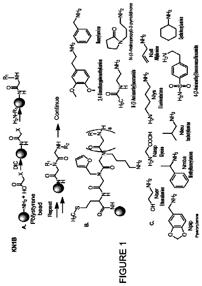
FIG. 1 is a schematic of the PROTOPLEX™ Immune Response Assay design with respect to the well-established Novex antigen detection immunoassay.
FIG. 2 is a graph of the immune response p-values and prevalence as calculated for each antigen (dots=known SLE markers; dots with concentric circle=novel markers). High reliability responses that displayed prevalence greater than 20% are within the box.
FIG. 3 is a table of 15 novel SLE antigens characterized for corresponding immune response using PROTOPLEX™ Immune Response Assay. Prevalence of response with SLE samples and controls were calculated. Significance was calculated using M statistics.
FIG. 4A-D is a series of graphs of sensitivity (y-axis) and specificity (x-axis) SLE biomarkers Ro52 (A), RNP complex (B), EIF2C1 (C), and HMGN1 (D). Area under the curve (AUC) was calculated for each marker.
FIG. 5A-C is a series of grids showing the sensitivity and specificity of new (A), established (B), and combined SLE markers (C).
FIG. 6A-E displays the distribution of serum cytokine levels: (A) IL-12, (B) IL2R, (C) IL-13, (D) Eotaxin, and (E) Rantes, among 177 human serum samples tested, healthy controls and individuals diagnosed with SLE.
FIG. 7A-7J displays the distribution of serum antibody levels against particular proteins among 177 human serum samples from healthy controls and individuals diagnosed with SLE. Proteins are FIG. 7(A) cardiolipin, FIG. 7(B) centromere Protein B (CENP B), FIG. 7(C) Jo-1, FIG. 7(D) transglutaminase, FIG. 7(E) Mi-2b, FIG. 7(F) myeloperoxidase, FIG. 7(G) Ro/SS-A, FIG. 7(H) smith-antigen (Sm), FIG. 7(I) thyroglobulin, and FIG. 7(J) thyroid peroxidase.
FIG. 8A-C is a series of matching matrices (also referred to as “confusion matrices”). These matrices highlight SLE predictive panels (A) cytokines; (B) antibodies to particular identified proteins; and (C) the combination of cytokines and antibodies to particular identified proteins). Sera of known diagnosis origin were analyzed for cytokine concentrations or antibody levels to specified antigens. True positive (upper left) and true negative (lower right) correspond to the noted predictive value of each panel.
Detailed description
The present disclosure provides biomarkers and a biomarker panel(s) highly indicative for systemic lupus erythematosus (SLE) and thereby permitting a more accurate assessment of whether an individual is suffering from SLE. Furthermore, the disclosed biomarker panel(s) can distinguish between an individual with SLE and another disease. For example, using the biomarker panel(s) disclosed herein, it is possible to distinguish between SLE and other diseases such as Crohn's disease, rheumatoid arthritis and multiple sclerosis. Therefore, the disclosed biomarker panel(s) may be used in the diagnosis of SLE.
SLE is a systemic autoimmune disease characterized by the production of autoantibodies directed against nuclear self-antigens and circulating immune complexes. This results in damage to various organs or systems, including skin, joints, kidneys and the central nervous system. Clinical manifestations of SLE can be diverse, and can include glomerulonephritis, dermatitis, thrombosis, vasculitis, seizures and arthritis. Because of this heterogeneity, the accurate and early identification of SLE is challenging.
Accordingly, the instant disclosure provides, in part, kits, and methods of their use, combining affinity reagents for the capture and/or detection of one or more cytokines and one or more antibodies that recognize particular antigens in a biological sample. The combination of the detection of cytokine(s) and antibody(ies) that recognize particular antigens being highly correlative with SLE.
By “affinity reagent,” “binding ligand,” “capture binding ligand,” “capture binding species,” “capture probe” or “capture ligand” herein is meant a compound that is used to detect the presence of or to quantify, relatively or absolutely, a target analyte or target species and that will bind to the target analyte or target species. Binding ligands include proteins, particularly including antibodies or fragments thereof as discussed further below.
Generally, the capture binding ligand allows the attachment of a target species or target sequence to a solid support for the purposes of detection as further described herein. Attachment of the target species to the capture binding ligand may be direct or indirect.
In some embodiments, the instant disclosure provides kits, and methods of their use, combining a first and a second affinity reagent for the capture and/or detection of a cytokine and an affinity reagent for the capture and/or detection of an antibody that specifically binds a polypeptide, or a fragment of, disclosed in Table II or Table III.
A “cytokine” is any secreted polypeptide that affects the functions of other cells, and is a molecule that modulates interactions between cells in the immune or inflammatory response. Cytokines include, but are not limited to, monokines, chemokines and lymphokines regardless of the cells that produce them. For instance a monokine is generally referred to as being produced and secreted by a mononuclear cell, such as a macrophage and/or monocyte but many other cells produce monokines, such as natural killer cells, fibroblasts, basophils, neutrophils, endothelial cells, brain astrocytes, bone marrow stromal cells, epideral keratinocytes, and B-lymphocytes. Lymphokines are generally referred to as being produced by lymphocyte cells.
Examples of cytokines include, but are not limited to, Regulated and Normal T Cell Expressed and Secreted (RANTES), epidermal growth factor (HGF), interleukin-2 (IL-2), interleukin-15 (IL-15), human growth factor (HGF), interleukin-7 (IL-7), macrophage inflammatory protein-1 alpha (MIP-1α), eotaxin, interleukin-17 (IL-17), interferon-α (IFN-α), interleukin-8 (IL-8), macrophage inflammatory protein-1 beta (MIP-1β). Fibroblast growth factor-basic (bFGF), interleukin-4 (IL-4), interferon gamma-induced protein 10 (IP-10), interferon-gamma (IFN-γ), interleukin-10 (IL-10), granulocyte colony-stimulating factor (G-CSF), interleukin-5 monocyte chemotactic protein-1 (MCP-1), interleukin-12 (IL-12), tumor necrosis factor-alpha (TNF-α), granulocyte-macrophage colony-stimulating factor (GM-CSF), interleukin-6 (IL-6), monokine induced by gamma interferon (MIG), interleukin-1 beta (IL-1β), interleukin-13 (IL-13) and vascular endothelial growth factor (VEGF) and regulated and normal T cell expressed and secreted (RANTES).
In some embodiments, the cytokine specifically bound by one or more affinity reagents is RANTES. RANTES, also referred to as chemokine (C-C motif) ligand 5 (CCL5), is a protein which in humans is encoded by the cc15 gene. Schlecker et al. (2012). The polypeptide sequence of RANTES is provided in SEQ ID NO:1 and appears as GenBank accession NP_002976.2.
TABLE-US-00001 (RANTES) SEQ ID NO: 1 MKVSAAALAVILIATALCAPASASPYSSDTTPCCFAYIARPLPRAHIKEY FYTSGKCSNPAVVFVTRKNR QVCANPEKKWVREYINSLEMS
In some embodiments, the one or more affinity reagents are an antibody. “Antibody” refers to a polypeptide substantially encoded by an immunoglobulin gene or immunoglobulin genes, or fragments thereof, which specifically binds and recognizes an epitope. The recognized immunoglobulin genes include the kappa and lambda light chain constant region genes, the alpha, gamma, delta, epsilon and mu heavy chain constant region genes, and a myriad of immunoglobulin variable region genes. Antibodies exist, for example, as intact antibodies or as a number of well characterized fragments produced by digestion with various peptidases. This includes, for example, Fab′ and F(ab)′.sub.2 fragments.
Thus, in some embodiments, a kit and a method of its use are provided, encompassing a first and a second antibody, wherein the first and the second antibody specifically bind RANTES and a third antibody that specifically binds an antibody that specifically binds any one of the polypeptides, or fragments thereof, provided in Tables II and III.
The term antibody also includes polyclonal antibodies and monoclonal antibodies. The terms “polyclonal antibody” and “polyclonal antibodies” as used herein refer to a heterogeneous mixture of antibody molecules which is capable of binding to or reacting with several different epitopes on the same antigen. Polyclonal antibody preparations isolated from the blood, milk, colostrum or eggs of immunized animals typically include antibodies that are not specific for the target antigen in addition to antibodies specific for the target antigen. Thus, the term “polyclonal antibody” as used herein refers both to antibody preparations in which the antibody specific for a particular antigen has been enriched, for example, by antigen affinity chromatography, and to preparations that are not so enriched.
Accordingly, in some embodiments, one or more antibodies are a polyclonal antibody or antibodies preparation. In other embodiments, one or more antibodies are monoclonal antibodies. A “monoclonal” antibody refers to an antibody produced by a single hybridoma (or clone thereof) or other cell line, or by a transgenic animal such that the monoclonal antibody will typically recognize one epitope on the antigen. A monoclonal antibody composition displays a single binding specificity for a particular epitope.
The term antibody, also includes antibody fragments either produced by the modification of whole antibodies or those synthesized de novo using recombinant DNA methodologies. Antibodies can be a variety of structures, including, but not limited to, bispecific antibodies, minibodies, domain antibodies, synthetic antibodies (sometimes referred to herein as “antibody mimetics”), chimeric antibodies, humanized antibodies, antibody fusions (sometimes referred to as “antibody conjugates”), and fragments of each, respectively.
In some embodiments, an antibody is provided that specifically binds the fragment crystallizable region of an autoantibody. The “Fragment Crystallizable region,” “Fc region,” “Fc domain,” “Fc” portion of an antibody refers to that portion of an immunoglobulin heavy chain that comprises one or more heavy chain constant region domains, C.sub.H1, C.sub.H2 and C.sub.H3, but does not include the heavy chain variable region.
In other embodiments is provided an antibody that specifically binds the constant region of the antibody light chain (kappa and/or lambda).
“Autoantibody” refers to antibody produced by the immune system of a subject that is directed to, and specifically binds to, a naturally occurring epitope in the subject.
In some embodiments, the subject is a human and an antibody that specifically binds an epitope naturally occurring in humans is detected; that is, the detected antibody is an autoantibody and the antigen bound by the autoantibody is an autoantigen. Thus, in some embodiments, a kit and a method of its use are provided, encompassing a first and a second antibody, wherein the first and the second antibody specifically bind RANTES and a third antibody specifically binds a human autoantibody. In some embodiments, the third antibody specifically binds an antibody that specifically binds any one of the polypeptides, or fragments thereof, provided in Tables II and III.
The antibody hypervariable region, also referred to as the complementary determining region (CDR) contributes to the formation of the antigen-binding, or more specifically, epitope binding site of antibodies. “Epitope” refers to a determinant that interacts with a specific antigen binding site in the variable region of an antibody molecule known as a paratope. Epitopes are groupings of molecules such as amino acids or sugar side chains and usually have specific structural characteristics, as well as specific charge characteristics. A single antigen may have more than one epitope.
Epitopes can be either conformational or linear. A conformational epitope is produced by spatially juxtaposed amino acids from different segments of the linear polypeptide chain. A linear epitope is one produced by adjacent amino acid residues in a polypeptide chain.
Conformational and nonconformational epitopes may be distinguished in that the binding to the former but not the latter is lost in the presence of denaturing solvents. An epitope typically includes at least 3, and more usually, at least 5 or 8-10 amino acids in a unique spatial conformation. Antibodies that recognize the same epitope can be verified in a simple immunoassay showing the ability of one antibody to block the binding of another antibody to a target antigen, for example “binning.”
An “antigen” as used herein refers to molecules such as nucleic acids, lipids, carbohydrates, proteoglycans, ribonucleoprotein complexes, protein complexes, proteins, glycoproteins, polypeptides, peptides, lipids, glycolipids, and naturally occurring or synthetic (in vitro) modifications of such molecules against which an immune response involving B lymphocytes can be generated. For each antigen, there exists more than one epitope.
As used herein, “target antigen” refers to a protein, or to a portion, fragment, variant, isoform, processing product thereof having immunoreactivity of the protein, that is used to determine the presence, absence, or amount of an antibody in a sample from a subject. A “test antigen” is a protein evaluated for use as a target antigen. A test antigen is therefore a candidate target antigen, or a protein used to determine whether a portion of a test population has antibodies reactive against it.
Use of the terms “target antigen” and “test antigen” is meant to include the complete wild type mature protein, or can also denote a precursor, processed form (including, a proteolytically processed or otherwise cleaved form) unprocessed form, post-translationally modified, or chemically modified form of the protein indicated, in which the target antigen, test antigen, or antigen retains or possesses the specific binding characteristics of the referenced protein to one or more antibodies of a test sample. The protein can have, for example, one or more modifications not typically found in the protein produced by normal cells, including aberrant processing, cleavage or degradation, oxidation of amino acid residues, atypical glycosylation pattern, etc. The use of the terms “target antigen” or “test antigen” also include splice isoforms or allelic variants of the referenced proteins, or can be sequence variants of the referenced protein, with the proviso that the “target antigen” and the “test antigen” retain or possess the immunological reactivity of the referenced protein to one or more autoantibodies of a test sample. Use of the term “target antigen” and “test antigen,” encompasses fragments of a referenced protein (“antigenic fragments”) that have the antibody binding specificity of the reference protein.
As used herein, the word “protein” refers to a full-length protein, a portion of a protein, or a peptide. Proteins can be produced via fragmentation of larger proteins, or chemically synthesized. Proteins may, for example, be prepared by recombinant overexpression in a species such as, but not limited to, bacteria, yeast, insect cells, and mammalian cells. Proteins to be placed in a protein microarray of the invention, may be, for example, are fusion proteins, for example with at least one affinity tag to aid in purification and/or immobilization. In certain aspects of the invention, at least 2 tags are present on the protein, one of which can be used to aid in purification and the other can be used to aid in immobilization. In certain illustrative aspects, the tag is a His tag, a GST tag, or a biotin tag. Where the tag is a biotin tag, the tag can be associated with a protein in vitro or in vivo using commercially available reagents (Invitrogen, Carlsbad, Calif.). In aspects where the tag is associated with the protein in vitro, a Bioease tag can be used (Invitrogen, Carlsbad, Calif.).
As used herein, the term “peptide,” “oligopeptide,” and “polypeptide” are used interchangeably with protein herein and refer to a sequence of contiguous amino acids linked by peptide bonds. As used herein, the term “protein” refers to a polypeptide that can also include post-translational modifications that include the modification of amino acids of the protein and may include the addition of chemical groups or biomolecules that are not amino acid-based. The terms apply to amino acid polymers in which one or more amino acid residue is an analog or mimetic of a corresponding naturally occurring amino acid, as well as to naturally occurring amino acid polymers. Polypeptides can be modified, for example, by the addition of carbohydrate residues to form glycoproteins. The terms “polypeptide,” “peptide” and “protein” include glycoproteins, as well as non-glycoproteins.
A “variant” of a polypeptide or protein, as used herein, refers to an amino acid sequence that is altered with respect to the referenced polypeptide or protein by one or more amino acids. Preferably a variant of a polypeptide has at least 60% identity to the referenced protein over a sequence of at least 15 amino acids. More preferably a variant of a polypeptide is at least 70% identical to the referenced protein over a sequence of at least 15 amino acids. Protein variants can be, for example, at least 80%, at least 90%, at least 95%, or at least 99% identical to referenced polypeptide over a sequence of at least 15 amino acids. Protein variants of the invention can be, for example, at least 80%, at least 90%, at least 95%, or at least 99% identical to referenced polypeptide over a sequence of at least 20 amino acids. The variant may have “conservative” changes, wherein a substituted amino acid has similar structural or chemical properties (for example, replacement of leucine with isoleucine). A variant may also have “nonconservative” changes (for example, replacement of glycine with tryptophan). Analogous minor variations may also include amino acid deletions or insertions, or both.
“Specific binding” or “specifically binds to” or is “specific for” a particular antigen or an epitope means binding that is measurably different from a non-specific interaction. Specific binding can be measured, for example, by determining binding of a molecule compared to binding of a control molecule, which generally is a molecule of similar structure that does not have binding activity. For example, specific binding can be determined by competition with a control molecule that is similar to the target.
Specific binding for a particular antigen or an epitope can be exhibited, for example, by an antibody having a KD for an antigen or epitope of at least about 10.sup.−4 M, at least about 10.sup.−5 M, at least about 10.sup.−6 M, at least about 10.sup.−7M, at least about 10.sup.−8 M, at least about 10.sup.−9 M, alternatively at least about 10.sup.−10 M, at least about 10.sup.−11 M, at least about 10.sup.−12 M, or greater, where KD refers to a dissociation rate of a particular antibody-antigen interaction. Typically, an antibody that specifically binds an antigen will have a KD that is 20-, 50-, 100-, 500-, 1000-, 5,000-, 10,000- or more times greater for a control molecule relative to the antigen or epitope.
Also, specific binding for a particular antigen or an epitope can be exhibited, for example, by an antibody having a KA or Ka for an antigen or epitope of at least 20-, 50-, 100-, 500-, 1000-, 5,000-, 10,000- or more times greater for the epitope relative to a control, where KA or Ka refers to an association rate of a particular antibody-antigen interaction.
The phrase “selectively binds” to an antibody or “selectively immunoreactive with,” when referring to a protein or peptide, refers to a binding reaction that is determinative of the presence of the protein in a heterogeneous population of proteins and other biologics. Thus, under designated immunoassay conditions, the specified antibodies bind to a particular protein at least two times the background and do not substantially bind in a significant amount to other proteins present in the sample. For example, solid-phase ELISA immunoassays are routinely used to select antibodies specifically immunoreactive with a protein (see, for example, Harlow & Lane, Antibodies, A Laboratory Manual (1988), for a description of immunoassay formats and conditions that can be used to determine specific immunoreactivity). Typically a specific or selective reaction will be at least twice background signal or noise and more typically more than 10 to 100 times background.
In some embodiments, a kit and a method of its use are provided, encompassing a first and a second antibody, wherein the first and the second antibody specifically bind RANTES, a third antibody that specifically binds a human antibody that specifically binds anyone of the proteins, or fragments thereof provided in Tables II and III, and a purified antigen. In other embodiments, the human antibody is an autoantibody.
In other embodiments, a kit and a method of its use are provided, wherein the first and second antibodies that bind RANTES are monoclonal antibodies and wherein the first and second antibodies recognize distinct epitopes.
The terms “purified” and “isolated” as used herein, are synonymous, and refer to a material that is substantially or essentially free from other components. For example, in one embodiment, a recombinant protein is isolated or purified when it is free from other components used in the cloning reaction, or solid state synthesis, isolation or purity is generally determined using analytical chemistry techniques such as polyacrylamide gel electrophoresis, mass spectrometry, or high performance liquid chromatography (HPLC). In one embodiment, a polynucleotide, protein or peptide of the present invention is considered to be isolated when it is the predominant species present in a preparation. A purified protein, peptide or nucleic acid molecule of the invention represents greater than about 80% of the macromolecular species present, greater than about 90% of the macromolecular species present, greater than about 95% of the macromolecular species present, greater than about 96% of the macromolecular species present, greater than about 97% of the macromolecular species present, greater than about 98% of the macromolecular species present, greater than about 99% of the macromolecular species present in a preparation.
In some embodiments, a purified antigen is provided, wherein the purified antigen is transglutaminase. “Transglutaminase” refers to an enzyme family that catalyzes the formation of a covalent bond between a free amine group and the gamma-carboxamide group of protein or peptide bound glutamine. Members of the transglutaminase family include Factor XIII, keratinocyte transglutaminase, tissue transglutaminase, epidermal transglutaminase, prostate transglutaminase, TGM X, TGM Y, TGM Z and erythrocyte band 4.2. Korsgren et al. describe the amino acid sequences of erythrocyte band 4.2, guinea pig liver transglutaminase and the a subunit of human factor XIII and the nucleic acid sequence for erythrocyte band 4.2. Proc. Nat'l Acad. Sci. USA
87:613-617. The amino acid sequence of each of these is as follows:
TABLE-US-00002 (erythrocyte band 4.2) SEQ ID NO: 2 RRGSVPILRQWLTGRGRPVYDGQAWVLAAVACTVLRCLGIPARVVTTFAS AQGTGGRLLIDEYYNEEGLQNGEGQRGRIWIFQTSTECWMTRPALPQGYD GWQILDPSAPNGGGVLGSCDLVPVRAVKEGTVGLTPAVSDLFAAINASCV VWKCCEDGTLELTDSNTKYVGNNISTKGVGSDRCEDITQNYKYPEGSLQE KEVLERVEKEKMEREKDNGIRPPS (guinea pig liver transglutaminase) SEQ ID NO: 3 SPMSWIGSVDILRRWKDYGCQRVKYGQCWVFAAVACTVLRCLAIPTRVVT NFNSAHDQNSNLLIEYFRNESGEIEGNKSEMIWNFHSLLGGVVDDQAGPG AWVRGVQALDPTPQEKSEGTYCCGPVPVRAIKEGHLNVKYDAPFVFAEVN ADVVNWIRQKDGSLRKSINHLVVGLKISTKSVGRDEREDITHTYKYPEGS EEEREAFVRANHLNKLATKEEAQEETG (a subunit Factor XIII) SEQ ID NO: 4 GSVDILLEYRSSENPVRYGQCWVFAGVFNTFLRCLGIPARIVTNYFSAHD NDANLQMDIFLEEDGNVNSKLTKDSVWNYHCWNEAWMTRPDLPVGFGGWQ AVDSTPQENSDGMYRCGPASVQAIKHGHVCFQFDAPFVFAEVNSDLIYIT AKKDGTHVVENVDATHIGKLIVTKQIGGDGMMDITDTYKFQEGQEEERLA LETALMYGAKKPLNTEG
In some embodiments, a kit and a method of its use are provided, encompassing a first and a second antibody, wherein the first and the second antibody specifically bind RANTES, a third antibody that specifically binds a human antibody that specifically binds a transglutaminase. In some embodiments, the human antibody specifically binds guinea pig liver transglutaminase. In other embodiments, the human antibody specifically binds a guinea pig liver transglutaminase epitope.
Arrays
The present disclosure is based, in part, on the identification of antibodies with reactivities associated with SLE. Serum samples from healthy individuals as well as patients with autoimmune diseases, were profiled on PROTOARRAY™ human protein microarrays (Invitrogen Corporation, Carlsbad, Calif.), described in more detail below, to identify multiple SLE-specific biomarkers. The extensive content of the arrays, including lower abundance proteins, native conformation, and insect cell-derived post-translational modifications, enabled the identification of biomarkers not previously known to be associated with SLE.
Microarrays, or other assay formats, containing these biomarkers are able to detect the presence of antibodies in a patient sample that bind the biomarkers, enabling the diagnosis and monitoring of the disease. Microarrays or other assays can contain specific biomarkers or a specific group of biomarkers, such as those associated with SLE.
As used herein, the term “array” refers to an arrangement of entities in a pattern on a substrate. Although the pattern is typically a two-dimensional pattern, the pattern may also be a three-dimensional pattern. In a protein array, the entities are proteins. In certain embodiments, the array can be a microarray or a nanoarray. A “nanoarray” is an array in which separate entities are separated by 0.1 nm to 10 μm, for example from 1 nm to 1 μm. A “microarray” is an array in the density of entities on the array is at least 100/cm.sup.2. On microarrays separate entities can be separated, for example, by more than 1 μm.
The term “protein array” as used herein refers to a protein array, a protein microarray or a protein nanoarray. A protein array may include, for example, but is not limited to, a “PROTOARRAY™,” protein high density array (Invitrogen, Carlsbad, Calif., available on the Internet at Invitrogen.com). The PROTOARRAY™ high density protein array can be used to screen complex biological mixtures, such as serum, to assay for the presence of autoantibodies directed against human proteins. Alternatively, a custom protein array that includes autoantigens, such as those provided herein, for the detection of autoantibody biomarkers, can be used to assay for the presence of autoantibodies directed against human proteins.
The term “protein chip” is used in the present application synonymously with protein array or microarray.
Biomarkers
Protein biomarkers used in a protein array of the present invention may be the full protein or fragments of the full protein. Protein fragments are suitable for use as part of the protein array as long as the fragments contain the epitope recognized by the antibodies. The required epitope for a given full protein can be mapped using protein microarrays, and with ELISPOT or ELISA techniques. It is understood that the antigen biomarkers provided by the present invention are meant to encompass the full protein as well as fragments thereof comprising an epitope. Typically, suitable protein fragments comprise at least 5%, 6%, 7%, 8%, 9%; at least 10%, 11%, 12%, 13%, 14%, 15%, 16%, 17%, 18%, 19%; at least 20%, 25%, 30%, 35%, 45%; or at least 50%, 60%, 70%, 80%, 90% of the full length protein amino acid sequence. In one embodiment of the present invention, protein fragments contain at least 6 contiguous amino acids; at least 10 contiguous amino acids; at least 20 contiguous amino acids; at least 50 contiguous amino acids; at least 100 contiguous amino acids; or at least 200 contiguous amino acids of the full length protein.
“Biomarker” refers to a biochemical characteristic that can be used to detect, diagnose, prognose, direct treatment, or to measure the progress of a disease or condition, or the effects of treatment of a disease or condition. Biomarkers include, but are not limited to, the presence of a nucleic acid, protein, carbohydrate, or combination thereof, associated with the presence of a disease in an individual. Further examples of biomarkers include antibodies and cytokines.
As used herein, a “biomarker detection panel” or “biomarker panel” refers to a set of biomarkers that are provided together for detection, diagnosis, prognosis, staging, or monitoring of a disease or condition, based on detection values for the set (panel) of biomarkers.
One embodiment of the disclosure is a method of detecting one or more target antibodies in a test sample of an individual suspected of having an autoimmune disease, such as SLE, comprising: a) contacting the test sample from the individual with one or more target antigens of Table II or III, or a fragment thereof comprising an epitope; and b) detecting binding of the one or more target antigens, wherein the binding of the one or more target antigens detects the presence of the one or more target antibodies in the test sample.
In a further embodiment, the test sample is contacted with 1, 2, 3, 4, 5, 6, 7, 8, 9, 10, 11, 12, 13, 14, 15, 16, 17, 18, 19, 20, 21 or 22 of the antigens of Table III or fragments thereof comprising an epitope. In a further embodiment, the quantitative amount of antibodies that bind to each biomarker is determined.
In a further embodiment, at least 1, or any combination of 2, 3, 4, 5, 6, 7, 8, 9, 10, 11, 12, 13, 14, 15, 16, 17, 18, 19, 20, 21, 22 or more antigen biomarkers must be bound by an antibody from the test sample to indicate the presence of an autoimmune disease.
Another embodiment of the present disclosure is a method of diagnosing SLE in an individual comprising: a) contacting a test sample from the individual with one or more target antigens, each comprising an antigen of Table II or III, or a fragment thereof comprising an epitope; and b) detecting binding of the one or more target antigens to one or more antibodies in the test sample, wherein the presence of the one or more antibodies bound against the one or more target antigens is indicative of SLE.
In a further embodiment, the test sample is contacted with 1, 2, 3, 4, 5, 6, 7, 8, 9, 10, 11, 12, 13, 14, 15, 16, 17, 18, 19, 20, 21 or 22 of the antigens of Table II or III or fragments thereof comprising an epitope. In a further embodiment, the quantitative amount of antibodies that bind to each biomarker is determined.
In a further embodiment, at least 1, or any combination of 2, 3, 4, 5, 6, 7, 8, 9, 10, 11, 12, 13, 14, 15, 16, 17, 18, 19, 20, 21, 22 or more antigens of Table II or III or fragments thereof comprising an epitope are bound by an antibody from the test sample to indicate the presence of SLE.
Another embodiment of the disclosure is a mixture comprising one or more antigen(s) of Table II or III, or a fragment thereof comprising an epitope; and a test sample from an individual suspected of having SLE. The mixture optionally further comprises a control antibody against one or more of the target antigens. In a further embodiment, the mixture comprises 2, 3, 4, 5, 6, 7, 8, 9, 10, 11, 12, 13, 14, 15, 16, 17, 18, 19, 20, 21, or 22 of the antigens of Table II or III, or fragments thereof comprising an epitope. The test sample includes, but is not limited to, cells, tissues, or bodily fluids from an individual.
In other embodiments of the disclosure is provided a mixture comprising a test sample, a first affinity reagent, a second affinity reagent, a third affinity reagent and a purified antigen. In some embodiments, one or more of the affinity reagents is an antibody. In still other embodiments, the purified antigen is a transglutaminase.
In some embodiments of the disclosure is provided a mixture comprising a test sample, a first antibody that specifically binds RANTES, a second antibody that specifically binds RANTES, a third antibody that specifically binds human antibodies and a purified antigen, wherein the antigen is a transglutaminase. In other embodiments, the transglutaminase is guinea pig liver transglutaminase.
The disclosure identifies proteins that are selectively recognized by antibodies that represent an important pool of novel candidates for potential diagnostic markers or therapeutic targets.
Methods of Diagnosing
The present invention provides biomarkers for SLE that are antibodies present in the sera of subjects diagnosed with SLE. The biomarker antibodies in the present invention are the antibodies displaying increased reactivity in individuals with an autoimmune disease, most likely as a consequence of their increased abundance. These antibodies can be detected by contact with antigens.
The methods of the present invention are carried out on test samples derived from patients, including individuals suspected of having an autoimmune disease and those who have been diagnosed to have a disease. A “test sample” as used herein can be any type of sample, such as a sample of cells or tissue, or a sample of bodily fluid, preferably from an animal, most preferably a human. The sample can be a tissue sample, such as a swab or smear, or a pathology or biopsy sample of tissue, including tumor tissue. Samples can also be tissue extracts, for example from tissue biopsy or autopsy material. A sample can be a sample of bodily fluids, such as but not limited to blood, plasma, serum, sputum, semen, synovial fluid, cerebrospinal fluid, urine, lung aspirates, nipple aspirates, tears, or a lavage. Samples can also include, for example, cells or tissue extracts such as homogenates, cell lysates or solubilized tissue obtained from a patient. A preferred sample is a blood or serum sample.
By “blood” is meant to include whole blood, plasma, serum, or any derivative of blood. A blood sample may be, for example, serum.
A “patient” is an individual diagnosed with a disease or being tested for the presence of disease. A patient tested for a disease can have one or more indicators of a disease state, or can be screened for the presence of disease in the absence of any indicators of a disease state. As used herein an individual “suspected” of having a disease can have one or more indicators of a disease state or can be part of a population routinely screened for disease in the absence of any indicators of a disease state.
The individual from whom the test sample is taken can be any individual, healthy or suspected of having an autoimmune disease, more particularly, the individual is being screened for SLE.
By “an individual suspected of having an autoimmune disease,” is meant an individual who has been diagnosed with SLE or who has at least one indicator of autoimmune disease, or who is at an increased risk of developing autoimmune disease due to age, environmental and/or nutritional factors, or genetic factors.
The phrase “diagnosis” as used herein refers to methods by which the skilled artisan can estimate and/or determine whether or not a patient is suffering from a given disease or condition. The skilled artisan often makes a diagnosis on the basis of one or more diagnostic indicators, that is, a marker, the presence, absence, or amount of which is indicative of the presence, severity, or absence of the condition, physical features (lumps or hard areas in or on tissue), or histological or biochemical analysis of biopsied or sampled tissue or cells, or a combination of these.
The description continues in the full USPTO document.
 Lapsed, fee not paid11 drawings