 Lapsed, fee not paid14 drawings
Medical Devices · US 8,714,154 B2
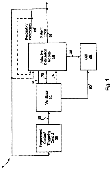
Systems and methods for automatic adjustment of ventilator settings
Various embodiments of the present disclosure provide systems, methods and devices for respiratory support.
US 8,714,742 B2 · Assignee: AMO Manufacturing USA, LLC · Inventors: Dai; Guang-Ming et al.
Sheet 1 of 45 from the published document. All sheets in the USPTO PDF
Methods and systems for changing a property of an eye. A property of the eye can be changed using an aspheric profile for increasing a depth of focus. Aspheric profile generation can include calculating a shape for increasing the focal depth to be rotationally symmetrical.
This invention generally relates to goal functions or visual function diagnostic metrics, and in particular provides methods, devices, and systems for mitigating or treating vision conditions such as presbyopia, often by determining a compound modulation transfer function. Presbyopia normally develops as a person ages, and is associated with a natural progressive loss of accommodation, sometimes referred to as "old sight." The presbyopic eye often loses the ability to rapidly and easily refocus on objects at varying distances. There may also be a loss in the ability to focus on objects at near distances. Although the condition progresses over the lifetime of an individual, the effects of presbyopia usually become noticeable after the age of 45 years. By the age of 65 years, the crystalline lens has often lost almost all elastic properties and has only limited ability to change shape. Res
1 of 45 drawing sheets so far from the published document, cropped to the drawing. Every sheet is in the USPTO PDF.
What the patent claimed, word for word. All of it is now free to use.
STATEMENT AS TO RIGHTS TO INVENTIONS MADE UNDER FEDERALLY SPONSORED RESEARCH OR DEVELOPMENT
Not Applicable
REFERENCE TO A "SEQUENCE LISTING," A TABLE, OR A COMPUTER PROGRAM LISTING APPENDIX SUBMITTED ON A COMPACT DISK
Not Applicable
This invention generally relates to goal functions or visual function diagnostic metrics, and in particular provides methods, devices, and systems for mitigating or treating vision conditions such as presbyopia, often by determining a compound modulation transfer function.
Presbyopia normally develops as a person ages, and is associated with a natural progressive loss of accommodation, sometimes referred to as "old sight." The presbyopic eye often loses the ability to rapidly and easily refocus on objects at varying distances. There may also be a loss in the ability to focus on objects at near distances. Although the condition progresses over the lifetime of an individual, the effects of presbyopia usually become noticeable after the age of 45 years. By the age of 65 years, the crystalline lens has often lost almost all elastic properties and has only limited ability to change shape. Residual accommodation refers to the amount of accommodation that remains in the eye. A lower degree of residual accommodation contributes to more severe presbyopia, whereas a higher amount of residual accommodation correlates with less severe presbyopia.
Known methods and devices for treating presbyopia seek to provide vision approaching that of an emmetropic eye. In an emmetropic eye, both distant objects and near objects can be seen due to the accommodation properties of the eye. To address the vision problems associated with presbyopia, reading glasses have traditionally been used by individuals to add plus power diopter to the eye, thus allowing the eye to focus on near objects and maintain a clear image. This approach is similar to that of treating hyperopia, or farsightedness.
Presbyopia has also been treated with bi-focal eyeglasses, where one portion of the lens is corrected for distance vision, and another portion of the lens is corrected for near vision. When peering down through the bifocals, the individual looks through the portion of the lens corrected for near vision. When viewing distant objects, the individual looks higher, through the portion of the bi-focals corrected for distance vision. Thus with little or no accommodation, the individual can see both far and near objects.
Contact lenses and intra-ocular lenses (IOLs) have also been used to treat presbyopia. One approach is to provide the individual with monovision, where one eye (usually the primary eye) is corrected for distance-vision, while the other eye is corrected for near-vision. Unfortunately, with monovision the individual may not clearly see objects that are intermediately positioned because the object is out-of-focus for both eyes. Also, an individual may have trouble seeing with only one eye, or may be unable to tolerate an imbalance between their eyes. In addition to monovision, other approaches include bilateral correction with either bi-focal or multi-focal lenses. In the case of bi-focal lenses, the lens is made so that both a distant point and a near point can be focused. In the multi-focal case, there exist many focal points between near targets and far targets.
Surgical treatments have also been proposed for presbyopia. Anterior sclerostomy involves a surgical incision into the sclera that enlarges the ciliary space and facilitates movement of the lens. Also, scleral expansion bands (SEBs) have been suggested for increasing the ciliary space. Problems remain with such techniques, however, such as inconsistent and unpredictable outcomes.
In the field of refractive surgery, certain ablation profiles have been suggested to treat the condition, often with the goal of increasing the range of focus of the eye, as opposed to restoring accommodation in the patient's eye. Many of these ablation profiles can provide a single excellent focus of the eye, yet they do not provide an increased depth of focus such that optimal distance acuity, optimal near acuity, and acceptable intermediate acuity occur simultaneously. Shapes have been proposed for providing enhanced distance and near vision, yet current approaches do not provide ideal results for all patients.
To evaluate the effectiveness of a refractive correction, such as with a spectacle lens, contact lens, intra-ocular lens, or laser refractive surgery procedure, it may be desirable to consider a merit function, or gauge of optical quality, that can determine such effectiveness. Gauges of optical quality are discussed in copending patent application Nos. 60/431,634, filed Dec. 6, 2002, 60/468,303, filed May 5, 2003, and 10/738,358 filed Dec. 5, 2003, the disclosures of which are hereby incorporated by reference. Merit functions may be used in evaluating post-corrective measurements, and in predicting the effect or outcome of a proposed corrective procedure. While the merit function may be objective, it may also desirable that the merit function have a good correlation with subjective test results such as visual acuity, contrast acuity, and the like. The following optical metrics can be or have been used as possible optical metrics or merit functions: high order (HO) root mean square (RMS) error; Strehl ratio; modulation transfer function (MTF) at specific spatial frequencies; volume under MTF surface up to a certain spatial frequency; compound MTF; encircled energy; and wavefront refractions. Other goal functions or visual function diagnostic metrics are available for characterizing lenses and other optical systems, including visual acuity such as log MAR, refractive error such as sphere and cylinder, and contrast sensitivity (CS). However, many of the currently used goal functions are difficult and cumbersome to implement with current clinical methods, and are insufficient in utilizing currently available clinical data and in providing guidance to the administration and diagnosis of reported visual difficulties.
In light of the above, it would be desirable to have improved methods, devices, and systems for treatment and/or mitigation of optical defects, based on improved goal functions such as a compound modulation transfer function. The goal functions should be easily implemented with existing clinical data, and with clinical data that is currently being generated by present measurement techniques. Optionally, it would be desirable to have improved methods, devices, and systems for treatment and/or mitigation of presbyopia and other optical defects. It may be desirable to provide improved prescriptions in the form of practical customized or optimized prescription shapes for treating or mitigating vision conditions such as presbyopia in a particular patient.
The present invention provides devices, systems, and methods that use improved goal functions for mitigating or treating vision conditions in a patient. The goal function can reflect optical quality throughout a vergence range. The goal function may also comprise a ratio of an optical parameter of the eye with a diffraction theory parameter. Relatedly, the goal function may also comprise at least one parameter selected from the group consisting of Strehl Ratio (SR), modulation transfer function (MTF), point spread function (PSF), encircled energy (EE), MTF volume or volume under MTF surface (MTFV), compound modulation transfer function (CMTF), and contrast sensitivity (CS).
In one aspect, the present invention provides a method for determining an optical surface shape that mitigates or treats a vision condition of an eye of a particular patient. The method can include determining an optical surface shape for the particular patient using a set of patient parameters for the specific patient with a compound modulation transfer function (CMTF). The compound modulation transfer function can include a combination of modulation transfer functions (MTF's) at a plurality of distinct frequencies. In some aspects, the CMTF is normalized to a diffraction limited MTF. In related aspects, the MTF's at the plurality of distinct frequencies can be combined in a linear combination. In some aspects, a CMTF can be calculated according to the following formula
.times..times..alpha..times. ##EQU00001## where n is the number of MTF curves, .alpha..sub.i is the reciprocal of the ith diffraction-limited MTF, and h.sub.i is the ith MTF curve. In related aspects, a CMTF can be calculated according to the following formula F(.nu.)=(.alpha..sub.1MTF.sub.1+.alpha..sub.2MTF.sub.2+.alpha..sub.3MTF.s- ub.3)/3 where MTF.sub.1, MTF.sub.2, and MTF.sub.3 comprise MTF values ranging from about 5 cycles/degree to about 20 cycles/degree, from about 15 cycles/degree to about 45 cycles/degree, and from about 30 cycles/degree to about 75 cycles/degree, respectively. In some aspects, MTF.sub.1, MTF.sub.2, and MTF.sub.3 comprise MTF values of 10 cycles/degree, 20 cycles/degree and 30 cycles/degree, respectively. In still other aspects, weighting coefficients .alpha..sub.1, .alpha..sub.2, .alpha..sub.3 can be chosen so that 1/.alpha..sub.1, 1/.alpha..sub.2, 1/.alpha..sub.3 are the diffraction-limited MTF at these spatial frequencies, respectively. In yet other aspects, one MTF at a spatial frequency can correspond to one angular extend of features of targets, and the compound MTF can be calculated as linear combination of MTF at different spatial frequencies normalized by a diffraction-limited MTF. In some aspects, the CMTF can be used to predict visual outcome.
In yet other aspects, the CMTF can be calculated according to the following formula
.function..times..times..alpha..times..function. ##EQU00002## where nu is visual vergence and .alpha..sub.i, is the reciprocal of the i-th diffraction-limited MTF. In some aspects, the CMTF can include three MTF curves at 10, 20 and 30 cycles per degree. In further aspects, the CMTF can have a value of about 1, which can be an ideal case. In related aspects, the CMTF can have a value ranging from about 0.2 to about 0.3. In still further aspects, the CMTF can be calculated over a vergence of 3 diopters. In still further related aspects, the MTF's at the plurality of distinct frequencies can include MTF's at 10, 20, and 30 cycles per degree. In other related aspects, the MTF's at the plurality of distinct frequencies can include MTF's at 15, 30, and 60 cycles per degree. In some related aspects, the MTF's at the plurality of distinct frequencies can include MTF's at 30, 45, and 60 cycles per degree. In yet another related aspect, the MTF's at the plurality of distinct frequencies can include at least one MTF ranging from about 5 cycles/degree to about 20 cycles/degree, at least one MTF ranging from about 15 cycles/degree to about 45 cycles/degree, and at least one MTF ranging from about 30 cycles/degree to about 75 cycles/degree. In some aspects, the CMTF can be used in an optimization routine as a goal function. In still other related aspects, MTF.sub.1, MTF.sub.2, and MTF.sub.3 can include MTF values of 10 cycles/degree, 20 cycles/degree and 30 cycles/degree, respectively, and the vision condition can include presbyopia.
In one aspect, the present invention provides a method for treating or mitigating a vision condition of an eye in a particular patient. The method can include selecting a gauge of optical quality appropriate for the vision condition of the eye; inputting a set of patient parameters specific for the particular patient; determining an optical surface shape for the particular patient using a set of patient parameters for the specific patient with a compound modulation transfer function (CMTF), the compound modulation transfer function comprising a combination of modulation transfer functions (MTF's) at a plurality of distinct frequencies; and mitigating or treating the vision condition of the eye in the patient by administering to the patient a procedure selected from the group consisting of: ablating a corneal surface of the patient to provide a corneal surface shape that corresponds to the optical surface shape; providing the patient with a contact lens or spectacle lens that has a shape that corresponds to the optical surface shape; and providing the patient with an intra-ocular lens that has a shape that corresponds to the optical surface shape. The gauge of optical quality can include a compound modulation transfer function (CMTF) parameter.
In one aspect, the present invention can provide a system for establishing an optical surface shape that mitigates or treats a vision condition of an eye in a particular patient. The system can include an input that accepts a set of patient parameters; and a module that determines an optical surface shape for the particular patient based on the set of patient parameters, using a gauge of optical quality appropriate for the vision condition of the eye. The gauge of optical quality can include a compound modulation transfer function (CMTF) parameter, the compound modulation transfer function parameter based on a CMTF comprising a combination of modulation transfer functions (MTF's) at a plurality of distinct frequencies.
In one aspect, the present invention provides a system for reprofiling a surface of a cornea of an eye of a particular patient from a first shape to a second shape having correctively improved optical properties. The system can include an input that accepts a set of patient parameters; a module that determines an optical surface shape for the particular patient based on the set of patient parameters, using a gauge of optical quality appropriate for a vision condition of the eye; a processor that generates an ablation profile; and a laser system that directs laser energy onto the cornea according to the ablation profile so as to reprofile a surface of the cornea from the first shape to the second shape, wherein the second shape corresponds to the determined optical surface shape. The gauge of optical quality can include a compound modulation transfer function (CMTF) parameter, the compound modulation transfer function parameter based on a CMTF comprising a combination of modulation transfer functions (MTF's) at a plurality of distinct frequencies.
The present invention also provides improved devices, systems, and methods for mitigating or treating presbyopia and other vision conditions. The present invention can establish a prescription that mitigates or treats presbyopia in a particular patient. In some embodiments, an optically optimized shape may be generated based on patient data input. Typically, the shape will represent a compromise between improved near vision and improved distance vision. These optimized shapes can be derived numerically using input patient parameters such as pupil size, residual accommodation, and desired vergence. Presbyopia-mitigating shapes may be scaled (or otherwise varied) in response to patient data such as one or more pupil diameters. Appropriate scaling may be determined at least in part from prior patient data from patients having differing pupil sizes and/or differing shapes. Advantageously, presbyopia-mitigating prescriptions may be derived from, scaled using, and/or optimized to provide at least one desired optical power (and/or manifest power), often to provide a plurality of optical powers at differing viewing conditions, thereby taking advantage of changes in pupil size when viewing objects under differing viewing conditions such as at differing distances and lighting conditions.
In a first aspect, the invention provides a method for treating existing or potential presbyopia of a patient. The patient has an eye with a pupil, a change in viewing distance with the eye inducing a change in pupil dimension. The method comprises measuring a first dimension of the pupil at a first viewing distance, and determining a first desired power for the eye at the first viewing distance. A prescription for the eye is determined such that the prescription provides the first desired power when the pupil has the first dimension, and such that the prescription effects a desired change in power in response to the change in pupil dimension, the desired change in power mitigating the presbyopia.
In many embodiments, a rate of the desired change in power for the change in pupil dimension comprises from about 0.25 D/mm to about 5.0 D/mm. When the patient is about 45 years old or less, and the rate may comprise from about 0.25 D/mm to about 1.0 D/mm. When the patient is about 60 years old or less the rate may comprise from about 1.0 D/mm to about 5.0 D/mm. A second desired optical power for the eye may be determined at a second viewing distance. At least a third desired optical power for the eye may also be determined, each optical power having an associated viewing condition, with a rate of an incremental desired change in power for an incremental change in pupil size varying within a pupil size range of the patient. The change in pupil dimension of the patient may be measured by measuring a second pupil dimension of the pupil at the second viewing distance, and/or the rate of the desired change in optical power for the change in pupil dimension may be assumed to be consistent for a plurality of patients.
The eye may have a residual accommodation range, and the first desired power for the eye may be determined so that the eye adjusts within the residual accommodation range when viewing at the first viewing distance with the first desired optical power. Optionally, particularly when the patient is about 60 years old or less, the first desired power for the eye and/or the desired change in power may be adjusted in response to an anticipated shrinkage of the pupil with age and/or anticipated reduction of residual accommodation.
The prescription may be determined at least in part by iteratively optimizing a goal function, by scaling a refractive shape, and/or by analytically or numerically deriving an optical shape providing a plurality of desired optical powers at an associated plurality of viewing conditions.
In a system aspect, the invention provides a system for treating existing or potential presbyopia of a patient. The patient has an eye with a pupil, a change in viewing distance with the eye inducing a change in pupil dimension. The system comprises a pupilometer for measuring a first dimension of the pupil while the eye is viewing at a first viewing distance. A prescription generating module has an input accepting a desired power for the eye and the first dimension. The module determines a prescription for the eye providing a first desired power when the pupil has the first dimension, the prescription effecting a desired change in power in response to the change in pupil dimension. The desired change in power mitigates the presbyopia.
The prescription generating module may comprise an optimizer module that determines the prescription based on the pupil diameter and the desired power using a goal function appropriate for the presbyopia; a scaling module that scales a central portion of a prescription shape based on the pupil dimension such that the prescription shape ameliorates presbyopia, and such that the central portion has a dimension between about 0.35 and about 0.55 of the pupil dimension; and/or a prescription calculating module calculating a presbyopia-mitigating prescription for the eye in response to the pupil dimension and the change in pupil dimension so that the eye has the first desired power suitable for the first viewing distance and so that the eye has a second desired power for a second viewing distance. Optionally, a laser may impose the prescription on the eye, typically by ablating corneal tissue.
In another aspect, the invention provides a method for determining a prescription that mitigates or treats presbyopia in a particular patient. The method comprises selecting a goal function appropriate for presbyopia of an eye, inputting a set of patient parameters specific for the particular patient, and determining an optical shape for the particular patient appropriate for differing viewing conditions based on the set of patient parameters per the goal function so as to mitigate or treat the presbyopia in the patient.
The goal function may also be based on geometrical optics. Similarly, the goal function can be determined using ray tracing. In this context, the phrase `ray tracing` has a meaning identical to `geometrical optics`. The set of patient parameters can include at least one parameter selected from the group consisting of pupil size, residual accommodation, power need, and vergence. In this context the phrase "power need" has a meaning identical to "vergence."
The prescription may comprise an optical shape determined by inputting a set of patient parameters specific for the particular patient into an optimizer. The shape is derived for the particular patient per a goal function so as to mitigate or treat the presbyopia in the patient. An initial optical shape can be input, the initial shape often being radially symmetric. Relatedly, the radially symmetric shape may be decomposed into a set of polynomials having at least two independent variables. Further, one of the at least two independent variables can be the ratio of the customized shape diameter to pupil diameter. The iterative optimization may be selected from the group consisting of Downhill Simplex method, Direction set method, and Simulated Annealing method, or the like. The set of patient parameters can include at least one parameter selected from the group consisting of pupil size, residual accommodation, and power need.
Optionally, the presbyopia may be treated by administering to the patient a procedure selected from the group consisting of ablating a cornea of the patient to provide a corneal shape that corresponds to the optical shape, providing the patient with a contact lens or spectacle lens that has a shape that corresponds to the optical shape, and providing the patient with an intra-ocular lens that has a shape that corresponds to the optical shape. The optical shape may be determined based at least in part on an expansion such as a regular polynomial (Even-Power-Term polynomials ("EPTP") or non-EPTP), a Zernike polynomial, a Fourier series, and a discrete shape entirety. The expansion may be a 3rd order or 4th order non-EPTP expansion, or a 6th or 8th order EPTP expansion. The optical shape may be determined based at least in part on a presbyopia-add to pupil ratio (PAR), the PAR ranging from about 0.2 to about 1.0.
In another system aspect, the present invention provides a system for establishing a prescription that mitigates or treats presbyopia in a particular patient, where the system includes an input that accepts a set of patient parameters, and a module that determines an optical shape for the particular patient based on the set of patient parameters, using a goal function appropriate for presbyopia of an eye.
The module may include data processing software and/or hardware, and may be optionally integrated with other data processing structures. The module may comprise an optimizer module that determines the prescription for the particular patient based on the set of patient parameters, using a goal function appropriate for presbyopia of an eye. A processor may generate an ablation profile, and a laser system can direct laser energy onto the cornea according to the ablation profile so as to reprofile a surface of the cornea from the first shape to the second shape, the second shape corresponding to the determined optical shape. Pupil diameters may be measured for input under one or more of the following conditions: when focusing on a near object; when focusing on a distant object; under photopic conditions; under mesopic conditions; under scotopic conditions. The prescription shape may be aspherical when the central portion of the prescription shape is aspherical; the prescription shape may be spherical when the central portion of the prescription shape is spherical; the prescription shape may be aspherical when the central portion of the prescription shape is spherical; and/or the prescription shape may be spherical when the central portion of the prescription shape is aspherical, with healing and LASIK flap effects and the like optionally varying the final shape of the eye. The dimension of the prescription shape central portion may comprise a diameter of the central portion and may remain within a range between about 0.4 and about 0.5 of the pupil diameter of the particular patient, or within a range between about 0.43 and about 0.46 of the pupil diameter of the particular patient; a power of the central portion is optionally between about 1.5 diopters and about 4.0 diopters (ideally being about 3.1 diopters).
In another aspect, the invention provides a method for treating presbyopia of an eye of a patient. The method comprises identifying a first pupil size of the eye under a first viewing condition. A second pupil size of the eye is identified under a second viewing condition. A presbyopia-mitigating prescription is calculated for the eye in response to the pupil sizes so that the eye has a first power suitable for the first viewing condition at the first size and so that the eye has a second power suitable for the second viewing condition at the second size.
Calculating the prescription may comprise determining a first effective power of the eye with the first pupil size and calculating a second effective power of the eye with the second pupil size. The first and second pupil diameters may be measured from the eye of the patient while the eye is viewing with the first and second viewing conditions, respectively. The prescription often comprises a prescription shape, and the method may include altering the refraction of the eye according to the prescription shape. The refraction of the eye can be altered using at least one of a laser, a contact lens, an intraocular lens, and a spectacle. One or more additional pupil diameters of the eye may be determined under one or more associated viewing condition, and the prescription can be calculated so that the eye has appropriate powers suitable for viewing at each additional viewing condition.
The prescription may be derived by determining at least one coefficient of a set of Zernike polynomials. Calculating the prescription often comprises determining a plurality of selected Zernike coefficients of spherical aberration at various orders. The eye at the first viewing condition may be viewing at a first viewing distance, and the eye at the second viewing condition may be viewing at a second viewing distance which is less than the first distance, with the second power being more negative than the first power. The eye at the first viewing condition can have a power between 0.25D and -0.25D, and the eye at the second viewing condition may have a power between -0.5D and -3.0D.
In another aspect, the invention may comprise a method for deriving a prescription for an eye. The method comprises determining a polynomial expansion from a wavefront of an eye, and calculating a plurality of effective powers based on a plurality of expansion coefficients of the polynomial expansion at different viewing pupil sizes. The prescription may be generated so as to provide a plurality of desired effective powers at said pupil sizes.
In yet another aspect, the invention provides a method for determining an effective power of an eye under a viewing condition. The method comprises determining a plurality of coefficients of a Zernike polynomial expansion from a wavefront of an eye while the eye has a first pupil size, and determining a second pupil size of the pupil under the viewing condition. The effective power of the eye is calculated from at least one of the coefficients of the Zernike polynomial from a relationship between effective power and pupil size.
In yet another aspect, the invention provides a system for correcting refraction of an eye, the system comprising at least one input for a first pupil size of the eye under a first viewing condition and a second pupil size of the eye under a second viewing condition. A prescription calculating module calculates a presbyopia-mitigating prescription for the eye in response to the pupil sizes so that the eye has a first power suitable for the first viewing condition at the first size and so that the eye has a second power suitable for the second viewing condition at the second size.
In another aspect, the invention provides a system for deriving a prescription for an eye, the system comprising a polynomial expansion module having an input for a wavefront of an eye and an output for a polynomial expansion. An effective power module has an input coupled to the output of the polynomial expansion module and an output. The effective power module determines an effective power from the polynomial expansion. A prescription module is coupled to the effective power module. The prescription module generates the prescription so as to provide a plurality of different desired effective powers at an associated plurality of different viewing pupil sizes.
In yet another aspect, the invention provides a system for determining an effective power of an eye under a viewing condition, the system comprising a first input for a plurality of coefficients of a Zernike polynomial expansion from a wavefront of an eye while the eye has a first pupil size. A second input accepts a second pupil size of the pupil under the viewing condition. An effective power calculating module calculates the effective power of the eye from at least one of the coefficients of the Zernike polynomial and a relationship between effective power and pupil size.
For a fuller understanding of the nature and advantages of the present invention, reference should be had to the ensuing detailed description taken in conjunction with the accompanying drawings.
FIG. 1 illustrates a laser ablation system according to an embodiment of the present invention.
FIG. 2 illustrates a simplified computer system according to an embodiment of the present invention.
FIG. 3 illustrates a wavefront measurement system according to an embodiment of the present invention.
FIG. 3A illustrates another wavefront measurement system according to an embodiment of the present invention.
FIG. 4A illustrates an example of the compound MTF (upper panel) versus its corresponding individual MTF curves at 15, 30, and 60 cycles per degree (lower panel).
FIG. 4B illustrate an example of the compound MTF (upper panel) versus its corresponding individual MTF curves at 10, 20, and 30 cycles per degree (lower panel).
FIG. 5 is a flow chart illustrating exemplary method steps for optimizing a optical prescription that treats or corrects a vision condition.
FIG. 6 illustrates a data flow process for shape optimization for correction or treatment of a vision condition.
FIG. 7 illustrates a comparison of Direction Set method and Downhill Simplex method.
FIGS. 8A and 8B illustrate alternative prescriptions optimized for an eye of a particular patient, and their characteristics.
FIG. 8C illustrates a comparison of optimizer values using even-term polynomials and all power term polynomials for pupil sizes of 4 mm, 5 mm, and 6 mm.
FIGS. 9A-D, show alternative presbyopia-mitigating prescriptions optimized for an eye of a particular patient.
FIG. 10 illustrates effects of random noise on prescriptions optimized for an eye of a particular patient.
FIGS. 11A-C compare optimized prescriptions to alternative treatments for differing pupil sizes.
FIGS. 12A-C compare optimized prescriptions to alternative treatments for a range of viewing distances.
FIG. 13 illustrates simulated viewing charts viewed at differing distances to compare optimized prescriptions to alternative treatments.
FIGS. 14-16 illustrate graphical interface computer screen displays for a prescription optimizer and system.
FIGS. 17 and 18 illustrate pupil sizes and changes at differing viewing conditions for a particular patient.
FIG. 19 graphically illustrates optimizer values for differing levels of residual accommodation.
FIG. 20 illustrates effects of pupil change and residual accommodation on optimized prescriptions for a particular patient.
FIGS. 21A-C illustrate effects of pupil change and residual accommodation on optimized prescriptions for a particular patient.
FIGS. 22-24 compare optical properties and results of eyes corrected with a optimized prescription to alternative treatments.
FIG. 25 schematically illustrates a system for determining a prescription for a particular patient and delivering that treatment using laser refractive surgery.
FIG. 26A illustrates a relationship between accommodation and pupil size when healthy eyes adjust to differing viewing distances.
FIG. 26B illustrates one exemplary relationship between effective power of an eye and pupil size for a patient, as can be provided from the presbyopia prescriptions of the present invention by generating an optical shape which effects desired changes in power with changes in pupil size of a particular patient under differing viewing conditions.
FIG. 26C illustrates a relationship between manifest power and pupil diameter, for example, as measured from patients having differing pupil diameters who have been successfully treated with a presbyopia-mitigating prescription. Such a relationship may be used to identify a desired change in optical power with changes in pupil diameter for a specific patient.
FIGS. 27A-B graphically illustrate optical properties of an eye relevant to presbyopia.
FIG. 28 schematically illustrates a presbyopia-mitigating shape having a central add region.
FIGS. 29 and 30 schematically illustrates residual accommodation and presbyopia treatments for increasing a focal range.
FIGS. 31-37 graphically illustrate results from presbyopia-mitigating treatments for a population of individual patients.
FIG. 38 graphically illustrates accommodation through a range of differing patient ages.
FIG. 39 schematically illustrates another system for determining a presbyopia-mitigating prescription for a particular patient and delivering that treatment using laser refractive surgery.
FIGS. 40 and 41 graphically illustrate a presbyopia-mitigating prescription derived so as to provide appropriate effective powers at two differing viewing conditions for a particular patient.
FIGS. 42 and 43 graphically illustrate a presbyopia-mitigating prescription derived so as to provide appropriate effective powers at three differing viewing conditions for a particular patient.
FIGS. 44 and 45 graphically illustrate a presbyopia-mitigating prescription derived so as to provide appropriate effective powers at four differing viewing conditions for a particular patient.
FIGS. 46A and 46B graphically illustrate different presbyopia-mitigating prescriptions which provide differing effective power variation characteristics during pupil size changes under differing viewing conditions.
FIGS. 47 and 48 graphically illustrate effects of different pupil sizes on derived presbyopia-mitigating prescriptions and their optical characteristics.
FIG. 49 illustrates simulated eye-chart letters as viewed with a presbyopic eye treated with a presbyopia-mitigating prescription derived for a particular patient.
FIGS. 50A and 50B illustrate an exemplary power/pupil correlation and corresponding presbyopia prescription.
Although the methods, devices, and systems of the present invention are described primarily in the context of a laser eye surgery system, it should be understood that the techniques of the present invention may be adapted for use in other eye treatment procedures and systems such as contact lenses, intra-ocular lenses, radial keratotomy, collagenous corneal tissue thermal remodeling, removable corneal lens structures, glass spectacles, corneal ring implants, and the like.
Turning now to the drawings, FIG. 1 illustrates a laser eye surgery system 10 of the present invention, including a laser 12 that produces a laser beam 14. Laser 12 is optically coupled to laser delivery optics 16, which directs laser beam 14 to an eye E of patient P. A delivery optics support structure (not shown here for clarity) extends from a frame 18 supporting laser 12. A microscope 20 is mounted on the delivery optics support structure, the microscope often being used to image a cornea of eye E.
Laser 12 generally comprises an excimer laser, ideally comprising an argon-fluorine laser producing pulses of laser light having a wavelength of approximately 193 nm. Laser 12 will preferably be designed to provide a feedback stabilized fluence at the patient's eye, delivered via delivery optics 16. The present invention may also be useful with alternative sources of ultraviolet or infrared radiation, particularly those adapted to controllably ablate the corneal tissue without causing significant damage to adjacent and/or underlying tissues of the eye. Such sources include, but are not limited to, solid state lasers and other devices which can generate energy in the ultraviolet wavelength between about 185 and 205 nm and/or those which utilize frequency-multiplying techniques. Hence, although an excimer laser is the illustrative source of an ablating beam, other lasers may be used in the present invention.
Laser system 10 will generally include a computer or programmable processor 22. Processor 22 may comprise (or interface with) a conventional PC system including the standard user interface devices such as a keyboard, a display monitor, and the like. Processor 22 will typically include an input device such as a magnetic or optical disk drive, an internet connection, or the like. Such input devices will often be used to download a computer executable code from a tangible storage media 29 embodying any of the methods of the present invention. Tangible storage media 29 may take the form of a floppy disk, an optical disk, a data tape, a volatile or non-volatile memory, RAM, or the like, and the processor 22 will include the memory boards and other standard components of modern computer systems for storing and executing this code. Tangible storage media 29 may optionally embody wavefront sensor data, wavefront gradients, a wavefront elevation map, a treatment map, a corneal elevation map, and/or an ablation table. While tangible storage media 29 will often be used directly in cooperation with a input device of processor 22, the storage media may also be remotely operatively coupled with processor by means of network connections such as the internet, and by wireless methods such as infrared, Bluetooth, or the like.
The description continues in the full USPTO document.
About 6,048 words. The USPTO PDF has it with every drawing.
Fees are due 3.5, 7.5 and 11.5 years after grant. This patent expired on May 6, 2026, so the fee marked "not paid" was the one that went unpaid.
Compound modulation transfer function for laser surgery and other optical applications
Filed Aug 2004 · published Nov 2005Compound modulation transfer function for laser surgery and other optical applications
Filed Aug 2004 · granted Jan 2008COMPOUND MODULATION TRANSFER FUNCTION FOR LASER SURGERY AND OTHER OPTICAL APPLICATIONS
Filed Nov 2007 · published Jun 2008Compound modulation transfer function for laser surgery and other optical applications
Filed Nov 2007 · granted Jan 2009COMPOUND MODULATION TRANSFER FUNCTION FOR LASER SURGERY AND OTHER OPTICAL APPLICATIONS
Filed Dec 2008 · published Apr 2009Compound modulation transfer function for laser surgery and other optical applications
Filed Dec 2008 · granted Jan 2011COMPOUND MODULATION TRANSFER FUNCTION FOR LASER SURGERY AND OTHER OPTICAL APPLICATIONS
Filed Nov 2010 · published Jun 2011Compound modulation transfer function for laser surgery and other optical applications
Filed Nov 2010 · granted Oct 2011COMPOUND MODULATION TRANSFER FUNCTION FOR LASER SURGERY AND OTHER OPTICAL APPLICATIONS
Filed Sep 2011 · published Feb 2012Compound modulation transfer function for laser surgery and other optical applications
Filed Sep 2011 · granted Jul 2012COMPOUND MODULATION TRANSFER FUNCTION FOR LASER SURGERY AND OTHER OPTICAL APPLICATIONS
Filed Jun 2012 · published Oct 2012Compound modulation transfer function for laser surgery and other optical applications
Filed Jun 2012 · granted Jan 2013COMPOUND MODULATION TRANSFER FUNCTION FOR LASER SURGERY AND OTHER OPTICAL APPLICATIONS
Filed Dec 2012 · published Sep 2013Compound modulation transfer function for laser surgery and other optical applications
Filed Dec 2012 · granted May 2014Earlier publications, parents and continuations. None of them can still be enforced, or this patent would not be listed.
No US citations on record.
Everything on this page comes from the documents linked above.