 Lapsed, fee not paid8 drawings
Medical Devices · US 8,696,905 B2
Fluid concentrator
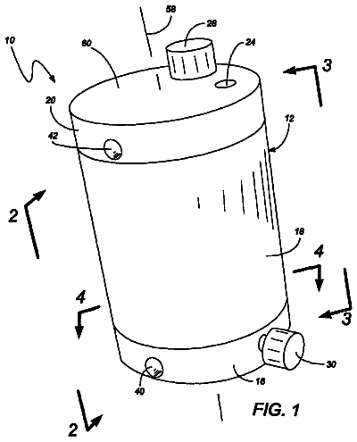
A concentrator is used for concentrating a fluid, particularly a plasma component out of blood, for treatment of a patient.
US 8,697,109 B2 · Assignee: Encore Health, LLC · Inventors: Garner; William et al.
Sheet 1 of 12 from the published document. All sheets in the USPTO PDF
Novel mercaptan compounds, particularly those including a photolabile protecting group, are described as well as methods of using the compounds for the prevention and treatment of ocular damage and disease.
As we age, our lenses undergo physiological changes that make it more difficult to focus on near objects. That is why nearly everyone requires reading glasses, even as early as age 35-40. The ability of the eye to change focal power, also known as accommodative amplitude, decreases significantly with age. The accommodative amplitude is 20 diopters in children and young adults, but it decreases to 10 diopters by age 25 and to <1 diopter by age 60. The age-related inability to focus on near objects is called presbyopia. All of us will develop presbyopia and will use corrective lenses unless a new treatment is found. In a healthy eye, the ciliary muscle can deform the lens via the suspensory ligaments to change the focal power of the eye. The lens takes on a different shape when the ciliary muscle is relaxed for near vision (FIG. 2A) than when the ciliary muscle is contracted for far vis
1 of 12 drawing sheets so far from the published document, cropped to the drawing. Every sheet is in the USPTO PDF.
What the patent claimed, word for word. All of it is now free to use.
As we age, our lenses undergo physiological changes that make it more difficult to focus on near objects. That is why nearly everyone requires reading glasses, even as early as age 35-40. The ability of the eye to change focal power, also known as accommodative amplitude, decreases significantly with age. The accommodative amplitude is 20 diopters in children and young adults, but it decreases to 10 diopters by age 25 and to <1 diopter by age 60. The age-related inability to focus on near objects is called presbyopia. All of us will develop presbyopia and will use corrective lenses unless a new treatment is found.
In a healthy eye, the ciliary muscle can deform the lens via the suspensory ligaments to change the focal power of the eye. The lens takes on a different shape when the ciliary muscle is relaxed for near vision (FIG. 2A) than when the ciliary muscle is contracted for far vision (FIG. 2B). When the ciliary muscle is relaxed, the central thickness is larger, and the equatorial circumference is smaller. Also, the lens nucleus is more posterior than central, and the space between iris and lens is larger. These changes involve the fibers of the lens cortex (C) because even by young adulthood, the lens nucleus (N) is incompressible.
The ciliary muscle, the suspensory ligaments, the posterior chamber of the aqueous humor, and the lens all must be considered when defining the etiology of presbyopia. Presbyopia may be caused, in part, by lens growth, oxidative stress, and disulfide bond formation.
The lens is a unique stratified epithelium where new fiber cells are laid down in shells throughout life. But the older fibers are not sloughed, so cross-sectional area, equatorial circumference, total volume, and weight increase all with age. The anterior part of each fiber cell must differ from that of the posterior because the anterior cortex is thicker than the posterior. Furthermore, elasticity decreases in the anterior cortex with age. Because of the change in lens size, the amount of force required to change its shape increases. The circumlental space decreases causing the posterior aqueous volume to decrease. This phenomenon is more pronounced in the temporal quadrant. There are also aging changes in the ciliary muscle and suspensory ligaments that contribute to the loss of lens deformability.
Although growth is a major contributor to the decreased deformability in the presbyopic lens, small changes in fiber membrane and/or cytoskeleton structure also play a role. Lens fiber plasma membranes are relatively stable and immobile due to the high levels of sphingomyelin and cholesterol that ranges from 50 percent in the cortex to 90 percent in the nucleus. While targeting lens membrane lipids may improve deformability, it may increase the risk for cataract because cataract is associated with decreased cholesterol.
The cytoskeleton is equally critical for fiber stability and elasticity. The lens fiber has actin microfilaments, a unique beaded intermediate filament, and microtubules, all of which are associated with the inner leaflet of the fiber plasma membrane. Disulfide bonds in intrinsic membranes and in membrane associated proteins increase with age in the non-cataractous human lens and in rodent lenses. Glutathione, believed to be the lens' major defense against oxidation, decreases with age and with distance from the lens surface. In other systems, glutathionylation of actin causes actin-microfilament depolymerization. Actin microfilaments are the most elastic of the cytoskeletal components.
Such disulfide bonds can also induce cataract. Oxidative stress can oxidize lens proteins, which destroys the balanced redox state required to maintain transparency. Thiolation of lens protein changes the tertiary structure of the protein, and more functional groups are exposed for further modification. The first line of defense, endogenously high levels of glutathione, fends off reactive oxygen species and keep lens proteins in a reduced state. As a second line of defense, intrinsic repair enzymes dethiolate the protein-thiol mixed disulfides or protein-protein disulfides induced by oxidative stress, thus keeping lens proteins thiols free and restoring lens proteins-enzyme function and activity.
With age, these protection and repair mechanisms against oxidative stress slowly deteriorate and become ineffective, resulting in a lens less able to counteract the effects of reactive oxygen species and other oxidants. Sulfhydrals are among groups most susceptible to oxidation. Sulfhydral groups may then undergo oxidation creating intra- and inter-molecular cross-links which increases with age in normal human lenses. These disulfide cross-links are present in water insoluble protein fractions. High molecular weight aggregates containing proteins and membrane particles with sizes over 5.times.10.sup.7 Da will scatter light. When a sufficient number of high molecular weight protein aggregates of this size or greater occur, transparency is lost and cataract occurs. Thus disulfide bond formation may be a cause of both presbyopia and cataract.
There is a need for compounds and methods for combating presbyopia and/or cataract. The compounds and methods described herein can be prophylactic and/or therapeutic for presbyopia and cataract by preventing or reducing disulfide bond formation in lens membranes and membrane associated proteins. The compounds may also affect one or more of lens growth, lens cystine and lipoic acid concentrations, cellular and lens fiber redox state, cellular elasticity, and lens transparency.
In one embodiment, a compound is provided that comprises an active agent X removably linked to at least one cage. The active agent can be lipoic acid or a derivative thereof. Specific exemplary active agents include 5-(1,2-dithiolan-3-yl)pentanoic acid; 5-(1,2-thiaselenolan-5-yl)pentanoic acid; 5-(1,2-thiaselenolan-3-yl)pentanoic acid; 6,8-dimercaptooctanoic acid; dihydrolipoate; 3,3'-disulanedylbis(2-aminopropanoic acid); 2-amino-3-mercaptopropanoic acid; 2-amino-3-hydroselenopropanoic acid; and salts and esters thereof.
The at least one cage can be, for example, a coumarin cage. In one embodiment, the cage is a 7-hydroxy-coumarin-4-ylmethyl-carboxyl group or a 6-bromo-7-hydroxy-coumarin-4-ylmethyl-carboxyl group. The at least one cage can be removably linked to at least one of a carboxylate group, an amino group (e.g., via --CO.sub.2), or a sulfur atom of the active agent.
In another embodiment, a pharmaceutical composition for ocular use comprises a compound as described above and a pharmaceutically acceptable carrier. In another embodiment, the pharmaceutical composition for ocular use comprises cystine or seleno-cystine, or a salt or ester thereof, optionally caged by a photolabile protecting group, and a pharmaceutically acceptable carrier. The pharmaceutical compositions can include, e.g., an emulsifier and a buffered carrier.
In another embodiment, a method comprises providing or administering a caged compound and uncaging the active agent, e.g., by applying light. A chemical energy source such as glucose of NADPH can optionally be additionally provided with the caged compound.
In one embodiment, the light is UVA light and has a wavelength of about 350 to 380 nm. The light can be applied to a localized region if desired.
The method can include administration to cells in vitro or in vivo and in either case, ocular cells. The compound can be administered via a topical ocular, subtenons, subconjunctival, intracameral, intravitreal, or iontophoresis route.
The method can be used to increase or maintain accommodative amplitude, as measured in diopters, to at least 2% greater than the accommodative amplitude expected in an untreated lens of about the same age. The method can increases accommodative amplitude by at least 0.25 diopters. The method can be used to increase or maintain lens elasticity, as measured in diopters or by elasticity E, to at least 2% greater than the elasticity expected in an untreated lens of about the same age. The method can be used to decrease or maintain lens opacity to at least 2% less than the opacity expected in an untreated lens of about the same age.
FIG. 1 is a pictorial step-wise depiction of an exemplary method as described herein. Step 1 represents delivery of a caged reducing agent by drops to the cornea. Step 2 represents allowing time to the agent to accumulate in the eye. Step 3 represents uncaging the agent at specific site(s) in the lens. Step 4 represents the agent reducing disulfide bonds in the specified site(s) in the lens. Step 5 represents allowing time for the remaining caged compound to disappear. Step 6 represents a softer lens that is better able to accommodate after site selective reduction.
FIG. 2 is a schematic of the changes in lens shape when the ciliary muscle is relaxed for near vision (FIG. 2A) and contracted for far vision (FIG. 2B).
FIG. 3 depicts the cellular transport mechanism of cystine (CSSC) and lipoate into lens epithelial cells and fibers using intracellular glutamate and the sodium gradient.
FIG. 4 depicts the glutathione concentration (FIG. 4A) and cystine concentration (FIG. 4B) in the eye.
FIG. 5 shows the products of photolysis for caged cystine (FIG. 5A) and caged lipoic acid (FIG. 5B). R.sub.1 is either H or Br.
FIG. 6 depicts the accommodative amplitude in diopters (D) of an untreated human lens as a function of age in years. Borja, D et al. 2008. Optical Power of the Isolated Human Crystalline Lens. Invest Opthalmol V is Sci 49(6):2541-8. Borja et al. calculated the maximum possible accommodative amplitude of each measured lens power data point (n=65). As shown, there is good agreement between the age-dependent loss of accommodation and the maximum amplitude of accommodation calculated from the isolated lens power.
FIG. 7 shows a trend graph of the shear modulus versus position in the lens and age. Weeber, H A et al. 2007. Stiffness gradient in the crystalline lens. Graefes Arch Clin Exp Opthalmol 245(9): 1357-66. The line at the bottom is the 20-year-old lens; the line at the top is the 70-year-old lens. The modulus increases with age for all positions in the lens. Measurements were taken up to 4.0 mm from the lens centre. The lines are extrapolated to a radius of 4.5 mm (lens diameter 9.0 mm).
FIG. 8 depicts the average opacity (opacimetry) of an untreated human lens as a function of age in years. Bonomi, L et al. 1990. Evaluation of the 701 interzeag lens opacity meter. Graefe's Arch Clin Exp Opthalmol 228(5):447-9. Lens opacity was measured in 73 healthy subjects between 10 and 76 years of age without slit-lamp evidence of cataract and with a visual acuity of 20/20. These subjects were classified into ten age groups. This study was carried out using the Interzeag Opacity Meter according to the procedure described by Flammer and Bebies (Flammer J, Bebie H.1987. Lens Opacity Meter: a new instrument to quantify lens opacity. Opthalmologica 195(2):69-72) and following the suggestions of the operating manual for the instrument.
FIG. 9 plots wavelength versus intensity (maximal absorption at 470 nm) for the two-photon uncaging of an exemplary compound.
FIG. 10 plots time versus intensity and describes the uncaging linear rate constant for the two-photon uncaging of an exemplary compound.
FIG. 11 depicts a scatter plot of the change in .DELTA.D (micrometers) in the absence (control) and presence of lipoic acid in lens organ culture experiments. The symbol .dagger-dbl. designates significantly larger changes in .DELTA.D when compared to controls. Statistical values are highly significant at p<0.00001 by unpaired t-test and by Kiruskal Wallis test, which compared medians of each data set. The relative change in Young's modulus (E) can be calculated as the cubic value derived from the .DELTA.D of the control divided by the .DELTA.D of the experimental or E fractional change=(.DELTA.D con/.DELTA.D exp)^3.
FIG. 12 depicts a scattergram of the percent of the total protein SH groups in disulfide bonds. Free SH groups were alkylated with 4-acetamido-4'-maleimidylstilbene-2,2'-sulfonic acid (c, 1 .mu.M, 5 .mu.M, 9.6 .mu.M, 50 .mu.M, 96 .mu.M) or 7-diethylamino-3-(4'maleimidylphenyl)-4-methyl coumarin (500 .mu.M, and 500 .mu.M c). Following removal of the first alkylating agent, the S--S bonds were reduced and alkylated with fluorescein-5-maleimide. Absorption spectra were used to calculated total protein (A280 nm), free protein SH (A322 or A384), and protein SS (A490) using the appropriate extinction coefficients. The symbol .dagger-dbl. indicates statistically significant difference of mean with mean of control (c, p<0.05). The symbol ** indicates means of 500 .mu.M lipoic acid and the 500 .mu.M control were significantly different from each other (p=0.027).
Compounds and methods are provided that can affect one or more of: disulfide bond formation in lens membranes and membrane associated proteins, lens growth, lens cystine and lipoic acid concentrations, cellular and lens fiber redox state, cellular elasticity, and lens transparency. These compounds and methods may thus effectively prevent or treat presbyopia and/or cataract.
In one embodiment, we provide a compound comprising an active agent X removably linked to at least one cage.
The active agent X is any agent capable of inducing a therapeutic effect as described above. Exemplary reducing agents include, but are not limited to, lipoic acid, cystine, glutathione, ascorbic acid, Vitamin E, tetraethylthiuram disulfyl, ophthalmic acid, inositol, beta-carbolines, reducing thiol derivatives, reducing sulfur derivatives, thiodisulfide exchange reaction agents such as dithiothreitol (DTT), trialkylphosphine, and tris[2-carboxyethyl]phosphine hydrochloride (TCEP.HCl), thioredoxin, and bis(mercaptoacetyl)hydrazine derivatives, as well as variations thereof. See, e.g., co-pending US 2008/0139990; see also U.S. Pat. Nos. 5,688,828 and 5,686,450. Preferably, the active agent is a reducing agent that is capable of reducing disulfide bonds, particularly disulfide bond formation in lens membranes and membrane associated proteins. Accordingly, particularly preferred active agents are capable of entering into the lens epithelial cells.
In one embodiment, the compound enters the lens epithelial cells using a naturally occurring transport mechanism. For example, lipoic acid and cystine enter lens cells via specific plasma membrane symporters and antiporters. The lens epithelium and fibers have transporters for lipoic acid and cystine that depend upon the sodium gradient for uptake into cells, where the agents are reduced to dihydrolipoic acid and cysteine, respectively (FIG. 3). By using lipoic acid- or cystine-based compounds, one can utilize a naturally occurring transport mechanism to deliver the agents to the lens cells.
The transport mechanism operates as follows: In the lens, there are three Na,K-ATPase isozymes that use the energy of ATP hydrolysis for the electrogenic exchange of three intracellular sodium ions (Na.sup.+) for two extracellular potassium ions (K.sup.+). The result is a gradient for Na.sup.+ where [Na.sup.+].sub.intracellular<[Na.sup.+].sub.extracellular and a gradient for K.sup.+ where [K.sup.+].sub.intracellular>[K.sup.+].sub.extracellular. The .alpha.2.beta.2 isozyme of Na,K-ATPase maintains the sodium gradient in lens fibers. The .alpha.1.beta.1 and .alpha.3.beta.2 Na,K-ATPase isozymes maintain the sodium gradient in the lens epithelium.
Excitatory amino acid transporters EAAT1-EAAT5 are responsible for uptake of glutamate in the lens. EAATs are high affinity sodium-dependent glutamate (GLU, FIG. 3) symporters whose efficiency is regulated by the sodium gradient's steepness, i.e., the steeper the Na.sup.+ gradient, the faster the uptake of extracellular glutamic acid. EAATs are highest in lens epithelium and cortical fibers and lowest, almost non-existent, in nuclear fibers.
The glutamate gradient created by the EAATs ([GLU].sub.intracellular>[GLU].sub.extracellular) is used for the exchange of extracellular cystine (CSSC, FIG. 3) via the glutamate-cystine antiporter (xCT). xCT is cytoplasmic in superficial lens cortical fibers and an integral membrane protein in lens nuclear fibers. This glutamate-cystine exchange is critical for the lens cell's oxidative status as demonstrated by mice deficient in xCT, which display redox imbalances. xCT requires the anionic deprotonated carboxyl groups of cystine for exchange.
The sodium-dependent multivitamin transporter (symporter), SC5A6 (SMVT), is responsible for lipoate influx into cells. Like the EAATs, SC5A6 is regulated by the steepness of the Na.sup.+ gradient. Again, the carboxylate anion is required for transport. Lens cortical fibers are one target for the lipoic acid-based compounds described herein because these cells express SC5A6 and its mRNA is found there.
In one embodiment, the active agent is lipoic acid, especially alpha-lipoic acid, or a derivative thereof. Lipoic acid-based active agents include, but are not limited to, 5-(1,2-dithiolan-3-yl)pentanoic acid (lipoic acid); 6,8-dimercaptooctanoic acid (dihydrolipoic acid); and dihydrolipoate. Lipoic acid not only advantageously utilizes a naturally occurring transport mechanism, but may also be effective to combat prevent and/or treat a wide variety of cell damage and/or disorder types.
Lipoic acid functions as an acyl group transferring factor in aerobic metabolism shuttling acyl groups between thiamine pyrophosphate (ThPP) and Coenzyme A (CoA). In the mitochondria, lipoic acid acts as a co-factor in the glycine cleavage, forming 2-oxoacid dehydrogenase, pyruvate dehydrogenase, branched chain oxoacid dehydrogenase and acetoin complexes. Lipoic acid is synthesized in the mitochondria. Lipoic acid levels are adequate in differentiating fibers containing mitochondria (superficial cortex), but fall precipitously in fibers of the deeper cortex where cellular organelles have been lost.
Exogenous lipoic acid, with a redox potential of -0.29V, acts as an antioxidant. In lenses treated with lipoic acid, the lens fibers in the process of losing mitochondria and those fibers devoid of mitochondria can benefit from the antioxidant properties of the agent.
Lipoic acid may also prevent symptoms associated with vitamin E deficiency, reverse H.sub.2O.sub.2-dependent inhibition of alpha-ketoglutarate dehydrogenase, and prevents reserpine-induced oxidative stress in the striatum, hyperglycemia-induced SH-group oxidation in erythrocyte membranes, and buthionine sulfoximine-induced cataract formation. Lipoic acid may also reverse or prevent age dependent memory loss and improve hemodynamic properties following ischemia-reperfusion in heart without improvement of cardiac electrophysiological properties. Lipoic acid changes cellular redox status, thus stimulating glucose uptake in adipocytes and skeletal muscle presumably by increasing plasma membrane levels of GLUT4. Finally, lipoic acid ameliorates insulin resistance in the Goto-Kakizaki rat model of type II diabetes.
In another embodiment, the active agent is cystine or a derivative thereof. Cystine-based active agents include, but are not limited to, 3,3'-disulanedylbis(2-aminopropanoic acid) (cystine) and 2-amino-3-mercaptopropanoic acid (cysteine). Like lipoic acid, cystine not only advantageously utilizes a naturally occurring transport mechanism, but may also be effective to combat prevent and/or treat a wide variety of cell damage and/or disorder types.
The cysteine/cystine redox couple (CSH/CSSC) plays multiple roles in oxidative stress response as well as acts as a source of cysteine for the synthesis of glutathione GSH. GSH is the electron-donating compound of the GSH/GSSG redox couple. In plasma, the redox state of the CSH/CSSC and GSH/GSSG couples vary diurnally. The redox states of these two couples do not vary concomitantly, i.e., each couple has a unique maximum and nadir. The intracellular redox potentials of GSH:GSSG and CSH:CSSC regulate cellular proliferation, differentiation, and programmed cell death. Total glutathione concentration (GSH+GSSG) is highest in the epithelium and superficial cortex as depicted by the darker area in FIG. 4A. Total Cys concentration (CSH+CSSC), on the other hand, is higher in the superficial cortex and lens nucleus than in the deep cortex, i.e., a bimodal distribution, as shown by the darker area in FIG. 4B. GSH is senses and protects against oxidative stress in the lens epithelium and superficial fibers. However, the low levels of GSH deeper in the cortex suggest a role for the GSH/GSSG redox couple in the regulation of mitochondrial disappearance during fiber differentiation. In the lens nucleus, the CSH/CSSC couple is most likely involved in protection against oxidative stress. The fibers of the deep cortex appear to be deficient in both the glutathione and cysteine couple.
The agents disclosed herein can be used to selectively increase lens cystine levels in deep anterior cortex, thereby beneficially changing the redox potential of extracellular and intracellular space, altering disulfide levels, decreasing lens stiffness, decreasing the potential for, or existence of, high molecular weight protein aggregates, and/or reducing cataract formation. These beneficial effects can be demonstrated in lens epithelial cell and lens organ culture models.
In another embodiment, the active agents include seleno-substituted agents. Without being bound by theory, it is believed that including selenium in the active agent can improve redox potential compared to the same agent without selenium. The selenium derivative can thus take advantage of the intracellular redox potential of the lens.
Accordingly, the active agent can be a lipoic acid or cystine derivative including selenium. In one embodiment, the active agent is a seleno-lipoic acid-based agent such as 5-(1,2-thiaselenolan-5-yl)pentanoic acid or 5-(1,2-thiaselenolan-3-yl)pentanoic acid. In another embodiment, the active agent is a seleno-cystine-based agent such as 2-amino-3-hydroselenopropanoic acid.
In one embodiment, the active agent is 5-(1,2-dithiolan-3-yl)pentanoic acid; 5-(1,2-thiaselenolan-5-yl)pentanoic acid; 5-(1,2-thiaselenolan-3-yl)pentanoic acid; 6,8-dimercaptooctanoic acid; dihydrolipoate; 3,3'-disulanedylbis(2-aminopropanoic acid); 2-amino-3-mercaptopropanoic acid; or 2-amino-3-hydroselenopropanoic acid.
In another embodiment, the active agent is 5-(1,2-dithiolan-3-yl)pentanoic acid; 5-(1,2-thiaselenolan-5-yl)pentanoic acid; 5-(1,2-thiaselenolan-3-yl)pentanoic acid; 6,8-dimercaptooctanoic acid; dihydrolipoate; 3,3'-disulanedylbis(2-aminopropanoic acid); or 2-amino-3-hydroselenopropanoic acid.
In one embodiment, the active agent is lipoic acid or a derivative thereof. For example, the active agent can be 5-(1,2-dithiolan-3-yl)pentanoic acid; 5-(1,2-thiaselenolan-5-yl)pentanoic acid; 5-(1,2-thiaselenolan-3-yl)pentanoic acid; 6,8-dimercaptooctanoic acid; or dihydrolipoate. In another embodiment, the active agent is 5-(1,2-dithiolan-3-yl)pentanoic acid or 6,8-dimercaptooctanoic acid. In yet another embodiment, the active agent is 5-(1,2-dithiolan-3-yl)pentanoic acid.
In another embodiment, the active agent is cystine or a derivative thereof. For example, the active agent can be 3,3'-disulanedylbis(2-aminopropanoic acid); 2-amino-3-mercaptopropanoic acid; or 2-amino-3-hydroselenopropanoic acid. In yet another embodiment, the active agent can be 3,3'-disulanedylbis(2-aminopropanoic acid) or 2-amino-3-hydroselenopropanoic acid.
The active agent can also be in a salt or ester form.
The active agent can be administered as a racemate or as an enantiomer. Lipoic acid and its derivatives are preferably administered to include the R form; cystine and its derivatives are preferably administered to include the L form. Synthetic methods to yield a racemate may be less expensive than stereo-specific processes including isolation/purification steps. On the other hand, administering a single enantiomer can lower the therapeutically effective amount, thus decreasing toxicity effects of both the active agent and the accompanying cage.
The active agent is removably linked to at least one cage. The active agent may be linked to one, two, three, or more cages depending on the particular structure of the active agent and the cage(s).
A "cage" as used herein means a photolabile protecting group. The cage is linked to the active agent and is capable of being removed from the active agent by the application of light energy. Preferably, the cage is linked to the active agent in such a way as to render the agent biologically inactive. The agent can be activated by applying light to remove the cage. Because the cage is readily removed by light, one can control the time and/or place of the agent's activity. Exemplary cages and synthetic methods useful for the compounds herein include, but are not limited to those disclosed by U.S. Pat. No. 6,472,541 and Kao, J P. 2006. Caged Molecules: Principles and Practical Considerations. Curr Protoc Neurosci. Ch. 6:Unit 6.20. Specific exemplary cages include, but are not limited to, (6-nitrocoumarin-7-yl)methyl; N-(o-nitromandelyl)oxycarbonyl; p-hydroxyphenacyl; 7-Nitroindolinyl; 4-methoxy-7-nitroindolinyl; .gamma.-(.alpha.-carboxy-2-nitrobenzyl); 6-bromo-7-hydroxycoumarin; naphthalene groups, e.g., (6-hydroxy-3-oxo-3,4-dihydronaphthalen-1-yl)methyl); quinoline-2-one; xanthene; thioxanthene; selenoxanthene; anthracene; and nitroso groups.
In one embodiment, the cage is a coumarin group. Coumarin cages can be advantageous in that they exhibit low toxicity. Also, the coumarin cages may be removed with light in the UVA range, as opposed to many other photolabile cages that can only be removed with UVB light. In particular embodiments, the cage has the formula:
wherein Y is --H, --Br, --OCH.sub.3, --OCH.sub.2CO.sub.2H, --OCH.sub.2CO.sub.2CH.sub.2CH.sub.3, --NH.sub.2, --SO.sub.3H, or --CH.sub.2CO.sub.2H; and
Z is --H, --OH, --OCH.sub.3, --O.sub.2CCH.sub.3, --O.sub.2CCH.sub.2CH.sub.3, --CH.sub.2CO.sub.2H, --N(CH.sub.3).sub.2, --N(CH.sub.2CH.sub.3).sub.2, --NH.sub.2, or --SO.sub.3H.
In a particular embodiment, the cage is a 7-hydroxy-coumarin-4-ylmethyl-carboxyl group (HC) or a 6-bromo-7-hydroxy-coumarin-4-ylmethyl-carboxyl group (BHC).
A cage can be attached to any charged or polar substituent of the agent. In one embodiment, a cage is attached to a carboxylate group, an amino group, or a sulfur atom of the active agent. When the cage is attached to an amino group of the active agent, it is preferably linked via a carboxylate group.
In one embodiment, the active agent is one of:
5-(1,2-dithiolan-3-yl)pentanoic acid (lipoic acid);
5-(1,2-thiaselenolan-5-yl)pentanoic acid;
5-(1,2-thiaselenolan-3-yl)pentanoic acid;
6,8-dimercaptooctanoic acid (dihydrolipoic acid);
3,3'-disulanedylbis(2-aminopropanoic acid) (cystine);
2-amino-3-mercaptopropanoic acid (cysteine);
2-amino-3-hydroselenopropanoic acid;
and a cage is linked to a carboxylate group on the active agent. In this embodiment, the lipoic acid- and cystine-based compounds are inhibited from uptake into the cells while the carboxyl group is caged. Once uncaged, the ionized form of the carboxyl group facilitates transport across the lens cell membrane.
In another embodiment, the active agent is one of:
2-amino-3-mercaptopropanoic acid,
2-amino-3-hydroselenopropanoic acid,
3,3'-disulanedylbis(2-aminopropanoic acid),
and a cage is linked to an amino group of the active agent via --CO.sub.2.
In another embodiment, the active agent is 2-amino-3-mercaptopropanoic acid or dihydrolipoate, and a cage is linked to a sulfur atom of the active agent.
Specific exemplary caged compounds include, but are not limited to:
As the compounds described herein may have therapeutic uses as described in further detail below, it is preferable to select an active agent/cage combination with low toxicity. Furthermore, because the compound will be present in both the caged and uncaged form, it is preferable to select a combination such that any of its three forms--the caged compound, the active agent alone, and the cage alone--all exhibit low toxicity. For example, the coumarin cages described above have already been demonstrated to exhibit acceptable toxicology thresholds. For the 7-hydroxy-coumarin-4-ylmethyl-carboxyl group (HC), the median lethal dose (LD.sub.50) is 30 mM. In fact, the cage can be cytostatic at lower concentrations. Without being bound by theory, it is believed that coumarin cages may exhibit a cytostatic effect on lenses to reduce lens cell growth, reduce whole lens growth, and/or increase GSH/GSSG ratios. Thus, the cage component itself may contribute to improving accommodative amplitude and/or postponing the onset of presbyopia.
Other biologically acceptable components (including each of active agent, cage, and caged compound) can be selected by in vitro toxicology testing. See, e.g., Example 5.
The compounds described herein can be formulated with a pharmaceutically acceptable carrier to provide pharmaceutical compositions. The pharmaceutical composition may also contain one or more excipients as is well known in the art of pharmaceutical formulary. In one embodiment, the pharmaceutical composition is formulated for ocular use. That is, the pharmaceutically acceptable carrier and/or other excipients are selected to be compatible with, and suitable for, ocular use. Such carriers and excipients are well known in the art. The excipients may also be selected and/or formulated to improve the solubility of the compound. For example, the pharmaceutical composition can include one or more of emulsifiers, buffers, salts, preservatives, lubricants, polymers, solvents, and other known excipients for ocular pharmaceutical formulations. In one embodiment, the pharmaceutical composition includes an emulsifier and a buffered carrier such as Polysorbate 80 in HBSS (Hank's Balanced Salt Solution).
In one embodiment, a pharmaceutical composition can comprise cystine or a derivative thereof, optionally caged by a photolabile protecting group, and a pharmaceutically acceptable carrier. In another embodiment, the pharmaceutical composition contains caged cystine or a derivative thereof and a pharmaceutically acceptable carrier.
The compounds can also be administered with a chemical energy source, such as portion of glucose or NADPH, to facilitate reduction. The caged compound and chemical energy source can be co-formulated (e.g., prepared together in a single pharmaceutical formulation) or co-administered (administered simultaneously or consecutively in any order in individual formulations).
The compounds described herein can be employed in a method including the steps of: 1) providing a caged compound including an active agent removably linked to at least one cage, and 2) uncaging the active agent by applying light. The details of the caged compound are described above, while the details of light application are described below. Also, an exemplary uncaging method is provided below in Example 4.
The compounds described herein can be employed in a method for treating or preventing oxidation damage to cells. Such a method includes the steps of: 1) administering a caged compound including an active agent removably linked to at least one cage, and 2) uncaging the active agent by applying light.
The compounds can be administered to cells in vitro or in vivo. In one embodiment, the cells are in vivo. In either case, the cells can be ocular cells, e.g., lens cells. In one embodiment, the caged compound is administered to a lens, either in vitro or in vivo. Because oxidative damage has been implicated in other disorders including cancer, the caged compounds may prove useful for administration to any type of cell exhibiting or prone to oxidative damage.
The compounds can be administered to a lens by any route of administration including, but not limited to, topical, subtenons, subconjunctival, intracameral, intravitreal, or iontophoresis routes. In one embodiment, the compound can be delivered topically, e.g., via an eye drop, gel, ointment, or salve. In other embodiment, the compound can be delivered via an acute delivery system, e.g., using nanotubes, local injection, micro-injection, syringe or scleral deposition, or ultrasound.
The method can further include a step of waiting for accumulation, i.e., delaying activation of the compound for a period of time, called the "accumulation period," to allow the compound to migrate to the desired location of activity and/or vacate undesired locations of activity. For ocular applications, for example, the method can include waiting for accumulation as the compound migrates through the corneal boundary and diffuses into the interstitial space throughout the lens tissue. The compounds preferably accumulate in the anterior chamber, aqueous humor, and lens. The accumulation period can be, e.g., about 1, 5, 10, 15, 20, 30, 40, 45, 50, 60 minutes or more, or even a matter of days, such as about 1 to about 10 days depending on the method of administration. In another embodiment, the accumulation period is about 30 to about 60 minutes, about 10 to about 30 minutes, about 5 to about 15 minutes, about 1 to about 10 minutes, or about 1 to about 5 minutes.
Once the compound has accumulated at the desired location of activity, the compound is uncaged (activated) by applying light. Additionally or alternatively, the light can be applied to only a localized area of the target. In some embodiment, light is applied using an LED or laser source, which advantageously enables spatial specificity to deliver light to a localized region. Additionally or alternatively, other optical tools for creating and/or improving spatial specificity can be used with the methods described herein. The light can be targeted to particular areas, e.g., areas exhibiting inelasticity, opacity, and/or proliferation, while leaving other areas unaffected. In one embodiment, the compound and/or light can be localized to the anterior central portion of the lens or along the cylindrical optical axis.
Light application releases the active agent within the "activation volume" to change the flexibility of the lens so that the restoring force of the lens capsule is able to form the lens to a maximal spherical shape with increased curvature. The "activation volume" would be limited only by the available dilation of the patient papillary area although a smaller area may suffice to restore accommodative amplitude.
The light need not achieve 100% uncaging, but it should achieve a high enough degree of uncaging to achieve a therapeutic effect. Preferably, upon the application of light, at least about 50% of the caged compound portion administered is uncaged. More preferably, the caged compound becomes at least about 60, 70, 75, 80, 90, 95, 97, or 99 percent uncaged. Exemplary photolysis products are shown in FIG. 5.
The light should be strong enough to uncage the compound (that is, to remove the photolabile protecting group from the active agent), but mild enough to minimize collateral damage to surrounding cells and/or tissues. The light can be light of any wavelength including ultraviolet (less than about 400 nm), visible (about 380-750 nm), or infrared wavelengths (above about 750 nm).
In one embodiment, the compound can be uncaged using 1-photon or 2-photon photolysis. See, e.g., Example 4 and U.S. Pat. No. 6,472,541 Example 4. The photolabile 7-hydroxy-coumarin-4-ylmethyl-carboxyl- (HC) and 6-bromo-7-hydroxy-coumarin-4-ylmethyl-carboxyl (BHC) cages have large cross-sections for two-photon activation with a Ti:sapphire laser.
In another embodiment, the light is UVA light, e.g., a black light. The UVA light has a wavelength in the range of 315 to 400 nm, preferably about 325 to about 380 nm, about 330 to about 370, about 350 to about 375 nm, about 350 to about 380, or about 365 nm. In one embodiment, the UVA light has a wavelength of 365 nm.+-.5, 10, 15, 20, 25, or 30 nm. UVA light is particularly advantageous at least in part because the less damaging wavelengths decrease the likelihood of collateral tissue damage. Also, from a practical standpoint, UVA light is inexpensive, especially compared to laser 2-photon sources. With these advantages in mind, coumarin cages may prove especially useful in the compounds and methods herein because the coumarin cages can be removed using UVA light. Other cages, in contrast, often require UVB light for removal. Because UVB light uses more damaging wavelengths (e.g., 280-315 nm), UVB light may increase the likelihood of collateral, cytotoxic tissue damage.
The method can further include a step of waiting for clearance, i.e., waiting for the remaining caged compound and/or its components to disappear after activation. This "clearance period" can be, e.g., about 1, 5, 10, 15, 20, 30, 40, 45, 50, 60 minutes or more, more preferably about 30 to about 60 minutes, about 10 to about 30 minutes, about 5 to about 15 minutes, about 1 to about 10 minutes, or about 1 to about 5 minutes.
The methods preferably utilize a therapeutically effective amount of the compound. The term "therapeutically effective amount" means an amount that is capable (in the active form) of preventing, reducing, reversing, and/or slowing the rate of oxidative damage. For ocular applications, a therapeutically effective amount may be determined by measuring clinical outcomes including, but not limited to, the elasticity, stiffness, viscosity, density, or opacity of a lens.
Lens elasticity decreases with age, and is a primary diagnostic and causative factor for presbyopia. Lens elasticity can be measured as accommodative amplitude in diopters (D). FIG. 6 depicts the average elasticity in diopters of an untreated human lens as a function of age in years. The lower the value of D, the less elastic the lens. In one embodiment, the compounds described herein (in the active form) can decrease and/or maintain D at a value that is greater than the D value exhibited by an untreated lens of about the same age. In other words, the compounds can keep accommodative amplitude "above the line" (the solid line mean accommodative amplitude) depicted in FIG. 6. In one embodiment, D is increased and/or maintained at a value about 2, 5, 7, 10, 15, 25, 50, 100, 150, or 200 percent above the line. However, as individual lenses may differ with respect to average values, another embodiment provides any increase in accommodative amplitude, maintenance of accommodative amplitude, or reduction in the rate of decline of accommodative amplitude (i.e., reduction in the rate of decrease in diopters) for an individual lens compared to the accommodative amplitude of the same lens before treatment. Accordingly, in another embodiment, the methods provide an increase in accommodative amplitude of about 0.25 to about 8 diopters, or at least about 0.1, 0.2, 0.25, 0.5, 1, 1.2, 1.5, 1.8, 2, 2.5, 3, 5, or 8 diopters compared to the same lens before treatment.
The description continues in the full USPTO document.
About 5,759 words. The USPTO PDF has it with every drawing.
Fees are due 3.5, 7.5 and 11.5 years after grant. This patent expired on April 15, 2026, so the fee marked "not paid" was the one that went unpaid.
MERCAPTAN AND SELENO-MERCAPTAN COMPOUNDS AND METHODS OF USING THEM
Filed Nov 2008 · published May 2009Caged mercaptan and seleno-mercaptan compounds and methods of using them
Filed Nov 2008 · granted Apr 2014Earlier publications, parents and continuations. None of them can still be enforced, or this patent would not be listed.
Prior art cited by the examiner or applicant. Useful when you check your own idea for novelty.
Everything on this page comes from the documents linked above.


