Background
Although the rate of cancer incidence has declined since 1990, the number of people in the U.S. who are expected to die in 2015 from cancer is still expected to exceed half a million. The five most prevalent types of cancer in the U.S., ranked by the estimated number of new cases for the year 2015 (excluding base and squamous cell cancers of the skin), are as follows: prostate, female breast, lung and bronchus, colon and rectum, and urinary bladder. Breast cancer is the leading cause of cancer in U.S. women, with approximately 232,000 new cases diagnosed and 40,000 deaths per year.
Several modalities, including radiation, chemotherapy, and surgery, either alone or in combination, are being used for the treatment of cancer. Because of these treatments, most patients with skin cancer, and about half the people treated for internal cancers, are completely freed of their disease. However, the therapies now available for internal cancers often give rise to side effects so harmful that they compromise the benefits of treatment, and existing therapies for such internal cancers often fail in many cases. Radiation and surgery are limited in that they cannot treat widespread metastases that eventually form full-fledged tumors at numerous sites. In the 1960's it was discovered that chemotherapy could cure some cancers when several drugs were given in combination. Unfortunately, the most common cancers (breast, lung, colorectal, and prostate cancer) are not yet curable with chemotherapy alone.
Enzyme prodrug therapy was proposed in the mid-1980's as a means of restricting the action of cytotoxic drugs to tumor sites, thereby increasing their efficacy and reducing their normal tissue toxicity. Enzyme prodrug therapy is a two-step approach. In the first step, a drug-activating enzyme is targeted to the tumor cells. In the second step, a nontoxic prodrug, a substrate of the exogenous enzyme that is not expressed in tumors, is administered systemically. The net gain is that a systemically administered prodrug can be converted to high local concentration of an active anticancer drug in tumors. The enzyme should be either of nonhuman origin or a human protein that is absent or expressed only at low concentrations in normal tissues. The enzyme prodrug systems developed to-date have used antibodies to target the enzyme to the tumor, and this therapy has been called antibody-directed enzyme prodrug therapy (ADEPT). Drawbacks of ADEPT include poor accessibility of the enzyme/antibody conjugate to the tumor, the cost and difficulties with development and purification of antibodies, and immunogenicity of both the antibody and the enzyme. In other prodrug therapies, high doses of the prodrug and/or targeting enzymes are necessary.
Therefore, there is a need in the art for new and improved methods of targeting anticancer agents specifically to the surface of cancer cells, or specifically to the surface of blood vessels supplying the cancer cells. It is to such methods of targeting anticancer agents to the surface of cancer cells or blood vessels supplying the cancer cells, thereby requiring significantly lower dosages of anticancer agents than current methods, and thus overcoming the disadvantages and defects of the prior art, that the presently disclosed inventive concepts is directed.
Brief description of the drawings
This patent or application file contains at least one drawing executed in color. Copies of this patent or patent application publication with color drawing(s) will be provided by the Office upon request and payment of the necessary fee.
FIG. 1 shows preliminary results of enzyme prodrug combination therapy with rapamycin. An L-methioninase-annexin V (Met-AV) conjugate was administered daily by intraperitoneal (IP) injection at 10 mg/kg followed 12 h later by the administration of selenomethionine (Sel) at 5 mg/kg IP. Upon strong tumor growth despite enzyme prodrug treatment, daily rapamycin co-treatment was initiated at 5 mg/kg IP. Data is presented as mean±SE (n=6-9). Statistical significance is indicated by * (p<0.05). The combination therapy with rapamycin produces antitumor effect on large human MDA-MB-231 breast tumors (implanted orthotopically) in SCID mice.
FIG. 2 shows binding stability of a mouse cystathione-gamma-lyase-Annexin I conjugate (mCGL-AI), a mouse cystathione-gamma-lyase-Annexin V conjugate (mCGL-AV), and Met-AV on MDA-MB-231 cells for 3 days. Biotinylated mCGL-AI (x), mCGL-AV (open squares), and Met-AV (.box-tangle-solidup.) were incubated on MDA-MB-231 cells for 2 h at 37° C., and unbound protein was washed away. Streptavidin enzyme conjugated peroxidase was used to determine protein present on the three following days and is presented as a percentage of protein present immediately after the initial wash. Data is mean±SE (n=3). mCGL is a protein with the amino acid sequence as set forth in SEQ ID NO:3.
FIG. 3 shows live-cell confocal micrographs confirming membrane binding of mCGL-AI and mCGL-AV. (a) mCGL-AI and (b) mCGL-AV were conjugated to DyLight® 680 fluorescent dye (Pierce Biotechnology, Inc., Rockford, Ill.) (red) and incubated with MDA-MB-231/GFP cells (green). After 2 h at 37° C., cells were washed with culture medium to remove excess protein and imaged using a Leica SP8 confocal microscope with HyD detectors. Cells were kept at 37° C. using a Peltier stage, and viability was confirmed through the lack of nucleic acid staining despite inclusion of membrane impermeable Hoechst 33258 in the imaging medium.
FIG. 4 is a comparison of cytotoxic effects of mCGL-AI, mCGL-AV, and Met-AV enzyme prodrug therapy on MDA-MB-231 cells. Groups that received enzyme conjugate (fusion protein) were treated on day 0. Selenomethionine was administered daily. Viability was determined by the Alamar Blue assay on days 1, 2, and 3 (black, gray, and white bars, respectively), and each sample was represented as a percentage of untreated control on each day. Statistical analysis was performed with a one-way ANOVA test with data presented as mean±SE (n=3). Statistical significance vs. untreated control on the same day is denoted by *(p<0.001).
FIG. 5 is a comparison of cytotoxic effects of mCGL-AI, mCGL-AV, and Met-AV enzyme prodrug therapy on mouse 4T1 breast cancer cells. Groups that received enzyme conjugate were treated on day 0. Selenomethionine was administered daily. Viability was determined by the Alamar Blue assay on days 1, 2, and 3 (black, gray, and white bars, respectively), and each sample was represented as a percentage of untreated control on each day. Statistical analysis was performed with a one-way ANOVA test with data presented as mean±SE (n=3). Statistical significance vs. untreated control on the same day is denoted by *(p<0.001).
FIG. 6 is a comparison of efficacy of AV- and AI-targeted mCGL enzyme prodrug therapy with selenomethionine on 4T1 tumors implanted orthotopically in BALB/c mice. mCGL-AV and mCGL-AI were administered daily (10 mg/kg IP). Selenomethionine (5 mg/kg IP) was administered 10 h post fusion protein administration. Treatment began on day 11 and continued until day 18 as indicated by the arrow. Statistical significance vs. untreated is indicated by * (p<0.001). No significant difference was observed between treatment groups. No negative effects were observed with either treatment. Data is presented as mean volume±SE (n=6).
FIG. 7 demonstrates clearance of mCGL-AV from the circulation of SCID mice in <10 h. An ELISA assay for mCGL-AV was performed on serum samples at intervals following intraperitoneal administration of mCGL-AV at 10 mg/kg. Data is presented as mean±SE (n=3).
FIG. 8 shows the effects of combination therapy on 4T1 tumor volume in BALB/cJ mice (tumors implanted orthotopically). mCGL-AV and Met-AV were administered daily (10 mg/kg IP). Selenomethionine (5 mg/kg IP) was administered 10 h post fusion protein administration. Rapamycin (5 mg/kg IP) and cyclophosphamide (10 mg/kg IP) were administered daily. Treatment began on day 10 and continued until day 30 as indicated by the arrow. Statistical significance vs. untreated is indicated by * (p<0.001). Data is presented as mean volume±SE (n=5-10 initially, though does drop to as low as 2 as survival decreases towards the conclusion of the study).
FIG. 9 shows Kaplan-Meier survival curves for combination therapies which demonstrate the effects of combination therapy on survival in BALB/cJ mice (tumors implanted orthotopically). mCGL-AV and Met-AV were administered daily (10 mg/kg IP). Selenomethionine (5 mg/kg IP) was administered 10 h post fusion protein administration. Rapamycin (5 mg/kg IP) and cyclophosphamide (10 mg/kg IP) were administered daily. Treatment began on day 10 and continued until day 30 as indicated by the arrow. Statistical significance vs. untreated is indicated by * (p<0.001). Data is presented as mean volume±SE (n=5-10 initially, though does drop to as low as 2 as survival decreases towards the conclusion of the study).
FIG. 10 shows representative immunohistochemical images taken after the treatments of FIG. 9 .
FIG. 11 shows results demonstrating that enzyme prodrug treatment increases staining of apoptosis marker activated caspase-3. A Nikon Eclipse E800 compound microscope was used to capture 15 fields of view of tumor sections from 3 mice per group (necrotic tumor cores were excluded). Immunostaining for activated caspase-3 was quantified as percent of cells (hematoxylin counterstain) with DAB and is presented as mean±SE. Statistical significance between groups is indicated by +(p<0.01) or * (p<0.001).
FIG. 12 shows results demonstrating that enzyme prodrug treatment causes decreased staining of proliferation marker ki-67. A Nikon Eclipse E800 compound microscope was used to capture 15 fields of view of tumor sections from 3 mice per group (necrotic tumor cores were excluded). Immunostaining staining for ki-67 was quantified as percent of cells (hematoxylin counterstain) with DAB and is presented as mean±SE. Statistical significance between groups is indicated by * (p<0.001).
FIG. 13 demonstrates that enzyme prodrug treatments with cyclophosphamide included reduces the number of lung metastasis in 4T1-TdTomato BALB/c mouse model. A Leica stereomicroscope with an automated ImageJ macro was used to quantify fluorescent nodules in the lung. Data is shown as individual nodules from the lungs of 3 mice per group after 3 weeks of treatment on a log-normal scale, as the nodule sizes were logarithmically distributed. Median nodule size on the log scale is marked. Total nodules per group (n=3 mice) is summed and shown.
FIG. 14 demonstrates the effects of combination enzyme prodrug treatments on regulatory T cell levels in the spleen. CD4+ CD25+ FoxP3+ regulatory T cell levels were quantified with flow cytometry and are presented as a percentage of spleen lymphocytes in BALB/c mice with 4T1 grafts after 3 weeks of treatment or healthy BALB/c mice with no tumor. Data is mean±SE (n=3 mice). Statistical significance was observed compared with healthy mice, but no statistical significance was observed between groups bearing tumors.
FIG. 15 shows that Rapamycin with enzyme prodrug treatment reduces percent necrosis in 4T1 tumor sections. Necrotic regions were determined from hematoxylin and eosin stained tumor sections and quantified from whole section images of mice sacrificed after 3 weeks of treatment. Data is presented as mean±SE (n=3 mice). Statistical significance between groups is indicated by +(p<0.01) or * (p<0.001).
FIG. 16 shows that Rapamycin with enzyme prodrug treatment reduces percent of tumor expressing hypoxia-inducible factor-1 alpha (HIF-1-α). Immunohistochemistry staining of HIF-1-α with DAB development was quantified from whole section images of mice sacrificed after 3 weeks of treatment. Data is presented as mean±SE (n=3 mice). Statistical significance between groups is indicated by +(p<0.01) or * (p<0.001).
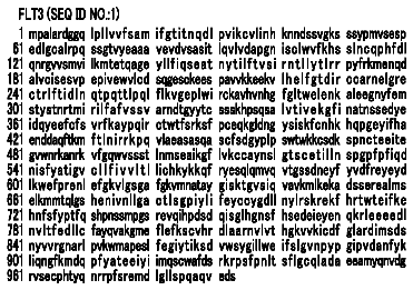
FIG. 17 shows the amino acid sequence of a wild-type mouse cystathione-gamma-lyase (SEQ ID NO:1).
FIG. 18 shows the amino acid sequence of a wild-type human cystathione-gamma-lyase (SEQ ID NO:2).
FIG. 19 shows the amino acid sequence of Annexin V (SEQ ID. NO:5).
FIG. 20 shows the amino acid sequence of Annexin I (SEQ ID. NO:6).
FIG. 21 shows the pre-cleavage amino acid sequence of the mCGL-AV fusion protein (SEQ ID NO: 8).
FIG. 22 shows the pre-cleavage amino acid sequence of the mCGL-AI fusion protein (SEQ ID NO: 9).
FIG. 23 shows the post-cleavage amino acid sequence of the mCGL-AV fusion protein (SEQ ID NO: 10).
FIG. 24 shows the post-cleavage amino acid sequence of the mCGL-AI fusion protein (SEQ ID NO: 11).
FIG. 25 shows the DNA sequence of the mCGL-AI fusion gene (SEQ ID NO:12) including sequencing primer (underlined).
FIG. 26 shows the DNA sequence of the mCGL-AV fusion gene (SEQ ID NO:13) including sequencing primer (underlined).
Detailed description
The presently disclosed inventive concepts are directed to methods of treating cancer utilizing an enzyme prodrug therapy, as well as methods of producing cancer cell-targeted enzyme conjugates including the enzyme utilized in the enzyme prodrug therapy. The presently disclosed inventive concepts are further related to compositions comprising said enzyme conjugate and prodrug, wherein the compositions may be utilized in the methods of treating cancer described herein. The presently disclosed inventive concepts are also related to kits that include said enzyme conjugate and prodrug.
In another embodiment, the presently disclosed inventive concepts are also directed to methods of treating cancer as described herein, wherein such methods further include the use of an immunostimulant, and/or a hypoxia-inducible factor-1 (HIF-1) inhibitor (for example, but not by way of limitation, a mammalian mechanistic target of rapamycin (mTOR) inhibitor), and/or a chemotherapeutic agent in such methods. The presently disclosed inventive concepts also include compositions that comprise the enzyme conjugate and prodrug as well as the immunostimulant, and/or an HIF-1 inhibitor, and/or chemotherapeutic agent. In addition, the presently disclosed inventive concepts are directed to kits that include the enzyme conjugate, prodrug, and/or an immunostimulant, and/or an HIF-1 inhibitor, and/or a chemotherapeutic agent.
Before further description of embodiments of the presently disclosed inventive concepts by way of exemplary drawings, experimentation, results, and laboratory procedures, it is to be understood that the presently disclosed inventive concepts are not limited in application to the details of compositions and methods set forth in the following description or illustrated in the drawings, experimentation and/or results. The presently disclosed inventive concepts are capable of other embodiments or of being practiced or carried out in various ways. As such, the language used herein is intended to be given the broadest possible scope and meaning; and the embodiments are meant to be exemplary—not exhaustive. Also, it is to be understood that the phraseology and terminology employed herein is for the purpose of description and should not be regarded as limiting.
Unless otherwise defined herein, scientific and technical terms used in connection with the presently disclosed inventive concepts shall have the meanings that are commonly understood by those of ordinary skill in the art. Further, unless otherwise required by context, singular terms shall include pluralities and plural terms shall include the singular. Generally, nomenclatures utilized in connection with, and techniques of, cell and tissue culture, molecular biology, and protein and oligo- or polynucleotide chemistry and hybridization described herein are those well known and commonly used in the art. Standard techniques are used for recombinant DNA, oligonucleotide synthesis, and tissue culture and transformation (e.g., electroporation, lipofection). Enzymatic reactions and purification techniques are performed according to manufacturer's specifications or as commonly accomplished in the art or as described herein. The foregoing techniques and procedures are generally performed according to conventional methods well known in the art and as described in various general and more specific references that are cited and discussed throughout the present specification. See e.g., Sambrook et al. Molecular Cloning: A Laboratory Manual (2nd ed., Cold Spring Harbor Laboratory Press, Cold Spring Harbor, N.Y.
and Coligan et al. Current Protocols in Immunology (Current Protocols, Wiley Interscience (1994)), which are incorporated herein by reference. The nomenclatures utilized in connection with, and the laboratory procedures and techniques of, analytical chemistry, synthetic organic chemistry, molecular and cellular biology, and medicinal and pharmaceutical chemistry described herein are those well known and commonly used in the art. Standard techniques are used for chemical syntheses, chemical analyses, pharmaceutical preparation, formulation, and delivery, and treatment of patients.
All publications, published patent applications, and issued patents mentioned in the specification are indicative of the level of skill of those skilled in the art to which the presently disclosed inventive concepts pertain. All publications, published patent applications, and issued patents are explicitly incorporated by reference herein to the same extent as if each individual publication, published patent application, or issued patent was specifically and individually indicated to be incorporated by reference.
The use of the word “a” or “an” when used in conjunction with the term “comprising” in the claims and/or the specification may mean “one,” but it is also consistent with the meaning of “one or more,” “at least one,” and “one or more than one.” The use of the term “or” in the claims is used to mean “and/or” unless explicitly indicated to refer to alternatives only or the alternatives are mutually exclusive, although the disclosure supports a definition that refers to only alternatives and “and/or.” Throughout this application, the term “about” is used to indicate that a value includes the inherent variation of error for the device, the method being employed to determine the value, or the variation that exists among the study subjects.
As used in this specification and claim(s), the words “comprising” (and any form of comprising, such as “comprise” and “comprises”), “having” (and any form of having, such as “have” and “has”), “including” (and any form of including, such as “includes” and “include”) or “containing” (and any form of containing, such as “contains” and “contain”) are inclusive or open-ended and do not exclude additional, unrecited elements or method steps.
The term “or combinations thereof” as used herein refers to all permutations and combinations of the listed items preceding the term. For example, “A, B, C, or combinations thereof” is intended to include at least one of: A, B, C, AB, AC, BC, or ABC, and if order is important in a particular context, also BA, CA, CB, CBA, BCA, ACB, BAC, or CAB. Continuing with this example, expressly included are combinations that contain repeats of one or more item or term, such as BB, AAA, AAB, BBC, AAABCCCC, CBBAAA, CABABB, and so forth. The skilled artisan will understand that typically there is no limit on the number of items or terms in any combination, unless otherwise apparent from the context.
All of the compositions and/or methods disclosed and/or claimed herein can be made and executed without undue experimentation in light of the present disclosure. While the compositions and methods of the presently disclosed inventive concepts have been described in terms of particular embodiments, it will be apparent to those of ordinary skill in the art that variations may be applied to the compositions and/or methods and in the steps or in the sequence of steps of the methods described herein without departing from the concept, spirit and scope of the inventive concepts. All such similar substitutes and modifications apparent to those skilled in the art are deemed to be within the spirit, scope and concept of the inventive concepts as described herein.
According to the presently disclosed inventive concepts, enzyme conjugates for use in a prodrug cancer therapy treatment protocol include a ligand having the ability to specifically and stably bind to an external receptor and/or binding site on an outer surface of a tumor vasculature endothelial cell and/or cancer cell, wherein the external receptor and/or binding site is specific for tumor vasculature endothelial cells and/or cancer cells (i.e., is uniquely expressed or overexpressed on a luminal surface of the tumor vasculature endothelial cell or cancer cell). The enzyme conjugate further includes an enzyme that is operatively attached to the ligand, wherein the enzyme is able to convert a prodrug into an active anticancer drug. In at least one embodiment, the enzyme conjugate is maintained on the outer surface of the tumor vasculature endothelial cell and/or cancer cell with substantially no internalization of the enzyme conjugate.
The ligand of the enzyme conjugate of the presently disclosed inventive concepts may be any protein or composition which binds to the receptor or other targeting molecule uniquely present on the surface of cancer cells or cells in the tumor vasculature (i.e., an aminophospholipid). When the ligand is a protein, the ligand may contain the entire protein that binds to the desired receptor or other targeting molecule, or the ligand may contain only a portion of the protein. For example, it may be desirable to remove a portion of the protein that has an undesirable biological activity, or it may be desirable to remove a portion of the protein to enable attachment of the anticancer agent. When a portion of the protein is present as the ligand in the enzyme conjugate, the only requirement is that the portion of the protein substantially retains the protein's receptor or targeting molecule binding activity. In addition, if the protein contains a portion that targets the protein for internalization, such portion should be removed so that the enzyme conjugate of the presently disclosed inventive concepts is stably bound to the outer surface of the cancer cell or blood vessel supplying the tumor. In one embodiment, the enzyme conjugate is maintained on the outer surface of the cancer cell or blood vessel with substantially no internalization thereof. The terms “portion” and “fragment” are used herein interchangeably.
Likewise, the enzyme conjugate may contain a variant or mutant of the ligand. For example, it may be desirable to modify a portion of the ligand that has an undesirable biological activity, or it may be desirable to modify a portion of the ligand to enable attachment of the anticancer agent. When a variant of the ligand is present in the enzyme conjugate, the only requirement is that the ligand variant substantially retains the ligand's receptor or targeting molecule binding activity. Also, sequences may be added to, or inserted within, the ligand during modification, as long as the modified ligand substantially retains the ligand's receptor binding activity. Therefore, it is to be understood that the term “ligand variant” includes both substitutions (including but not limited to conservative and semi-conservative substitutions) as well as additions and insertions to the native ligand's sequence that do not substantially affect the ligand's receptor binding activity. Such variations may occur at the nucleic acid level during construction of the construct from which the enzyme conjugate is expressed, or the variations may be produced by other posttranscriptional or posttranslational means known to those or ordinary skill in the art, including but not limited to, mutations and chemical modifications.
Examples of receptors that may be targeted by enzyme conjugates in accordance with the presently disclosed inventive concepts include, but are not limited to, urokinase receptor, epidermal growth factor (EGF) receptor, insulin-like growth factor receptor, interleukin-4 (IL-4) receptor, interleukin 6 (IL-6) receptor, keratinocyte growth factor (KGF) receptor, platelet-derived growth factor (PDGF) receptor, fibroblast growth factor (FGF) receptor, laminin receptor, vascular endothelial growth factor (VEGF) receptor, transferrin receptor, phosphatidylserine (PS), fibronectin, and the like, as well as portions thereof, and variants thereof, that substantially maintain the ability to bind to the ligand of the enzyme conjugate of the presently disclosed inventive concepts and/or maintain the enzyme conjugate on the surface of the cell with substantially no internalization thereof.
As stated above, the ligand portion of the enzyme conjugate specifically binds to the external receptor or binding site on the outer surface of the cell. In one embodiment, the ligand may be selected from the group consisting of annexins; antibodies to a receptor or aminophospholipid that is uniquely expressed or overexpressed on a surface of a tumor vasculature endothelial cell or cancer cell; RGD-motif peptides (Receptor: integrins alpha-v-beta 3 and alpha-v-beta 5); NGR-motif peptides (Receptor: aminopeptidase N, also known as CD13); F3, a 34-amino acid basic peptide from HMGN2 (Receptor: cell surface nucleolin); HWGF-motif peptides (selective inhibitors of matrix metalloproteinase-2 and matrix metalloproteinase-9, also known as gelatinase A and gelatinase B); the synthetic peptide CTTHWGFTLC (SEQ ID NO:7), which targets angiogenic blood vessels, inhibits the migration of human endothelial cells and tumor cells, and also prevents tumor growth and invasion in animal models and improves survival of mice bearing human tumors; amino-terminal fragment (ATF) of urokinase (which binds to the urokinase receptor, but, unlike full length urokinase, is not internalized); and fragments or variants thereof which substantially retain the ability to bind to the receptor or binding site. In one embodiment, the ligand may be a phosphatidylserine-binding protein.
Where used herein, the terms “specifically binds to,” “specific binding,” “binds specifically to,” and “binding specificity” refer to the ability of a ligand (e.g., an annexin) or other agent to detectably bind to a receptor or a binding epitope while having relatively little detectable reactivity with other proteins, epitopes, or receptor structures presented on cells to which the ligand or other agent may be exposed.
Where used herein the term “annexin” refers to any of annexins 1-11 and 13, which are more particularly designated as annexins A1, A2, A3, A4, A5, A6, A7, A8, A9, A10, A11, and A13. Annexin I and annexin V where used herein refer to Annexin A1 and Annexin A5, respectively, for example. The annexins contemplated herein further include non-human cognate orthologs of A1-A11 and A13 from non-human vertebrates, including but not limited to, non-human primates, dogs, cats, horses, livestock animals and zoo animals, which may be used for treatment in said non-human mammals in the methods contemplated herein. The annexins contemplated for use herein are discussed in further detail in V. Gerke and S. E. Moss (Physiol. Rev., 82:331-371 (2002)), the entirety of which is expressly incorporated by reference herein.
Anionic phospholipids are largely absent from the surfaces of resting mammalian cells under normal conditions. Phosphatidylserine (PS) is the most abundant anionic phospholipid of the plasma membrane and is tightly segregated to the internal side of the plasma membrane in most cell types. Recently, it has been discovered that PS is expressed on the outside surface of the endothelial cells that line the blood vessels in tumors in mice but is not expressed on the outside surface of the vascular endothelium in normal organs. In addition, anionic phospholipids have been shown to be expressed on the outside surface of cancer cells.
The tumor vasculature is increasingly recognized as a target for cancer therapy. Angiogenesis, the formation of new capillaries from existing blood vessels, is essential for the growth of solid tumors beyond 1-3 mm in size. Damage to the endothelial cells that line the blood vessels results in the induction of the coagulation cascade, causing intratumoral vessel occlusion and subsequent tumor necrosis. Targeting the tumor vasculature has the advantage that the delivery vehicle, once in the bloodstream, has direct access to the target endothelial cells. Other advantages of targeting the tumor vasculature rather than the tumor cells themselves include a potentiation effect, because one blood vessel nourishes hundreds of tumor cells. There have, however, been no studies reported of targeting enzyme/prodrug therapy to the tumor vasculature.
In one embodiment of the enzyme conjugate of the presently disclosed inventive concepts, human annexin V, a member of the annexin family of Ca.sup.2+-dependent anionic phospholipid binding proteins (others are noted above), is used as the ligand and is operatively attached to or otherwise physically associated with an enzyme for targeting the tumor vasculature endothelial cells. Annexin V is a member of a class of widely distributed proteins which bind to anionic phospholipids and membranes in a Ca.sup.2+-dependent manner. Annexin V is a monomeric protein, which has been crystallized and shown to consist of four tandem repeats of similar structure. Structural evidence shows that the N-terminus of annexin V is located at the surface of the protein and faces away from the membrane-binding side of the molecule. It was later found that the attachment of prourokinase at the N-terminus of annexin V did not alter its affinity for cell membranes in which PS was exposed on the membrane surface, which is consistent with the previous structural evidence.
Annexin V (and other annexins) binds with very high affinity to PS-containing phospholipid bilayers. Annexin V may be obtained, for example, as described in U.S. Pat. No. 7,393,833, issued to Lind et al. on Jul. 1, 2008, the entire contents of which are hereby expressly incorporated by reference.
Examples of other PS-binding proteins that can be used in substitution include those in the Annexin family (listed above), lactadherin, domains found in proteins known to bind PS, such as Factor V/Va, Factor X/Xa, Factor II/II, Factor VII/VIIa, Factor IX/IXa, Factor VIII/IIIa, Spectrin, Class B Scavenger receptor type I, Protein Kinase C, and proteins containing the C2 domains of protein kinase C (this includes synaptotagmins), Rabphilin family members, the PS receptor, endothelial lectin-like OxLDL receptor-1 (LOX-1), antibodies to PS, phosphatidylserine decarboxylase, MARCKS (myristoylated, alanine-rich protein kinase C substrate), PS-p68, Myosin, Erythrocyte protein 4.1, hemoglobin, Calponin family members, S100A, S100B, calcyclin-binding protein family members, milk membrane-glycoprotein, MFG-E8 (milk fat globule-EGF factor 8), and other PS-binding motifs known to those of ordinary skill in the art.
Alternatively, the ligand of the enzyme conjugate of the presently disclosed inventive concepts may be an anionic phospholipid-specific antibody, such as (but not limited to) a PS-specific monoclonal antibody. Non-limiting examples of PS-specific monoclonal antibodies include those described in U.S. Pat. Nos. 6,312,694; 6,406,693; 6,783,760; 6,818,213; and 7,067,109. The ligand may be a non PS-binding moiety which binds to another tumor specific feature, such as (but not limited to) those described in U.S. Pat. Nos. 6,451,312; 6,093,399; 6,004,555; and 6,051,230. The ligands of the presently disclosed inventive concepts may be targeted to other tumor/cancer specific external receptors other than anionic phospholipids. Such receptors include, for example, those described in U.S. Pat. Nos. 6,818,213; 6,783,760; 6,451,312; and 6,406,693. As noted above, all of the patents, published patent applications, and publications listed herein are hereby expressly incorporated herein by reference in their entireties.
The modification of one of the receptor-binding ligands described herein above to provide a fragment or variant thereof that substantially maintains the receptor binding ability of the native receptor-binding ligand is fully within the skill of a person in the art and therefore is also within the scope of the presently disclosed inventive concepts. The term “substantially maintains the receptor-binding ability of the native receptor-binding ligand” means that the protein fragment or variant maintains at least 50% of the native ligand's receptor-binding ability, at least 75% of the native ligand's receptor-binding ability, at least 90% of the native ligand's receptor-binding ability, or at least 95% of the native ligand's receptor-binding ability.
The enzyme attached to the ligand of the enzyme conjugate may include any enzyme capable of converting a prodrug into an active anticancer drug that can function in accordance with the presently disclosed inventive concepts. The enzyme can either be (i) of nonhuman origin, (ii) a human protein that is absent or expressed only at low concentrations in normal tissues, or (iii) a variant that is non-immunogenic. Examples of enzymes that may be utilized in enzyme conjugates in accordance with the presently disclosed inventive concepts include, but are not limited to, cystathione-gamma-lyase, L-methioninase, nitroreductase, cytochrome P450, purine-nucleoside phosphorylase, thymidine kinase, alkaline phosphatase, β-glucuronidase, glycosidase, carboxypeptidase, carboxyesterase, penicillin amidase, β-lactamase, and cytosine deaminase, and effective variants (mutants) thereof.
In one embodiment, the enzyme may be L-methioninase (also known as methionine γ-lyase). In certain embodiments the enzyme is a non-L-methioninase that has methioninase activity, such as (but not limited to) a variant of a wild type mammalian cystathione-gamma-lyase (CGL). In at least one embodiment, the enzyme is a variant of wild type mouse CGL or of wild type human CGL. A non-limiting example of one such variant is the mutant of mouse CGL having the amino acid sequence as set forth in SEQ ID NO:3, which is like wild type mouse CGL (SEQ ID NO:1) except for having substitutions in positions 58, 118, and 338 (see Table 5). Other variants may contain substitutions in only one or two of positions 58, 118, and 338. In another variant, position 58 is substituted with valine rather than asparagine. Another non-limiting example of such a variant is the mutant of human CGL having the amino acid sequence as set forth in SEQ ID NO:4, which is like wild type human CGL (SEQ ID NO:2) except for having substitutions in positions 59, 119, and 339 (see Table 5). Other variants may contain substitutions in only one or two of positions 59, 119, and 339. In another variant position 59 is substituted with valine rather than asparagine. The amino acids included in the substitutions in said variant may include amino acids others than those identified in Table 5, including conservative amino acid substitutions described hereinbelow and those identified in U.S. Pat. No. 8,709,407 (for example, in columns 2, 3, and 32 therein). Other variants include variants (with mutations in the homologous positions) of any other primate CGL (e.g., see U.S. Pat. No. 8,709,407, and Stone et al., “De Novo Engineering of a Human Cystathione-gamma-Lyase for Systemic L-Methionine Depletion Cancer Therapy,” ACS Chem. Biol. 2012, 7, 1822-1829), or other mammalian CGL, such as (but not limited to) dog, cat, and horse. Other variants include mutant CGLs which have additional substituted amino acids such that they have at least 80% identity to the CGL sequences listed above, or at least 81% identity thereto, or at least 82% identity thereto, or at least 83% identity thereto, or at least 84% identity thereto, or at least 85% identity thereto, or at least 86% identity thereto, or at least 87% identity thereto, or at least 88% identity thereto, or at least 89% identity thereto, or at least 90% identity thereto, or at least 91% identity thereto, or at least 92% identity thereto, or at least 93% identity thereto, or at least 94% identity thereto, or at least 95% identity thereto, or at least 96% identity thereto, or at least 97% identity thereto, or at least 98% identity thereto, or at least 99% identity thereto, wherein “% identity” is defined in at least one embodiment as the percentage of amino acids (or nucleotides) which are identical at corresponding positions in two amino acid (or nucleic acid) sequences of a protein (or nucleic acid). Said variants of CGL described herein have L-methioninase activity.
The proteins of the presently disclosed inventive concepts may be produced using any nucleotide sequence which encodes the desired amino acid sequence. The proteins may include, for example (but not by way of limitation), conservative substitutions of the amino acid residues of the CGL sequence described herein, wherein such amino acid substitutions do not substantially reduce the L-methioninase activities of the encoded enzyme variant. Examples of conservative amino acid substitutions include, but are not limited to, glycine:alanine substitutions; valine:isoleucine:leucine substitutions; asparagine:glutamine:histidine substitutions; aspartic acid:glutamic acid substitutions; serine:threonine:methionine substitutions; lysine:arginine:histidine substitutions; and phenylalanine:tyrosine:tryptophan substitutions. Other examples of conservative and semi-conservative amino acid substitutions that may be utilized in accordance with the presently disclosed inventive concepts are shown in Table 2. Other types of substitutions, variations, additions, deletions, and derivatives that result in functional CGL variants are also encompassed by the presently disclosed inventive concepts, and one of ordinary skill in the art would readily know how to make, identify, or select such variants or derivatives, and how to test for methioninase activity of those variants.
In one embodiment, the CGL enzyme may itself also have an anticancer activity. Examples of enzyme/anticancer agents that may be utilized in accordance with the presently disclosed inventive concepts include, but are not limited to, L-methioninase and fragments and variants thereof which substantially retain the ability to degrade methionine.
The enzyme and the ligand of the enzyme conjugate may be directly coupled together (e.g., via a covalent bond) or indirectly coupled together via a linker, such as (but not limited to) via a linker peptide. In addition, the enzyme may be conjugated to polyethylene glycol (PEG), or the enzyme conjugate may be encapsulated in a liposome.
In one embodiment, the enzyme conjugate includes
an amino acid sequence as set forth in SEQ ID NO:3 or 4, or mutants (variants) thereof as described elsewhere herein, and
a ligand such as (but not limited to) an annexin (e.g., one of annexins 1-13) or functional variants thereof.
The presently disclosed inventive concepts include a purified nucleic acid segment encoding an enzyme-ligand conjugate described herein, a recombinant vector comprising said nucleic acid segment, and a recombinant host cell comprising said recombinant vector.
The description continues in the full USPTO document.
 Lapsed, fee not paid5 drawings