 Lapsed, fee not paid3 drawings
Consumer Products · US 9,964,287 B2
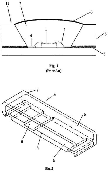
LED support, LED and backlight module
The invention discloses a LED support comprising an opaque housing.
US 9,964,472 B2 · Assignee: The Procter & Gamble Company · Inventors: Xie; Sancai et al.
Sheet 1 of 14 from the published document. All sheets in the USPTO PDF
One or more methods for sampling gingival metabolites and biomarkers.
Periodontal diseases, such as gingivitis and periodontitis, involve chronic inflammation in the gingival tissue caused by microbial communities and host immune responses. They are one of the most ubiquitous diseases worldwide, and remain the most common cause of tooth loss in the world today, and can affect up to 90% of the population worldwide. Gingivitis is defined per the FDA monograph (12 CFR Part 356, Vol. 68, No. 103 (2003)) as “An inflammatory lesion of the gingiva that is most frequently caused by dental plaque. Gingivitis is characterized by tissue swelling and redness, loss of stippling (a normal state in which the surface of healthy gingiva is comprised of small lobes), glossy surface, and increased tissue temperature. The gingiva also may bleed upon gentle provocation, such as tooth brushing or may bleed spontaneously. Gingivitis is usually not painful.” In healthy gingiva, t
1 of 14 drawing sheets so far from the published document, cropped to the drawing. Every sheet is in the USPTO PDF.
What the patent claimed, word for word. All of it is now free to use.
The invention is related to methods of collecting gingival samples non-invasively at a specific site, extracting and analyzing metabolites; using metabolites, such as citrulline, ornithine, succinic acid, to monitor the health status of gingivae.
Periodontal diseases, such as gingivitis and periodontitis, involve chronic inflammation in the gingival tissue caused by microbial communities and host immune responses. They are one of the most ubiquitous diseases worldwide, and remain the most common cause of tooth loss in the world today, and can affect up to 90% of the population worldwide. Gingivitis is defined per the FDA monograph (12 CFR Part 356, Vol. 68, No. 103 (2003)) as “An inflammatory lesion of the gingiva that is most frequently caused by dental plaque. Gingivitis is characterized by tissue swelling and redness, loss of stippling (a normal state in which the surface of healthy gingiva is comprised of small lobes), glossy surface, and increased tissue temperature. The gingiva also may bleed upon gentle provocation, such as tooth brushing or may bleed spontaneously. Gingivitis is usually not painful.” In healthy gingiva, the microbial community is in a homeostatic equilibrium with the host, and host immune systems limit bacterial overgrowth and neutralize toxic products, such as lipopolysaccharides (LPS) and lipoteichoic acids (LTA). The intricate balance between host and bacteria is disrupted as bacteria overgrow in the gingival margins or in the subgingival crevice. Recent data from metagenomics studies showed that bacterial species were increased in supragingival and subgingival plaques, such as Prevotella pallens, Prevotella intermedia, Porphyromonas gingivalis , and Filifactor alocis . Although the etiology of gingivitis and periodontitis remains elusive, one thing is clear; the composition of the dental plaques is significantly different in healthy sites compared with clinically defined disease sites. This observation, together with advances in characterizing the host and bacterial interactions using the newly developed tools in genomics, proteomics and metabonomics, has led to the notion that gingivitis and periodontitis are the result of disrupted homeostasis between host and polymicrobial communities (Lamont R J and Hajishengallis G. Polymicrobial synergy and dysbiosis in inflammatory disease. G Trends Mol Med. 2015; 21:172-83).
Polymicrobial communities in the dental plaques produce various virulence factors; for example, many bacteria produce digestive enzymes, such as hyaluronidases to breakdown polysaccharides that glue the host cell together, fibrinolytic enzymes that lyse the fibrins of blood clots, and collagenases that degrade collagens in the connective tissues. Gram negative bacteria secrete endotoxins, also called LPS, while Gram positive bacteria produce LTA and peptiglycans. Furthermore, one pathogen bacterium can generate multiple virulence factors; for example P. gingivalis has been reported to generate multiple virulence factors that are involved in the inflammatory and destructive events of periodontal tissues. These influence factors include the capsule, outer membrane, its associated LPS, fimbriae, proteinases, and selected enzymes.
LPS is an integral component of all Gram negative bacteria and is found in the outer membrane layer. P. gingivalis LPS possesses significant amounts of heterogeneity containing tetra- and penta-acylated structures. Several of them have been purified. LPS 1690 is highly toxic, while LPS 1435/1449 is relatively mild. Chemically, LPS consists of a hydrophilic polysaccharide and a hydrophobic lipid moiety referred to as lipid A. The latter is the actual toxic moiety of the LPS molecule and contains phosphate groups shown to be essential for its proinflammatory activity. Mechanistically, LPS first binds to LPS-binding protein (LBP), then the LBP-LPS complex is transferred to membrane-bound CD14, thereby enabling interactions with Toll-like receptor (TLR) 4 on cell membranes. Binding of LPS to TLR4 on the cell membrane activates both TIRAP-MyD88-dependent NFkB and TRAM-TRIF-dependent IRF3 or IRF7 signaling pathways, and subsequently stimulate production of proinflammatory cytokines and chemokines, such as interferon (IFN) gamma, tumor necrosis factor-α (TNFα), interleukin (IL)-1β, IL-6, IL-8, and IL-12. Also, induced is production of nitric oxide, prostaglandins, leukotrienes, and proteolytic enzymes. Importantly, LPS has been reported to cause periodontitis in mouse and rats.
P. gingivalis also secretes exotoxins and enzymes that exert damage on the host following their release. These enzymes include proteases, coagulases, and fibrinolysins. Noticeably, P. gingivalis generates peptidylarginine deiminase that can modify free or peptide-bound arginine to citrulline. The citrullinated proteins are especially harmful since they cause auto-immune responses, and are hypothesized to be the culprit of rheumatoid arthritis. In addition, P. gingivalis also produces two types of gingipains, lysine specific (Kgp) and arginine specific (Rgps). Gingipains play a major role in stirring up inflammation and tissue destruction in the periodontium.
Peptidoglycan is the cell wall component common to all Gram-negative and Gram-positive bacteria. It is a polymer consisting of sugars and amino acids that form a mesh-like layer outside the plasma membrane. The sugar component consists of alternating residues of β-(1, 4) linked N-acetylglucosamine and N-acetylmuramic acid. A peptide chain of three to five amino acids is cross-linked to the N-acetylmuramic acid. The peptide chain can also be cross-linked to the peptide chain of another strand of peptidoglycans to weave into a 3D mesh-like layer. The peptidoglycan layer is substantially thicker in Gram-positive bacteria (20 to 80 nanometers) than in Gram-negative bacteria (7 to 8 nanometers). Peptidoglycan accounts for around 90% of the dry weight of Gram-positive bacteria but only about 10% of Gram-negative strains. Thus, presence of high levels of peptidoglycan is the primary determinant of the characterization of bacteria as Gram-positive.
Both peptidoglycans and LTA have been shown to act as inflammatory mediators by activating TLR2 on the cell membrane of host innate immune cells and intracellular signaling receptors, such as nucleotide-binding oligomerization domain or NOD 1 and NOD 2. Binding to TLR2 activates the NF-κB signaling pathway, subsequently leading to production and release of proinflammatory cytokines and chemokines, such as IL-1α, IL-1β, IL-6, IL-8, IFN y, and TNF-α. Both Gram-negative and positive bacteria and the virulence factors (LPS, peptidoglycans and LTA) induce production of the inducible isoform of nitric oxide synthases. The latter catalyze the production of nitric oxide (NO) from L-arginine. NO is an important cellular signaling molecule, that promotes vascular dilation and many cellular functions. NO is also a free radical with an unpaired electron and is reported to kill bacteria. The inducible isoform of nitric oxide synthases is induced by LPS and other bacterial toxins, and is a part of innate immune responses.
As L-arginine is converted into NO by nitric oxide synthases, a byproduct, citrulline is regenerated. Citrulline is an amino acid that is not encoded in the genetic codes, so that is not incorporated in proteins during translation processes. Its name is derived from citrullus, the Latin word for watermelon. Citrulline is also a key intermediate in the urea cycle, the pathway by which mammals excrete ammonia. Citrulline is synthesized from ornithine and carbamoyl phosphate in the urea cycle, in which urea is produced in a series of reactions. Some of the reactions are carried out in the mitochondrial matrix and others in the cytosol.
The main metabolites of the urea cycle reactions are free amino acids, such as arginine, ornithine, citrulline, and arginisosuccinate. Arginine is the key intermediate in the urea cycle, and in NO production. It is cleaved by the cytosolic enzyme arginase, generating urea and ornithine. Ornithine, formed in the cytosol, is transported to the mitochondrial matrix via the action of ornithine translocase. In the mitochondria, ornithine transcabamoylase (OTC) catalyzes the condensation of ornithine with carbamoyl phosphate, producing citrulline. Concomitant with ornithine transport into the mitochondria is the export of citrulline to the cytosol where the remaining reactions of the cycle take place. Subsequently, citrulline is condensed with aspartate to form arginosuccinate, catalyzed by cytosolic argininosuccinate synthetase. Arginine and fumarate are produced from argininosuccinate by the cytosolic enzyme argininosuccinate lyase (also called argininosuccinase). The fumarate is reconverted to aspartate for use in the argininosuccinate synthetase reaction. In the final step of the urea cycle, arginases break arginine into urea and ornithine. The regenerated cytosolic ornithine is transported to the mitochondrial matrix for another round of urea synthesis. There are two arginase genes in humans, identified as the ARG1 and ARG2 genes. The ARG1 encoded isoform of arginase is a cytosolic enzyme primarily expressed in the liver and functions as the urea cycle enzyme. The ARG2 encoded arginase (arginase-2) is a mitochondrially localized enzyme expressed in non-hepatic tissues, primarily the kidney. The arginase-2 isoform is thought to be involved in nitric oxide and polyamine metabolism, however, the precise role of this enzyme is not clearly defined. More broadly, the biological functions of ornithine, citrulline, arginisosuccinate and arginine are not well understood yet in periodontal health.
Assessing the severity of gingivitis and periodontitis in a person is currently achieved with clinical measures such as gum redness, gum bleeding or pocket depth. While the measures are based on professionally developed scales, the actual values can vary due to examiner differences. There exists a need to quantify how severe gingivitis is and how effective treatments from oral hygiene products are in reducing the inflammatory response. It is desirable to have objective readings from an instrument that is free of human errors. Transcriptomics, proteomics and metabonomics measurements in saliva have been used to diagnose gingivitis, and to monitor progresses in treatment. But there is a disadvantage associated with saliva, in that the composition of saliva will be varied dependent upon the time of collection. As should be apparent, this field has a need for a more sensitive, accurate and consistent test whenever the volunteers appear in a dentist office, or in a clinical setting, or at home.
A method for non-invasive collection of gingival samples is provided that comprises using a collection device oriented parallel to the gum line of a specific tooth; and a portion of the collection device is detached from the collection device and placed into a container.
A method for extraction from gingival brush samples is provided that comprises using a collection device oriented parallel to the gum line of a specific tooth; a portion of the collection device is detached from the collection device and placed into a container containing extraction buffer; removing the collection device from the container; and extracting a biomarker.
FIG. 1 shows an illustration regarding collection of gingival and buccal samples.
FIG. 2 shows a line graph depicting clinical measurements.
FIG. 3 Changes in the amount of bacterial and host DNA in the supragingival plaques during 6 week of regimen treatment.
FIG. 4 comprises line graphs showing decreases in bacterial abundance in supragingival plaques.
FIG. 5 is a line graph showing decreasing citrulline concentrations in Buccal-brush samples.
FIG. 6 is a line graph showing a decrease in protein bound ornithine in Buccal brush samples.
FIG. 7 shows a drawing outlining the enzymes in the ornithine, citrulline and arginine cycle.
FIG. 8 shows point graphs illustrating changes in expression of genes that synthesize and ornithine, citrulline and arginine.
FIG. 9 shows line graphs illustrating increased concentrations of citrulline in Buccal-brush samples.
FIG. 10 shows line graphs illustrating decreased protein-bound citrulline in Buccal brush samples.
FIG. 11 shows line graphs illustrating increased concentrations of protein-bound ornithine in Buccal-brush samples.
FIG. 12 shows line graphs illustrating increased concentrations of total citrulline in Buccal-brush samples.
FIG. 13 shows line graphs illustrating decreased concentrations of protein bound arginine in Buccal-brush samples.
FIG. 14 shows line graphs illustrating decreased concentrations of total arginine in Buccal-brush samples.
The present invention includes methods of determining the status of epithelial health, particularly gingiva and oral mucosa. A method may comprise determining the levels of a set of biomarkers in gingivae. The set of biomarkers may include one or more metabolites, proteins, or messenger RNA (mRNA). Those metabolites have been shown to change in abundance at particular stages of treatment periods, or in in vitro models treated with different virulence factors, or human dental plaques. Accordingly, the set of metabolite biomarkers may be quantified to determine whether gingivae have inflammation, whether gingivae are under oxidative stresses or energy imbalance, and whether gingivae have cellular damage or injuries.
The present invention demonstrates a role for metabolite biomarkers to serve as indicators of gingivitis at different stages, and indicators for gingivae damage resulting from differing insults, such as oxidative stresses, high bacterial load, proinflammatory insults, energy imbalance or cellular injuries. The methods described herein demonstrate that either elevated or decreased levels of multiple metabolites can be used as a tool for accurately characterizing the quality of gingivae, such as gingivitis.
As used herein, the term “biomarker” means a substance that is measured objectively and evaluated as an indicator of normal biologic processes, pathogenic processes, treatment responses to chemical agents, or mechanical instruments. As used herein, biomarkers include, but are not limited to metabolites, proteins and messenger RNA (mRNA).
As used herein, the term “metabolite” means a substance that is measured objectively and evaluated as an indicator of normal biologic processes, pathogenic processes, treatment responses to chemical agents, or mechanical instruments; wherein said metabolites include, but are not limited to, a compound generated by lipid metabolism, protein metabolism, amino acid metabolism, carbohydrate metabolism, nuclear acid metabolism, or oxidative phosphorylation.
As used herein, the term “protein” means a substance that is measured objectively and evaluated as an indicator of normal biologic processes, pathogenic processes, treatment responses to chemical agents, or mechanical instruments; wherein the protein is a polymer consisting of more than three amino acids, including, but not limiting to, cytokines, chemokines, growth factors, cellular and extracellular proteins.
As used herein, the term “mRNA” means a substance that is a polymer of four ribonucleotides (adenine, uracil, guanine, cytosine), messenger RNA (mRNA) molecules convey genetic information from DNA to the ribosome, where they specify the amino acid sequence of the protein products of gene expression.
As used herein, the term “sample” means biological material isolated from an individual; wherein gingival samples are isolated from gingivae, and buccal samples are isolated from oral mucosa.
As used herein, the term “gum sensitivity” is a sensorial feeling, caused by activating transient receptor potential channel (TRP) V1 and/or TRPA1 on sensory neurons. Gum sensitivity is a common complaint due to inflammation, and can affect the area covering one or more teeth. Gum sensitivity is often noted when one eats or drinks something hot, cold, sweet, or sour; and can be experienced as a dull or sharp pain. The pain can begin suddenly and be felt deeply in the nerve endings of the tooth. Certain polyunsaturated fatty acids (PUFA), such as linoleic acid, arachidonic acid, hydroxyoctadecadienoic acid (HODE), and hydroxyeicosatetraenoic acid (HETE), are known to activate or sensitize TRPV1 and TRPA1. Certain oxidized lipids also activate TRPV1 and TRPA1 on sensory neurons, such as hydroxyoctadecadienoic acid (HODE) and hydroxyeicosatetraenoic acid (HETE), Prostaglandins, prostacyclins, and thromboxanes (Ruparel et al. Released lipids regulate Transient Receptor Potential Channel (TRP)-dependent oral cancer pain. Mol Pain. 2015; 11: 30).
As used herein, the term “oxidative stress” is a threshold criteria based on volunteers exhibiting an imbalance between the production of free radicals and the ability of the body to counteract or detoxify the reactive intermediates or to repair the resulting damage.
As used herein, the term “energy imbalance” or the term “mitochondrial dysfunction” means an imbalance of energy homeostasis. Mitochondria are found in every nucleated cell of the human body, and convert the energy of carbohydrate and fat into the ATP that powers most cellular functions. Both the citric acid cycle and β-oxidation of fatty acids are carried out in mitochondria. In gingivitis where gingivae are inflamed or damaged, AMP levels are high, meaning ATP production is impaired. Similarly, carnitine is a cofactor that helps carry fatty acid into mitochondria. Deoxycarnitine is an immediate precursor of carnitine.
As used herein, the term “barrier function” means the defense function of epithelium against the environment, such as heat, dust, and microbes.
As used herein, the term “immunoassay” means any assay based on antibody-binding-to-specific targets, including, but not limiting to, ELISA (enzyme-linked immunosorbent assay) and immunoblotting. The targets can include, but are not limited to, proteins, peptides, fatty acids, carbohydrates, metabolites, and nucleic acids.
Certain embodiments of the present invention provide a method for collection of gingival samples. Gingival tissues are taken around the tooth, or around the connecting areas between the gingiva and the tooth. In one or more embodiments, a collection device, such as an interdental gum brush or buccal brush is used to collect gingival samples by swabbing back and forth multiple times with the brush-head oriented parallel to the gum line. A portion of the collection device that contacted the connecting areas between the gingiva and tooth may be detached and placed into a container; for example a brush head may be clipped off with a pair of sterile scissors and placed into a container, which may contain a buffer solution or an RNAlater solution.
Included in the present invention are the findings that some protein biomarkers are decreased over time in gingivitis treatment, suggesting those protein biomarkers are associated with gingivitis, inflammation, sensitivity, energy imbalance, mitochondrial dysfunction, and oxidative stresses. Examples of protein biomarkers include ICAM-1, IL-1α, IL-β, TNF-β, IL-12p12, IL-13, IL-4, IL-5, CRP, eotaxin, GM-CSF, IFN-y, IL-10, IL-15, IL-16, IL-6, IL-7, IL-8, MCP-1/CCL2, MDC, SAA, tie-2, VCAM-1, VEGF, VEGF-2, VEGF-D, VEGF-C, and TARC/CCL17.
This application also identifies metabolites in the gingivae that are associated with gingivitis. Changes in metabolites 13-HODE, 9-HODE, 1-arachidonoylglycerophosphoethanolamine, 1-oleoylglycerophosphoethanolamine, citrulline and ornithine occur as the health status of gingival tissue improves or deteriorates. Subgingival bacteria in gingivitis sites stimulated production of ornithine, citrulline, arginine, succinate, fumarate and malate in human peripheral blood mononuclear cells (PBMC). Similarly, endotoxins and LTA also stimulated their production in PBMC to some degree. Those bacteria virulence factors are known to cause inflammation, oxidative stresses, and cellular damages.
Various lipid metabolites have been implicated in different inflammatory diseases. Although much is known about prostaglandins, much less attention has been given to other lipid metabolites. 13-HODE and 9-HODE are stable oxidation products of linoleic acid, the generation of which is increased where oxidative stress, inflammation, and epithelium damage are increased. 13-HODE is produced in large quantities during mitochondrial degradation steps in RBC maturation, whereas 13-hydroxy-linoleic acid, a related molecule, has been linked to airway hyperresponsiveness. 13-HODE is considered as a potential link between mitochondrial dysfunction, epithelial injury, and inflammatory disease (Ulaganathan Mabalirajan et al. Linoleic acid metabolite drives severe asthma by causing airway epithelial injury. Scientific Reports 3, Article number: 1349
doi:10.1038/srep01349).
This invention also describes in the gingivae an increase of adenosine 5′-monophosphate (AMP), 2-methylbutyrylcarnitine (C5), deoxycarnitine, and propionylcarnitine, which is indicative of mitochondrial disfunciton. Bacterial endotoxins, such as LPS, induce the ‘Warburg effect’ of aerobic glycolysis (Tannahill et al., Succinate is an inflammatory signal that induces IL-1b through HIF-1a, Nature 496, 238-242, 2013).
In certain embodiments, this invention communicates the decrease of 2-methylbutyrylcarnitine (C5), deoxycarnitine, and propionylcarnitine in gingivae during treatment over a time period, such as a six week period. As shown in FIG. 2 , gingivitis symptoms of bleeding and inflammation are diminished during the treatment period. The decrease of 2-methylbutyrylcarnitine (C5), deoxycarnitine, and propionylcarnitine is indicative of improvement of energy production or mitochondrial dysfunction. Carnitine, which is derived from deoxycarnitine transports long-chain acyl groups from fatty acids into the mitochondrial matrix, so they can be broken down through β-oxidation to acetyl CoA to produce ATP via the citric acid cycle. Supply of deoxycarnitine caused an increase of carnitine. The latter treatment also produced a transient but significant diminution of L-carnitine in heart, skeletal muscle and kidney (Sartorelli et al., Carnitine and deoxycarnitine concentration in rat tissues and urine after their administration. Biochim Biophys Acta. 1989 Nov. 6; 1006:15-8.). Increase of deoxycarnitine in gingivitis likely reduces L-carnitine levels, thus disturbing the β-oxidation and mitochondrial functions. Increase of deoxycarnitine in gingivitis likely changes L-carnitine levels, thus disturbing the β-oxidation and mitochondrial functions.
In gingivitis there is an increase in propionylcarnitine. Propionylcartitine is a byproduct of a branched-chain amino acid (BCAA). BCCA is an amino acid having aliphatic side-chains with a branch (a central carbon atom bound to three or more carbon atoms). Among the proteinogenic amino acids, there are three BCAAs: leucine, isoleucine and valine. Degradation of BCCA involves the branched-chain alpha-keto acid dehydrogenase complex. A deficiency of this complex leads to a buildup of the branched-chain amino acids (leucine, isoleucine, and valine) and their toxic by-products in the blood and urine, giving the condition the name maple syrup urine disease. Accumulation of propionylcartnitine is indicative of disturbed metabolism of BCAA.
One of the most observed symptoms of gingivitis is bleeding, which is partially the result of a damaged vasculature and epithelial barrier. ICAM-1 also known as CD54 (Cluster of Differentiation 54) is a cell surface glycoprotein that is typically expressed on endothelial cells and cells of the immune system. This protein is an intercellular adhesion molecule continuously present in low concentrations in the membranes of leukocytes and endothelial cells. Upon cytokine stimulation, the concentrations greatly increase in the vascular endothelium, macrophages, and lymphocytes. ICAM-1 is a ligand for LFA-1 (integrin), a receptor found on leukocytes. When activated, leukocytes bind to endothelial cells via ICAM-1/LFA-1 and then transmigrate into tissues. Because of these associations with immune responses, it has been hypothesized that ICAM-1 could function in signal transduction. ICAM-1 ligation produces proinflammatory effects such as inflammatory leukocyte recruitment. Importantly, ICAM-1 has antagonistic effects on the tight junctions, which forms the blood-testis barrier. ICAM-1 has been implicated in subarachnoid hemorrhage. Levels of ICAM-1 are shown to be significantly elevated in patients with subarachnoid hemorrhage over control volunteers in many studies.
The VCAM-1 gene contains six or seven immunoglobulin domains, and is expressed on both large and small blood vessels upon being stimulated by proinflammatory cytokines. It is alternatively spliced into two known RNA transcripts that encode different isoforms in humans. VCAM-1 is a cell surface sialoglycoprotein that mediates the adhesion of lymphocytes, monocytes and basophils to vascular endothelium. Human brain microvascular endothelial cells that form the blood-brain barrier release soluble vascular cell adhesion molecule-1 (sVCAM-1) under inflammatory conditions. Similarly, human brain endothelium also expresses integrin α-4/β-1, which binds to sVCAM-1. Binding of integrin α-4/β-1 to sVCAM-1 directly impairs blood-brain barrier function by triggering intracellular signaling events. Application of recombinant sVACM-1 to cultured primary brain endothelial cells increased permeability to the soluble tracer dextran as a result of damaged tight-junctions between endothelial cells (Haarmann et al. Soluble VCAM-1 impairs human brain endothelial barrier integrity via integrin α-4-transduced outside-in signaling, Acta Neuropathologica. May 2015, Volume 129, pp 639-652). ICAM-1 and VCAM-1 are high in gingivae having gingivitis.
Changes in bound arginine, ornithine and citrulline are indicative of damages in barrier function during gingivitis in gingiva tissues. Profilaggrin is the major component of the keratohyalin granules within epidermal granular cells in epidermis and gingivae. During epithelial terminal differentiation, the profilaggrin polyprotein is dephosphorylated and rapidly clipped by serine proteases into monomeric filaggrin. Filaggrin binds to the keratin cytoskeleton and thereby contributes to the squame biogenesis. Within the squames, filaggrin is citrullinated by peptidylarginine deiminase where an arginine residue is converted into a citrulline residue. Arginine is positively charged, while citrulline is neutral in charge. Positively charged arginine forms salt bonds with neighboring negatively charge amino acid residues to stabilize filaggrin proteins. Modification of positively charged arginine into a neutral amino acid residue, such as citrulline or ornithine, will diminish the salt bonds within a protein, thus leading to destabilizing and unfolding proteins, and finally degrading into hygroscopic amino acids. Those amino acids constitute one element of natural moisturising factors. As a result, citrullination is a required process in epidermal differentiation and barrier formation. Strong barrier functions protect the body from the entry of foreign environmental substances. (Sandilands et al., Filaggrin in the frontline: role in skin barrier function and disease. J Cell Sci. 2009 May 1; 122(Pt 9):1285-94. doi: 10.1242/jcs.033969).
In certain embodiments the present invention involves one or more methods for determining the expression of genes for the citric acid cycle, β-oxidation, and oxidative phosphorylation The citric acid cycle, also known as the tricarboxylic acid cycle, is a series of chemical reactions inside a cell to generate energy through the oxidation of acetyl-CoA derived from carbohydrate, fat and proteins into carbon dioxide and chemical energy in the form of ATP. In addition, the cycle provides the reducing agent NADH that is used in numerous other biochemical reactions. β-oxidation is the catabolic process by which fatty acid molecules are broken down in the mitochondria to generate acetyl-CoA, which enters the citric acid cycle, and NADH and FADH.sub.2, which are co-enzymes used in the electron transport chain. Oxidative phosphorylation (or OXPHOS in short) is the metabolic pathway in which the mitochondria in cells use their structure, enzymes, and energy released by the oxidation of carbohydrate, fatty acid and amino acid to form ATP.
In certain aspects, this invention describes an increase in degradation of macromolecules in gingivitis, such as proteins. There are more dipeptides in gingivitis at the baseline stage, such as threonylphenylalanine, threonylleucine, lysylleucine, lysylphenylalanine, leucylleucine, arginylphenylalanine, and arginylleucine. This degradation of proteins is indicative of increased production of proteases in inflammation.
The term “free” means a substance that is measured in supernatants directly as described in the EXAMPLES.
The term “total” means a substance that is measured in both supernatants and pellets as described in the EXAMPLES.
The term “bound” means a substance that is incorporated into proteins as described in the EXAMPLES. For example, bound citrulline means a citrulline molecule is incorporated into a protein. The bound citrulline is numerically equal to the total citrulline minus the free citrulline. Example 1—a Method to Collect Host Gingival Tissue from Above Individual Teeth to Assess Changes in Gingivitis-Related Molecular Markers
Assessing the degree of gingivitis in a person is typically achieved with clinical measures such as gum redness, gum bleeding or pocket depth. While the measures are based on professionally developed scales, the actual values can vary due to examiner differences. It is desirable to have objective readings free from human errors. This sample collection method enabled the taking of samples for objective measurements non-invasively and site-specifically.
Non-invasive gingival sample collection: Brush samples were taken from the upper, front gums and buccal surface of 4 volunteers, all female, ages 43-48. Interdental Gum Brushes (Sunstar America Inc, Chicago, Ill.), or A MasterAmp™ Buccal Brush (Catalog #MB100SP; Epicentre Technologies Corp., Madison, Wis.) brushes were used to sample 6 marginal gingiva, as shown in FIG. 1 , and 6 buccal areas, one brush per sample site. At each sample site a brush was swabbed back-forth 10 times with the brush-head horizontally oriented parallel to the gum line. Each brush head was clipped off with sterile scissors and placed into a 15 ml conical tube with 800 μl DPBS (Dulbecco's phosphate-buffered saline; Lifetechnologies, Grand Island, N.Y.) containing protease inhibitors, including AEBSF (4-(2-Aminoethyl) benzenesulfonyl fluoride hydrochloride) 2 mM, aprotinin 0.3 μM, Bestatin 130 μM, EDTA (Ethylenediaminetetraacetic acid) 1 mM, E-64 1 μM, and leupeptin 1 μM.
Metabolites and protein extraction from gingival samples: All gingival swabs from a given volunteer were pooled into the same collection tube. Similarly all buccal swabs from a given volunteer were pooled into a separate, single collection tube. All collection tubes were vigorously shaken on a multi-tube vortexer for 15 min at 4° C. to extract materials, including metabolites and proteins, from the harvested gingival and buccal samples. Using sterile tweezers the brush heads were dabbed to the side of the tube to collect as much lysate as possible and subsequently discarded. The extracted materials were then centrifuged at 5000 RPM (revolutions per minute) in a Refrigerated at 4° C. table top centrifuge Sigma 4k15 (SIGMA Laborzentrifugen GmbH P.O. Box 1713-37507 Osterode/Germany) to separate the soluble and insoluble fractions. The separated samples were stored at −80° C. in a freezer.
Upon analysis of the samples for total protein the samples appeared to have sufficient protein for further analysis, such as proteomics or metabonomics. Interdental gum brushes appeared to collect enough gingival tissue for further quantifiable molecular analysis. Example 2—Gingivitis-Enriched Bacteria were Reduced in Abundance in Six Week of Gingivitis Treatment
A randomized, parallel group clinical study was conducted with 69 volunteers (35 in the negative control group and 34 in the test regimen group). Volunteers were 39 years old on average, ranging from 20 to 69, and 46% of the volunteers were female. Treatment groups were well balanced, since there were no statistically significant (p≥0.395) differences for demographic characteristics (age, ethnicity, gender) or starting measurements for Gingival Bleeding Index (GBI); mean=29.957 with at least 20 bleeding sites, and Modified Gingival Index (MGI); mean=2.086. All 69 volunteers attended each visit and completed the research. The following treatment groups were compared over a 6-week period: Test regimen: Crest® Pro-Health Clinical Plaque Control (0.454% stannous fluoride) dentifrice, Oral-B® Professional Care 1000 with Precision Clean brush head and Crest® Pro-Health Refreshing Clean Mint (0.07% CPC) mouth rinse. Control regimen (negative control): Crest® Cavity Protection (0.243% sodium fluoride) dentifrice and Oral-B® Indicator Soft Manual toothbrush.
The test regimen group demonstrated significantly (p<0.0001) lower mean bleeding (GBI) and inflammation (MGI) relative to the negative control group at Weeks 1, 3 and 6 as shown in FIG. 2 .
Dental plaques were also collected from the same volunteers in the test regimen in this clinical study. A supragingival sample was taken from each volunteer with a sterile curette at the tooth/gum interface, using care to avoid contact with the oral soft tissue. Plaques were sampled from all available natural teeth (upper arch only) until no plaque was visible. Following sampling, plaques were released from the curettes by shaking with into a pre-labeled (volunteer ID, sample initials, visit, and date) Eppendorf tube 1.5 ml with 1 ml of PBS/Glycerol buffer (20% glyceroal) and about 30 sterile 1 mm glass beads stored on ice until all samples were collected. The samples were then transferred to a −70° C. freezer for storage until further processing. Genomic DNA was isolated from supragingival plaque samples using QIAamp® genomic DNA kits (Qiagen, Valencia, Calif.) following manufacturer's instruction. Metasequencing was carried out in BGI Americas Corporation (Cambridge, Mass.). All data was analyzed at Global Biotech of Procter & Gamble Company in Mason, Ohio.
The Amount of bacterial and host DNA was changed in the supragingival plaques in the 6 week treatments as shown in FIG. 3 . Certain bacteria, such as Porphyromonas sp oral taxon 279 and Prevotella pallens , were decreased in weeks 1 and 3 ( FIG. 4 ). The amount of each bacterial species was plotted over the four time periods of the treatment. The amount of certain bacteria, such as Peptostreptococcus stomatis and Prevotella intermedia was reduced from baseline to week 3. The amount of Prevotella intermedia was not statistically different at week 6 from the baseline in relative percentage abundance, but the absolute abundance of Prevotella intermedia was far lower at week 6 than at baseline since the total amount of bacterial DNA decreased dramatically at week 6 ( FIGS. 3 and 4 ). Example 3—Production of Cytokines, Chemokines and Other Bioactive Proteins Decreased as Gingivitis Symptoms were Alleviated During 6 Weeks of Treatment
Gingival-brush samples were collected using the procedures described in EXAMPLE 1, from the same volunteers as in EXAMPLE 2. Before sampling, volunteers rinsed their mouths for 30 seconds with water. A dental hygienist then sampled the area just above the gumline using a buccal swab brush (Epicentre Biotechnologies, Madison, Wis., cat. #MB100SP). The swab was immediately placed into 1 ml extraction buffer [PBS, 0.25M NaCl, 1× Halt™ Protease Inhibitor Single-Use Cocktail (Lifetechnologies, Grand Island, N.Y.)] in a 1.5 ml Eppendorf tube vortexed for 30 seconds, and immediately frozen on dry ice and stored in a −80 C freezer until analysis. The samples were taken out of the freezer, thawed and extracted by placing the samples on a tube shaker for 30 minutes at 4° C. The tubes were centrifuged at 15000 RPM for 10 min in Eppendorf Centrifuge 5417R (Eppendorf, Ontario, Canada) to pellet any debris. The extract (800 μl) was analyzed for protein concentrations using the Bio-Rad protein assay (BioRad, Hercules, Calif.).
Forty proteins were measured in the gingival samples using V-PLEX Human Biomarker 40-Plex Kit (Meso Scale Diagnostics Rockville, Md.). The assay was performed following the manufacturer's instruction.
V-PLEX Human Biomarker 40-Plex Kit was divided into four panels, or four 96-well plates. Among the proteins measured in the gingival samples, most proteins had significant changes in their abundance during the 6-week treatment (TABLE 1). Those include FN-γ, IL-1β, IL-2, IL-4, IL-6, IL-8, IL-10, IL-12p70, IL-13, TNF-α, GM-CSF, IL-5, IL-16, IL-7, IL-12/IL-23p40, IL-1α, VEGF-A, IL-17A, IL-15, TNF-β, IL-8 (HA), MCP-1, MCP-4, Eotaxin, IP-10, MDC, Eotaxin-3, TARC, MIP-1α, MIP-1β, VEGF-C, VEGF-D, Tie-2, Flt-1/VEGFR1, P1GF, FGF (basic), SAA, CRP, VCAM-1, and ICAM-1. ICAM-1 and VCAM-1 are high in gingivae having gingivitis, as shown in TABLE 1.
TABLE-US-00001 TABLE 1 Changes in abundance of proteins in the gingival-brush samples. Mean α = 0.05 Baseline Week 1 Week 3 Week 6 Baseline Week 1 Week 3 Week 6 ICAM-1 16.035 12.209 10.090 9.767 A B B, C C IL-1α 3.554 2.331 2.181 1.891 A A, B B, C C IL-1β 53.666 35.575 24.295 24.440 A B C C TNF-β 0.0013 0.0010 0.0008 0.0007 A B C C IL-12p70 0.172 0.148 0.118 0.127 A A, B C B, C IL-13 0.805 0.762 0.624 0.648 A A, B C B, C IL-4 0.127 0.115 0.090 0.096 A A, B C B, C IL-5 0.004 0.003 0.002 0.003 A B C B, C CRP 15.637 12.743 12.385 5.809 A A A B Eotaxin 0.077 0.064 0.059 0.059 A A, B B B GM-CSF 0.010 0.008 0.008 0.008 A B B B IFNγ 0.530 0.446 0.378 0.386 A A, B B B IL-10 0.875 0.490 0.423 0.244 A A, B B B IL-15 0.005 0.003 0.003 0.003 A B B B IL-16 0.466 0.345 0.342 0.295 A B B B IL-6 0.196 0.192 0.168 0.150 A A A, B B IL-7 0.004 0.003 0.003 0.003 A B B B IL-8 856.276 652.066 567.361 572.602 A B B B MCP-1 0.053 0.047 0.039 0.039 A A, B B B MDC 0.399 0.407 0.345 0.339 A A B B SAA 7.039 6.905 6.092 5.162 A A A, B B Tie-2 0.273 0.239 0.267 0.221 A A, B A B VCAM-1 4.971 3.706 3.156 2.892 A B B B VEGF 0.625 0.511 0.478 0.480 A B B B VEGF 2 0.772 0.661 0.620 0.582 A B B B VEGF-D 0.057 0.052 0.051 0.045 A A, B A, B B VEGF-C 0.145 0.149 0.125 0.137 A, B A B A, B TARC 0.020 0.029 0.019 0.019 A B A A bFGF 0.020 0.015 0.012 0.013 A A A A Eotaxin-3 0.095 0.108 0.091 0.094 A A A A Flt-1 0.390 0.518 0.433 0.415 A B A, B A IL-12p40 0.039 0.031 0.028 0.031 A A A A IL-2 0.166 0.199 0.210 0.162 A A A A IL-8 (HA) 47.508 44.362 41.260 39.119 A A A A IP-10 0.540 1.688 0.740 0.606 A A A A MCP-4 0.023 0.023 0.020 0.022 A A A A MIP-1α 0.091 0.091 0.084 0.080 A A A A MIP-1β 0.091 0.100 0.110 0.094 A A A A TNFα 2.009 2.067 2.021 1.670 A A A A Example 4—One Hundred Seventy Metabolites were Identified in Gingival Samples
The description continues in the full USPTO document.
About 5,871 words. The USPTO PDF has it with every drawing.
Fees are due 3.5, 7.5 and 11.5 years after grant. This patent expired on May 8, 2026, so the fee marked "not paid" was the one that went unpaid.
METHODS FOR SAMPLING GINGIVAL METABOLITES
Filed Jun 2016 · published Jan 2018Methods for sampling gingival metabolites
Filed Jun 2016 · granted May 2018Earlier publications, parents and continuations. None of them can still be enforced, or this patent would not be listed.
Prior art cited by the examiner or applicant. Useful when you check your own idea for novelty.
Everything on this page comes from the documents linked above.