Reference to sequence listing
The present application is being filed along with a Sequence Listing in electronic format. The Sequence Listing is provided as a file entitled “PCT Seq list TLR3-4_ST25”, created May 29, 2013, which is 46 KB in size. The information in the electronic format of the Sequence Listing is incorporated herein by reference in its entirety.
Field of the invention
The present invention relates to antibodies (e.g. monoclonal antibodies), antibody fragments, and derivatives thereof that bind and inhibit TLR3 signaling. The invention also relates to cells producing such antibodies; methods of making such antibodies; fragments, variants, and derivatives of the antibodies; pharmaceutical compositions comprising the same; methods of using the antibodies to diagnose, treat or prevent diseases, e.g. autoimmune diseases, inflammatory diseases and the like.
Background
Drosophila toll proteins control dorsal-ventral patterning and are thought to represent an ancient host defense mechanism. In humans, TLRs are believed to be an important component of innate immunity. Human and Drosophila Toll protein sequences show homology over the entire length of the protein chains. The family of human Toll-like receptors is comprised of ten highly conserved receptor proteins, TLR1-TLR10. Like Drosophila toll, human TLRs are type I transmembrane proteins with an extracellular domain consisting of a leucine-rich repeat (LRR) domain that recognizes pathogen-associated molecular patterns (PAMPs), and a cytoplasmic domain that is homologous to the cytoplasmic domain of the human interleukin-1 (IL-1) receptor. Similar to the signaling pathways for both Drosophila toll and the IL-1 receptor, human Toll-like receptors signal through the NF-κB pathway.
Although the different mammalian TLRs share many characteristics and signal transduction mechanisms, their biological functions are very different. This is due in part to the fact that four different adaptor molecules (MyD88, TIRAP, TRIF and TRAF) are associated in various combinations with the TLRs and mediate different signaling pathways. In addition, different ligands for one TLR may preferentially activate different signal transduction pathways. Furthermore, the TLRs are differentially expressed in various hematopoietic and non-hematopoietic cells. Accordingly, the response to a TLR ligand depends not only on the signal pathway activated by the TLR, but also on the nature of the cells in which the individual TLR is expressed.
Toll-like receptor 3 (TLR3) has received considerable attention as a therapeutic target as TLR3 signaling has been implicated in inflammatory and autoimmune conditions. Patent application WO98/50547 provides the nucleic acid and amino acid sequence of the hTLR3 protein. De Bouteiller et al.
J. Biol. Chem. 280(46): 38133-38145) disclose use of an anti-TLR3 antibody to bind cell surface TLR3. Antibody C1130 is stated to be activatory toward TLR3 and has been described in WO 2007/051164. Polyclonal antibodies that inhibited TLR3 were described in Cavassani et al.
J. Exp. Med. 205: 2609-2621. WO 03/106499 and Matsumoto et al.
J. Immunol. 171:3154-3162 describes an antibody corresponding to antibody clone TLR3.7 (eBioScience Inc., San Diego) reported to bind and inhibit cell surface TLR3 but not cell compartment TLR3 or in myeloid-lineage DC. WO 06/060513 describes an antibody C1068 which is reported to inhibit cytokine production in epithelial cells, which are reported to express TLR3 on the cell surface. PCT patent application WO2010/051470 provides further anti-TLR3 antibodies. Other anti-TLR3 antibodies for research use include polyclonal anti-TLR3 antibodies from R&D Systems Corp., antibody 40C1285 from Abcam and antibodies 619F7, 713E4, 716G10, IMG-5631 and -IMG-5348, all from lmgenex Corp.
However, among currently available anti-TLR3 antibodies, they are not optimally suited for use as therapeutic agents, e.g. to modulate TLR3 in vivo. For example, many suffer from lack of efficacy or affinity to their epitopes. There is therefore a need to provide improved antibodies directed to TLR3.
Summary of the invention
The present invention arises from the discovery of novel compositions comprising, and methods of using monoclonal antibodies, including but not limited to antibody fragments, and derivatives that specifically bind to and inhibit the function of human TLR3.
The present invention provides antibodies with new properties useful for targeting TLR3 in vivo. Since TLR3 binds its natural ligand (dsRNA) and signals exclusively in the endosome in macrophages and dendritic cells (DCs) (at acidic pH), antibodies have previously been selected based on high affinity at endosomal pH where signalling occurs. However, little remains known about the mechanism by which anti-TLR3 antibodies enter cells. The present invention provides antibodies that have strong binding to TLR3 exposed at the cell surface and in a pH neutral environment and which display improved potency in TLR3 inhibition. Optimizing binding of cell-surface expressed TLR3 may therefore be an important criteria for cellular (endosomal) uptake by a cell which may condition downstream (or overall) biological activity. The present antibodies show strong binding to human cell surface TLR3, as observed in an assay where TLR3 is expressed exclusively at the cell surface in neutral pH conditions. In this way, antibodies having improved cell surface binding were selected. The inhibitory activity of anti-TLR3 antibodies may therefore be governed by the cycling back to the cell surface of the endosomal TLR3 polypeptides involved in endocytosis once the receptors separated from their ligands.
In one aspect the invention provides an antibody that inhibits TLR3-mediated signalling in a TLR3-expressing cell, wherein the antibody specifically binds a human TLR3 polypeptide expressed solely at the surface of a cell, optionally at neutral pH. Optionally, the antibody has an EC.sub.50 of no more than 0.3 μg/ml, optionally no more than 0.2 μg/ml, optionally no more than 0.1 μg/ml, for binding to cells expressing TLR3 solely at the cell surface.
In one aspect the invention provides antibodies that bind the N-terminal portion of the TLR3 protein at least partly (or primarily or exclusively) on the glycan-free lateral surface of the TLR3 polypeptide (the face bound by dsRNA), and optionally furthermore at least partly within the N-terminal dsRNA binding site of a human TLR3 polypeptide. Optionally, the antibody further binds TLR3 at least partly within the backbone of the N-terminal portion of the TLR3 polypeptide.
While some previous epitopes on TLR3 have been shown to be useful for efficacious inhibition of TLR3, epitopes have not necessarily remained present in non-human primates. In one aspect the invention provides antibodies that inhibit TLR3 polypeptide activity by binding to the N-terminal portion of a human TLR3 protein, and that also bind non-human primate TLR3. In one aspect the invention provides antibodies that bind the N-terminal portion of the TLR3 protein and that do not compete with dsRNA for binding to human TLR3, wherein the antibodies also bind to a of non-human primate TLR3 polypeptide (in a non-human primate-TLR3-expressing cell). In one aspect the invention provides antibodies that bind the N-terminal portion of the TLR3 protein and that competes with dsRNA for binding to the N-terminal dsRNA binding site of a human TLR3 polypeptide, wherein the antibodies also bind to a non-human primate TLR3 polypeptide (in a non-human primate-TLR3-expressing cell). In one embodiment, the antibodies at least partly (or primarily or exclusively) on the glycan-free lateral surface of the TLR3 polypeptide (the face bound by dsRNA). In one embodiment, the antibodies bind to TLR3 at least partly within the N-terminal dsRNA binding site of a human TLR3 polypeptide. In one embodiment, the non-human primate is macaca fascicularis . In one embodiment, the non-human primate TLR3 polypeptide comprises an amino acid sequence shown in NCBI accession number BAG55033 (SEQ ID NO: 2).
In one aspect the invention provides antibodies that bind to the N-terminal portion of a human TLR3 protein, notably within residues 41 to 251, optionally at least partly within residues 41 to 139, 41 to 120 or residues 41 to 89, of human TLR3 of SEQ ID NO: 1.
In one aspect the invention provides antibodies that bind to the N-terminal portion of a human TLR3 protein, wherein the antibody has reduced binding to a TLR3 polypeptide having a mutation in its N-terminal portion in the segment corresponding to residues 41-139 of SEQ ID NO: 1, relative to binding between the antibody and a wild-type TLR3 polypeptide of SEQ ID NO: 1.
Optionally, the antibodies of the invention interfere with binding of dsRNA to the N-terminal dsRNA binding site of a human TLR3 polypeptide. Optionally, the antibodies bind one or more amino acid residues within the glycan-free lateral surface of the TLR3 polypeptide that is involved in binding of the TLR3 polypeptide to dsRNA, and/or residues adjacent thereto.
Optionally, the antibodies bind an epitope comprising residues 64 and/or residue 65 of SEQ ID NO: 1, and/or have reduced binding to a TLR3 polypeptide having a mutation at residues 64 and/or residue 65 of SEQ ID NO: 1. Optionally, the antibodies bind an epitope comprising residues 86 and/or residue 89 of SEQ ID NO: 1, and/or have reduced binding to a TLR3 polypeptide having a mutation at residues 86 and/or residue 89 of SEQ ID NO: 1. Optionally, the antibodies bind an epitope comprising residues 117 and/or residue 120 of SEQ ID NO: 1, and/or have reduced binding to a TLR3 polypeptide having a mutation at residues 117 and/or residue 120 of SEQ ID NO: 1. Optionally, the antibodies bind an epitope comprising residues 137 and/or residue 139 of SEQ ID NO: 1, and/or have reduced binding to a TLR3 polypeptide having a mutation at residues 137 and/or residue 139 of SEQ ID NO: 1. Optionally, the antibodies bind an epitope comprising residues 112, 113 and/or 115 of SEQ ID NO: 1, and/or have reduced binding to a TLR3 polypeptide having a mutation at residues 112, 113 and/or 115 of SEQ ID NO: 1.
In one embodiment, the antibodies have reduced binding to a TLR3 polypeptide having a mutation at residues 117 and/or residue 120 of SEQ ID NO: 1, and reduced binding to a TLR3 polypeptide having a mutation at residues 137 and/or residue 139 of SEQ ID NO: 1. Optionally, the antibodies have reduced binding to a TLR3 polypeptide having a mutation at residues 117 and/or residue 120 of SEQ ID NO: 1, and do not have reduced binding to a TLR3 polypeptide having a mutation at residues 137 and/or residue 139 of SEQ ID NO: 1.
In one embodiment, the antibodies have reduced binding to a TLR3 polypeptide having a mutation at residues 64 and/or residue 65 of SEQ ID NO: 1, and reduced binding to a TLR3 polypeptide having a mutation at residues 137 and/or residue 139 of SEQ ID NO: 1. Optionally, the antibodies have reduced binding to a TLR3 polypeptide having a mutation at residues 86 and/or residue 89 of SEQ ID NO: 1. and reduced binding to a TLR3 polypeptide having a mutation at residues 137 and/or residue 139 of SEQ ID NO: 1. Optionally, the antibodies do not have reduced binding to a TLR3 polypeptide having a mutation at residues 117 and/or residue 120 of SEQ ID NO: 1. Optionally, the antibodies do not have reduced binding to a TLR3 polypeptide having a mutation at residues 112, 113 and/or 115 of SEQ ID NO: 1.
Optionally, the antibodies have reduced binding to a TLR3 polypeptide having a mutation at residues 64 and/or residue 65 of SEQ ID NO: 1, a TLR3 polypeptide having a mutation at residues 86 and/or residue 89 of SEQ ID NO: 1 and a TLR3 polypeptide having a mutation at residues 137 and residue 139 of SEQ ID NO: 1. As evidenced by binding to TLR3 mutants, the antibodies differ in their epitope from previously described antibodies.
In one embodiment, the antibodies have reduced binding to a TLR3 polypeptide having a mutation at residues 112, 113 and/or 115 of SEQ ID NO: 1, and reduced binding to a TLR3 polypeptide having a mutation at residues 137 and/or residue 139 of SEQ ID NO: 1. In one embodiment, the antibodies have reduced binding to a TLR3 polypeptide having a mutation at residues 112, 113 and/or 115 of SEQ ID NO: 1, and reduced binding to a TLR3 polypeptide having a mutation at residues 117 and/or residue 120 of SEQ ID NO: 1. In one embodiment, the antibodies have reduced binding to a TLR3 polypeptide having a mutation at residues 112, 113 and/or 115 of SEQ ID NO: 1, reduced binding to a TLR3 polypeptide having a mutation at residues 117 and/or residue 120 of SEQ ID NO: 1, and reduced binding to a TLR3 polypeptide having a mutation at residues 137 and/or residue 139 of SEQ ID NO: 1. In one embodiment, the antibodies have reduced binding to a TLR3 polypeptide having a mutation at residues 112 and/or 113 of SEQ ID NO: 1, and reduced binding to a TLR3 polypeptide having a mutation at residue 137 of SEQ ID NO: 1. Optionally, the antibodies do not have reduced binding to a TLR3 polypeptide having a mutation at residues 86 and/or residue 89 of SEQ ID NO: 1. Optionally, the antibodies do not have reduced binding to a TLR3 polypeptide having a mutation at residues 64 and/or residue 65 of SEQ ID NO: 1.
Optionally, in any of the embodiments herein, the antibodies maintain binding (do not have reduced binding) to a TLR3 polypeptide having a mutation at residues 116, 145, 182, 196 and/or residue 171 of SEQ ID NO: 1.
In one aspect, the invention provides antibodies that interfere with binding of dsRNA to a human TLR3 polypeptide. In one aspect, the invention provides antibodies that interfere with binding of dsRNA to the N-terminal dsRNA binding site of a human TLR3 polypeptide. Optionally, the antibodies compete with dsRNA for binding to human TLR3 polypeptide, e.g, to the N-terminal dsRNA binding site of a human TLR3 polypeptide. Competition can be assessed using standard methods, e.g. Biacore assays to assess whether antibodies bind to immobilized TLR3 in the presence of dsRNA, and/or whether dsRNA binds to immobilized TLR3 in the presence of antibodies, under acidic conditions.
Optionally, the antibodies compete with dsRNA for binding to human TLR3 polypeptide in an in vitro assay comprising the steps of: (i) contacting an anti-TLR3 antibody with a TLR3 polypeptide so as to obtain antibodies bound to TLR3 polypeptide, and (ii) contacting the antibody bound TLR3 polypeptide of step (i) with dsRNA and assessing whether dsRNA decreases binding of TLR3 polypeptide (to the antibody, wherein a decrease of binding indicates competition with dsRNA for human TLR3 polypeptide. Optionally, the antibodies compete with dsRNA for binding to human TLR3 polypeptide in an in vitro assay comprising the steps of: (i) contacting a TLR3 polypeptide with dsRNA so as to obtain dsRNA bound to TLR3 polypeptide, and (ii) contacting the dsRNA bound TLR3 polypeptide of step (i) with an anti-TLR3 antibody and assessing whether the TLR3 polypeptide binds the dsRNA-TLR3 polypeptide complex, wherein lack of substantial binding indicates competition with dsRNA for human TLR3 polypeptide. Optionally, the antibodies compete with dsRNA for binding to human TLR3 polypeptide in an in vitro assay comprising the steps of: (i) attaching an anti-TLR3 antibody to a solid support (e.g., via a constant domain), (ii) contacting said antibody with a TLR3 polypeptide so as to obtain antibodies bound to TLR3 polypeptide, and (iii) contacting the antibody bound TLR3 polypeptide of step (ii) with dsRNA and assessing whether dsRNA decreases binding of TLR3 polypeptide to the antibody, wherein a decrease of binding indicates competition with dsRNA for human TLR3 polypeptide.
In one aspect of any of the embodiments of the invention, the antibody binds to a human TLR3 polypeptide expressed at the surface of a cell, optionally as assessed in a cell expressing TLR3 exclusively at the cell surface under neutral pH, internalizes into a cell that expresses TLR3, and inhibits TLR3 signaling in a cell (e.g. a TLR3-expressing human dendritic cell).
In one aspect, the antibodies bind human TLR3 polypeptides under neutral conditions, and in particular under conditions representative of that encountered in the cell cytosol. Such neutral conditions are generally characterized by a pH between 6.6 and 7.4, for example a slightly alkaline pH of 7.2 found in the cell cytosol. Optionally, the antibody has a K.sub.D of no more than 10.sup.−9M, optionally less than 10.sup.−10M, optionally less than 10.sup.−11M for binding to a TLR3 polypeptide at neutral pH. Optionally, the binding at neutral conditions is of better affinity than under acid conditions, e.g. where the K.sub.D for binding to TLR3 at neutral compared to acidic conditions is lower by at least 0.5-, 1.0-, 1.5- or 2.0-log.sub.10.
The present invention further provides specific antibodies have increased activity over previously reported antibodies. In one aspect of any of the embodiments of the invention, the antibody competes for binding to a TLR3 polypeptide (e.g. a human TLR3 polypeptide comprising an amino acid sequence of SEQ ID NO: 1) with any one or any combination of monoclonal antibody 11E1, 7G11, 31F6, 32C4 and 37B7, optionally under acid and/or neutral conditions. In one embodiment, an antibody of the invention competes for binding to a TLR3 polypeptide, optionally under acid and/or neutral conditions, with an antibody having respectively a VH and VL region of SEQ ID NOS: 3 and 4 (11E1), a VH and VL region of SEQ ID NOS: 14 and 15 (31F6), a VH and VL region of SEQ ID NOS: 25 and 26 (32C4), a VH and VL region of SEQ ID NOS: 36 and 37 (37B7) or a VH and VL region of SEQ ID NOS: 47 and 48 (7G11). In one aspect of any of the embodiments of the invention, the antibody may have a heavy and/or light chain having one, two or three CDRs of antibody 11E1, 7G11, 31F6, 32C4 or 37B7.
In one aspect, the antibody that specifically binds TLR3 has one or more (including any combination thereof, or all of) of the following properties: a. binds to a TLR3 polypeptide comprising an amino acid sequence of SEQ ID NO: 1 and/or 2; b. specifically binds a human TLR3 polypeptide expressed solely at the surface of a cell, wherein the antibody has an EC.sub.50 of no more than 0.3 μg/ml, optionally no more than 0.2 μg/ml, optionally no more than 0.1 μg/ml, for binding to cells expressing TLR3 solely at the cell surface; c. internalizes into a cell that expresses TLR3 on its surface; d. has a subnanomolar affinity for a TLR3 polypeptide at an neutral pH, e.g. a pH of about pH 7.2; e. has a subnanomolar affinity for a TLR3 polypeptide at an acidic pH, e.g. a pH less than about 6.5, or between about 4.5 to 6.5 or about pH 5.6; f. inhibits TLR3 signaling in the presence of a TLR3 ligand; g. inhibits TLR3 signaling in an inflammatory background, e.g. in the presence of inflammatory cytokines such as IFNα; h. competes for binding to a TLR3 polypeptide with antibody 11E1, 7G11, 31F6, 32C4 or 37B7; i. competes with dsRNA for binding to the N-terminal portion the TLR3 polypeptide; j. has reduced binding to a TLR3 polypeptide having a mutation at residues 64 and/or residue 65 of SEQ ID NO: 1, and/or reduced binding to a TLR3 polypeptide having a mutation at residues 86 and/or residue 89 of SEQ ID NO: 1; k. has reduced binding to a TLR3 polypeptide having a mutation at residues 117 and/or residue 120 of SEQ ID NO: 1, and/or has reduced binding to a TLR3 polypeptide having a mutation at residues 137 and/or residue 139 of SEQ ID NO: 1, and/or has reduced binding to a TLR3 polypeptide having a mutation at residues 112, 113 and/or 115 of SEQ ID NO: 1; and/or l. binds to at least one, two, three, four, five, six, seven or more residues in the segment corresponding to residues 41-251, 41-89, 41-120 or 41-139 of the TLR3 polypeptide of SEQ ID NO: 1.
In one aspect, the invention provides a monoclonal antibody that specifically binds to at least one, two, three, four, five, six, seven or more residues in the segment corresponding to residues 1-251, optionally 41-251, 41-89, 41-120 or 41-139 of the TLR3 polypeptide of SEQ ID NO: 1. Optionally, the antibody inhibits signaling by the TLR3 polypeptide. Optionally, the antibody does not bind residue 116, residue 145 and/or residue 182 of the TLR3 polypeptide of SEQ ID NO: 1. Optionally, the antibody does not bind residue 171, and/or residue 196 of the TLR3 polypeptide of SEQ ID NO: 1. Optionally, binding of the antibody to a TLR3 polypeptide having a mutation at residues 116, 145, 182 196 and/or residue 171 of the TLR3 polypeptide of SEQ ID NO: 1 is maintained (i.e., is not substantially reduced), in comparison to binding to a wild-type TLR3 polypeptide of SEQ ID NO: 1; preferably said mutation is a K145E, D116R, K182E, N196A and/or E171A mutation. Such antibodies can optionally further have any properties described herein, e.g. subnanomolar affinity for a TLR3 polypeptide at an acidic pH, inhibits TLR3 signaling in the presence of a TLR3 ligand or in an inflammatory background (e.g. in the presence of inflammatory cytokines such as IFNα), competes for binding to a TLR3 polypeptide with 11E1, 7G11, 31F6, 32C4 or 37B7; does not compete with dsRNA for binding to C-terminal portion the TLR3 polypeptide; or inhibits IP-10 secretion on DC (e.g. in human myeloid DC). Such antibodies can furthermore be used in any of the methods of the invention.
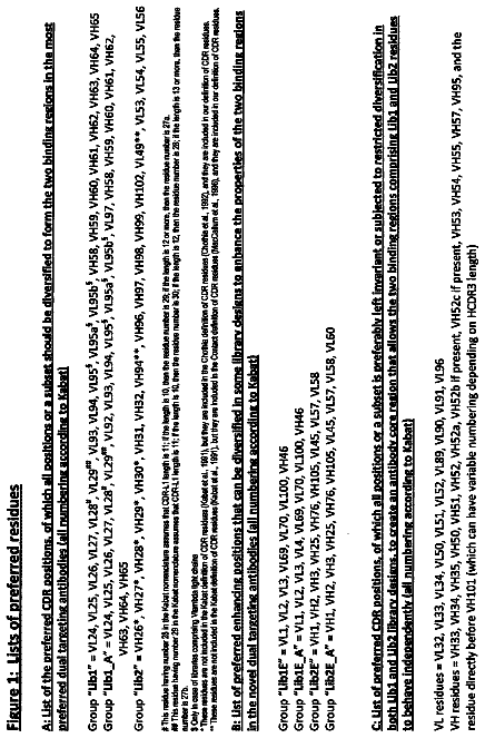
In one embodiment, the invention provides an antibody that binds a TLR3 polypeptide, wherein the antibody comprises (i) the heavy chain CDR 1, 2 and 3 (HCDR1, HCDR2, HCDR3) amino acid sequences as shown in SEQ ID NO: 5, 6 or 7 (HCDR1), 8 or 9 (HCDR2) and 10 (HCDR3), and (ii) the light chain CDR 1, 2 and 3 (LCDR1, LCDR2, LCDR3) amino acid sequences as shown in SEQ ID NO: 11, 12 and 13, respectively; wherein one, two, three, four, or five or more of the amino acids in any of said sequences may be substituted by a different amino acid. In one embodiment, the antibody comprises (i) the heavy chain CDR 1, 2 and 3 (HCDR1, HCDR2, HCDR3) amino acid sequences as shown in SEQ ID NO: 16, 17 or 18 (HCDR1), 19 or 20 (HCDR2) and 21 (HCDR3), and (ii) the light chain CDR 1, 2 and 3 (LCDR1, LCDR2, LCDR3) amino acid sequences as shown in SEQ ID NO: 22, 23 and 24, respectively; wherein one, two, three, four, or five or more of the amino acids in any of said sequences may be substituted by a different amino acid. In one embodiment, the antibody comprises (i) the heavy chain CDR 1, 2 and 3 (HCDR1, HCDR2, HCDR3) amino acid sequences as shown in SEQ ID NO: 27, 28 or 29 (HCDR1), 30 or 31(HCDR2) and 32 (HCDR3), and (ii) the light chain CDR 1, 2 and 3 (LCDR1, LCDR2, LCDR3) amino acid sequences as shown in SEQ ID NO: 33, 34 and 35, respectively; wherein one, two, three, four, or five or more of the amino acids in any of said sequences may be substituted by a different amino acid. In one embodiment, the antibody comprises (i) the heavy chain CDR 1, 2 and 3 (HCDR1, HCDR2, HCDR3) amino acid sequences as shown in SEQ ID NO: 38, 39 or 40 (HCDR1), 41 or 42 (HCDR2) and 43 (HCDR3), and (ii) the light chain CDR 1, 2 and 3 (LCDR1, LCDR2, LCDR3) amino acid sequences as shown in SEQ ID NO: 44, 45 and 46, respectively; wherein one, two, three, four, or five or more of the amino acids in any of said sequences may be substituted by a different amino acid. In one embodiment, the antibody comprises (i) the heavy chain CDR 1, 2 and 3 (HCDR1, HCDR2, HCDR3) amino acid sequences as shown in SEQ ID NO: 49, 50 or 51 (HCDR1), 52 or 53 (HCDR2) and 54 (HCDR3), and (ii) the light chain CDR 1, 2 and 3 (LCDR1, LCDR2, LCDR3) amino acid sequences as shown in SEQ ID NO: 55, 56 and 57, respectively; wherein one, two, three, four, or five or more of the amino acids in any of said sequences may be substituted by a different amino acid.
In another embodiment, the antibody of any of the embodiments herein is capable of being internalized by a cell that expresses TLR3 polypeptide on its surface.
In one embodiment, the antibody is chimeric, e.g. contains a non-murine, optionally a human, constant region. In one embodiment, the antibody is human or humanized. In another embodiment, the antibody is a mouse or rat antibody (e.g., comprises CDRs derived from a rat or rat gene or rat Ig locus gene segment). In another embodiment, the antibody does not substantially bind to other human TLRs (e.g. TLR4).
In one aspect of any of the embodiments of the invention, the isotype of the antibody is IgG, optionally IgG1 or IgG3. In one embodiment the antibody comprises an Fc domain or is of an isotype that is bound by FcγR.
In one aspect of any of the embodiments of the invention, the antibody is an antibody fragment selected from Fab, Fab′, Fab′-SH, F(ab′)2, Fv, diabodies, single-chain antibody fragment, or a multispecific antibody comprising multiple different antibody fragments. In one aspect of any of the embodiments of the invention, the antibody does not comprise an Fc domain or is of an isotype that is not substantially bound by FcγR (e.g. human CD16). In one embodiment, the antibody is of human IgG4 or IgG2 isotype. Human IgG4 isotypes or other IgG isotypes modified to reduce their FcγR binding can be used for their advantageous pharmacological properties such as serum half-life, while modulating TLR3 signaling, in e.g. a DC, without inducing the death of the cell. In one aspect of any of the embodiments of the invention, the anti-TLR3 antibody inhibits TLR3 signaling and comprises a constant region of human IgG4 or IgG2 isotype. In one aspect, of any of the embodiments of the invention, the anti-TLR3 antibody inhibits TLR3 signaling and comprises a constant region (heavy chain constant region) that does not substantially bind FcγRIIIa.
In one preferred embodiment, the anti-TLR3 antibody comprises a heavy chain of human IgG4 isotype. In one embodiment, the anti-TLR3 antibody comprises a human IgG4 heavy chain constant region and comprising a serine to proline mutation in residue 241, corresponding to position 228 according to the EU-index (Kabat et al., “Sequences of proteins of immunological interest”, 5.sup.th ed., NIH, Bethesda, Md., 1991). Compositions comprising such antibodies can be characterized as having less than about 15%, such as less than about 10% (e.g., about 5% or less, about 4% or less, about 3% or less, or even about 1% or less) of IgG4 “half-antibodies” (comprising a single heavy chain/light chain pair). Such IgG4 “half-antibody” by-products form due to heterogeneity of inter-heavy chain disulfide bridges in the hinge region in a proportion of secreted human IgG4 (see Angal et al., Molecular Immunology, 30(1):105-108, 1993 for a description of IgG4 “half-antibodies”, S241P mutation, and related principles). This effect is typically only detectable under denaturing, non-reducing conditions.
In another embodiment, the antibody is conjugated or covalently bound to a detectable or toxic moiety.
In one aspect, the antibodies optionally inhibit TLR3 signaling without blocking binding of a dsRNA TLR3 ligand to the principal (i.e. C-terminal) dsRNA binding site of the TLR3 polypeptide.
In one aspect, the antibodies also bind human TLR3 under acidic conditions, and in particular under conditions representative of that encountered in an acidified subcellular compartment of a cell (e.g. compartments of the endocytic pathway endosomic, lysosomal). Such acidic conditions are generally characterized by a pH lower than about pH 6.5, or between about pH 4.5 to 6.5, or about pH 5.6.
In one aspect of any of the embodiments herein, the antibodies modulate, optionally inhibit, TLR3 signaling in an acidified subcellular compartment of a cell (e.g. compartments of the endocytic pathway endosomic, lysosomal).
In one aspect of any of the embodiments herein, the antibodies modulate, optionally inhibit, TLR3 signaling in a dendritic cell (DC) (e.g. a myeloid DC, monocyte derived DC).
In other aspects of any of the embodiments herein, the antibodies' bivalent binding affinity for TLR3 under neutral and/or acidic conditions can optionally be characterized by a mean K.sub.D of no more than about (i.e. better affinity than) 100, 50, 10, 5, or 1 nanomolar, preferably sub-nanomolar or optionally no more than about 500, 200, 100 or 10 picomolar.
In other aspects of any of the embodiments herein, the antibodies inhibit TLR3 signaling by at least partly (or fully) blocking the binding of a TLR3 ligand to a TLR3 polypeptide. The TLR3 ligand will generally be a ligand other than an anti-TLR3 antibody and may be a naturally occurring or non-naturally occurring TLR3 ligand, optionally a dsRNA-based ligand such as polyAU (polyadenylic acid:polyuridylic acid) or polyIC (polyinosinic:polycytidylic acid).
In another aspect, the invention provides a method of identifying, screening and/or producing an antibody that specifically binds and inhibits a TLR3 polypeptide in a mammalian subject, said method comprising the steps of: a) providing a plurality of antibodies that bind human TLR3 polypeptide, optionally by a method comprising immunizing a non-human mammal with an immunogen comprising a TLR3 polypeptide, and (b) assessing the binding affinity of said antibodies for the TLR3 polypeptide in a cell which expresses human TLR3 solely at the cell surface. Optionally, the method further comprises selecting an antibody from said plurality that has an EC.sub.50 of no more than 0.3 μg/ml, optionally no more than 0.2 μg/ml, optionally no more than 0.1 μg/ml, for binding to cells expressing human TLR3 solely at the cell surface.
Any of the methods of the invention can further be characterized as comprising any step described in the application, including notably in the “Detailed Description of the Invention”). The invention further relates to an antibody obtainable by any of present methods. The invention further relates to pharmaceutical or diagnostic formulations of the antibodies of the present invention. The invention further relates to methods of using antibodies in methods of treatment or diagnosis, optionally in combination with a second therapeutic agent (e.g. a corticoid, a DMARD, an anti-cytokine or anti-cytokine receptor agent, an anti-TNFalpha agent, etc.).
These and additional advantageous aspects and features of the invention may be further described elsewhere herein.
Brief description of the drawings
FIGS. 1A, 1B, 1C and 1D show in vitro dose effect for binding to HEK293T cells transiently transfected with a TLR3 ECD construct and expressing TLR3 solely at their surface for antibodies 11E1, 7G11, 32C4 and 31F6, respectively. Each antibody is compared with reference antibody 31C3; binding to cell surface TLR3 is improved for antibodies 11E1, 7G11, 32C4 and 31F6.
FIGS. 2A, 2B, 2C, 2D and 2E show dose dependent inhibition of TLR3 signaling using a 293T-human TLR3 luciferase assay with the human anti-TLR3 antibodies. Each antibody is compared with reference antibody 31C3; inhibition of dsRNA-induced TLR3 signaling is improved for antibodies 11E1, 7G11, 32C4, 31F6 and 37B7.
FIGS. 2F, 2G, and 2H show view of the N-terminal end of the TLR3 polypeptide, showing amino acid residues mutated indicated in black and residues adjacent to residues which form part of the principle epitopes in grey. FIG. 2F shows a view of the glycan-free lateral surface of the TLR3 polypeptide, with the N-terminal end of the TLR3 polypeptide at the right of the image); FIG. 2G shows a view of the glycan-containing lateral surface of the TLR3 polypeptide and the backbone, with the N-terminal end of the TLR3 polypeptide in the foreground (at the left of the image). FIG. 2H shows a view of the glycan-free lateral surface of the TLR3 polypeptide and the backbone, with the N-terminal end of the TLR3 polypeptide in the foreground (at the right of the image).
FIG. 3 shows results of a rheumatoid arthritis mouse models. FIG. 3A shows the results of a preventive rheumatoid arthritis mouse model. FIG. 3B shows the results of a curative rheumatoid arthritis mouse model. FIG. 3C shows the results of a curative rheumatoid arthritis mouse model when mice are treated with PBS, a control antibody, 28G7 and an anti-TNFα antibody (Humira™).
FIG. 4 shows results of the mouse colitis model. FIG. 4A shows the wall thickness measurements for the mice treated with saline (black dots), with TNBS only (black squares) with an anti-TNFα antibody and TNBS (black triangles), with 28G7 and TNBS (open dots), and with a control Ab and TNBS (open squares). FIG. 4B shows the macroscopic damage score for the mice treated with saline (black dots), with TNBS only (black squares) with an anti-TNFα antibody and TNBS (black triangles), with 28G7 and TNBS (open dots), and with a control Ab and TNBS (open squares). The anti-TLR3 antibody according to the invention ameliorating the development of the disease, under stringent conditions. (*p<0.05, **p<0.01 vs saline).
FIG. 5 shows results of a COPD mouse model. FIG. 5A shows BAL differential cell counts for macrophages, eosinophils, neutrophils and lymphocytes. The anti-TLR3 antibodies strongly decreased the infiltration of neutrophils into the airways, while not substantially affecting macrophages eosinophils or lymphocytes. FIG. 5B shows venous blood saturated oxygen (in percent) for each of LPS/elastase alone and LPS/elastase in combination with anti-TLR3 antibodies or roflumilast. FIG. 5C shows IL17A in BAL fluid (BALF), where anti-TLR3 antibodies decreased IL17A (pg/ml) substantially, and as much as roflumilast. FIG. 5D shows IP-10 in BALF, where -TLR3 antibodies decreased IP-10 (pg/ml) substantially. FIG. 5E shows BAL differential cell counts for macrophages, neutrophils and lymphocytes for a second study comparing anti-TLR3 antibodies (28G7), roflumilast (Rofu) and the combination of roflumilast and anti-TLR3 antibodies (combo).
FIG. 6 shows results of a CLP (cecal ligation and puncture—sepsis) mouse model. In this acute model, mice experience an acute infection, mimicking septic shock.
FIGS. 7A and 7B show results of drug combinations with anti-human TLR3 mAbs (labeled IPH33.1) in combination with dexamethasone or Humira® respectively. Antibodies each substantially reduce in vitro IP-10 production by PBMC in response to polyIC further when combined with dexamethasone or Humira®.
Detailed description of the invention
Introduction
The present invention is based, at least in part, on the discovery of high affinity monoclonal antibodies that specifically and efficiently inhibit the TLR3 signaling pathway. The inventors also provide new epitopes present on human TLR3, including the epitope recognized by antibody 11E1, 7G11, 31F6, 32C4 and/or 37B7, which are particularly efficient in inhibiting TLR3 signaling, and inhibiting cytokine release in response to stimulation with a TLR3 ligand.
The antibodies can be used for treating an autoimmune or inflammatory disease in a subject in need thereof. The present invention also provides methods for treating relapses, attacks, or acute phases, occurring during the course of an inflammatory or autoimmune disease in a subject in need thereof using an anti-TLR3 antibody which inhibits TLR3 signaling. The present invention also provides novel methods for treating established inflammatory or autoimmune diseases in a subject in need thereof using an anti-TLR3 antibody which inhibits TLR3 signaling. The invention also provides treatment regimens and treatment combinations that can be used for the treatment of inflammatory or autoimmune disease in a subject in need thereof using an anti-TLR3 antibody which inhibits TLR3 signaling.
The antibodies of the present invention that bind TLR3 under acidic and neutral conditions will generally bind both cell surface TLR3 and endosomic TLR3 at high affinity, such that the antibodies will be useful in any situation (e.g. treatment or prevention of disease) where targeting (e.g. modulating) TLR3 is useful. TLR3 has been found in some cases of inflammation the surface of macrophages and blocking TLR3 upon chloroquine neutralization of endosomal acidification nevertheless exhibited some anti-inflammatory activity (Cavassani et al. 2008, supra). However, the antibodies of the invention will have the greatest advantage over other antibodies in the treatment or prevention of diseases where the modulating (e.g. inhibiting) the signaling by TLR3 in the cytosolic (e.g. endosomic) compartments is useful or required, and the relative importance of modulating signaling of such compartments TLR3 may depend on the disease. One example of such as disease is rheumatoid arthritis; endosomic compartment-expressed TLR3 is believed to play an important role in rheumatoid arthritis, since treatment with chloroquine, an inhibitor of endosomal acidification, inhibits TLR3 signaling and inhibits production of inflammatory cytokines from synovial cultures from patients having rheumatoid arthritis (Sacre et al.
J. Immunol. 181:8002-8009). Endosomic compartment-expressed TLR3 is believed to play an important role in a number of other diseases where DC (e.g. myeloid DC) are involved in exacerbating disease, as mDC have a well-documented capacity to take up antigens from apoptotic or necrotic cells including during tissue necrosis during acute inflammation.
Since the present antibodies are specific for TLR3, they can also be used for other purposes, including purifying TLR3 or TLR3-expressing cells, modulating (e.g. activating or inhibiting) TLR3 receptors in vitro, ex vivo, or in vivo, targeting TLR3-expressing cells for destruction in vivo, or specifically labeling/binding TLR3 in vivo, ex vivo, or in vitro, including for methods such as immunoblotting, IHC analysis, i.e. on frozen biopsies, FACS analysis, and immunoprecipitation.
Definitions
As used herein, “TLR3 ligand” refers to any compound that can specifically bind to and alter the activity of TLR3 in vitro, ex vivo, or in vivo. The compound can be a naturally occurring ligand, e.g., generally dsRNA or viral dsRNA, or a synthetic ligand such as polyIC or polyAU. The compound can be any type of molecule, including inorganic or organic compounds or elements, including proteins (such as antibodies), nucleic acids, carbohydrates, lipids, or any other molecular entity. Further, such compounds can modulate TLR3 receptors in any way, including activating or inhibiting, and by any mechanism, including by binding to the receptor and triggering or shutting off activity in a manner similar to a naturally occurring ligand, or by binding to the receptor and blocking access to other ligands. Preferably, the ligand activates the receptor, and as such can be used to induce the production of cytokines by TLR3-expressing cells.
The description continues in the full USPTO document.
 Lapsed, fee not paid5 drawings