Background
Prior methods and apparatus of non-invasively measuring subjects optically are less than ideal in at least some respects. For example, prior methods and apparatus of measuring blood of a subject can be less accurate than would be ideal. Also, alignment of the subject with the measurement apparatus can be challenging. Many of the prior art spectrometers are less than ideally suited to measure constituents non-invasively in vivo. Although prior methods and apparatus may measure blood spectroscopically in a controlled laboratory environment, these methods generally involve the removal of blood from the subject, which can be inconvenient and somewhat painful in at least some instances.
Summary
The specialized spectrometer methods and apparatus disclosed herein are particularly well suited for accurate non-invasive measurements of subjects. Although reference is made to measuring constituents of blood, the methods and apparatus disclosed herein can be used to non-invasively measure constituents in many tissues and bodily fluids including tears, saliva, or sweat.
In many embodiments, the specialized spectrometer comprises a light source to generate a monochromatic light beam to generate resonance Raman peaks from a region of the subject and a reference light source to illuminate the region with the reference light beam. The light energy from the resonance Raman peaks can be measured with a detector coupled to an optical wavelength separator. A portion of the reference beam scattered from the region can be measured with a reference detector. An amount of the constituent can be determined with a processor in response to the resonance Raman peaks measured with the detector and the portion of the reference beam measured with the reference detector. The illumination of the region with reference beam with the monochromatic beam allows accurate determination of the amount of the constituent, and the amount may comprise a concentration, such as an amount per unit volume. The constituent measured with resonance Raman spectroscopy may comprise a constituent of blood, such as a molecule, and the portion of scattered light measured with the reference detector may correspond to a second constituent of blood that that provides a measurable signal with the reference beam, for example hemoglobin. The spectrometer can be configured in many ways, such as with a table top configuration or wearable configuration carried by the subject.
The methods and apparatus disclosed herein can also provide improved safety. The processor can be configured with instructions to detect the presence of blood vessels with the reference beam in order to ensure that an appropriate region of the subject is aligned with the delivery system, and the processor configured with instructions to direct the monochromatic beam to the region in response to the portion of the reference beam measured with the detector. This approach can allow for increased amounts of light energy of the monochromatic beam when used near delicate structures such as an eye.
In one aspect, disclosed herein is a spectrometer to non-invasively measure a blood constituent through an epithelium and vessel walls of blood vessels at a region of a subject with light. The spectrometer comprises a light source to generate a monochromatic light beam, the monochromatic beam comprising one or more wavelengths having a frequency at an electronic excitation of the blood constituent to generate resonance Raman peaks. The spectrometer further comprises an optical system to deliver the monochromatic light beam through the epithelium and vessel walls to the blood constituent at the region, and receive light energy from the blood constituent through the epithelium and the vessel walls. The optical system may comprise an optical wavelength separator to selectively transmit the resonance Raman peaks. The spectrometer further comprises a detector coupled to the optical delivery system to measure the resonance Raman peaks from the blood constituent at the region. The optical system further comprises a reference light source to generate a reference beam to illuminate blood at the region. The optical system further comprises a reference detector to measure a portion of the reference beam backscattered from the blood at the region. The spectrometer further comprises a processor coupled to the detector. The processor may be configured with instructions to measure the resonance Raman peaks from the detector and the portion from the reference detector in order to determine an amount of the blood constituent from the resonance Raman peaks and the portion of the reference beam backscattered from the blood at the region.
The processor may be coupled to the light source and configured to transmit the monochromatic light beam to the region with an amount of energy greater than the reference beam. The processor may be configured to detect a presence of the blood vessels with the portion of the reference beam and transmit the monochromatic beam in response to the presence of the blood vessel.
The amount of energy of the monochromatic beam may exceed an eye safe threshold of the measurement beam, and the reference beam may not exceed an eye safe threshold of the reference beam. The processor may be configured to transmit the reference beam and not to transmit the monochromatic light beam when the presence of the blood vessels has not been detected.
The region may comprise a volume of blood and the reference beam and reference detector may be configured to measure a second constituent of blood, wherein the second constituent may comprise a greater amount by volume than the constituent of interest.
The optical system may be configured to focus the monochromatic light beam on a blood vessel beneath the epithelium. The optical system may be further configured to receive the resonance Raman peaks from the blood constituent through the epithelium and direct the resonance Raman peaks to the detector. The optical system may comprise a focusing element to collect the backscattered light from a region of the tissue. The focusing element may comprise one or more of a lens, an objective lens, a reflector, a curved reflector, a spherical reflector, or a curved reflector with an aperture, a concave reflector with an aperture, a convex reflector, a convex reflector aligned with an aperture of a concave reflector or Schwarzschild optics. The focusing element may be configured to receive light from the region and transmit the resonance Raman peaks in a substantially collimated configuration toward the detector.
The monochromatic light source may be transmitted toward the region in a substantially collimated configuration. The optical system may comprise an optical path along which the resonance Raman peaks and the portion are transmitted coaxially toward the detector and the reference detector.
The processor may be coupled to the reference detector and configured to determine the amount of the constituent in response to an intensity of the portion of the beam received from the tissue and an intensity of the resonance Raman peaks. The processor may be coupled to the reference detector and configured to determine a concentration of the constituent in blood in response to an intensity of the portion of the beam received from the tissue and an intensity of the resonance Raman peaks. The processor may be coupled to the reference detector and configured to determine a concentration of the constituent in response to an intensity of the portion of the beam received from the tissue and an intensity of the resonance Raman peaks, the concentration comprising the amount of the constituent per unit volume of the blood.
The wavelength separator may be configured to separate non-resonance Raman light energy from the resonance Raman peaks, wherein the non-resonance Raman light energy may comprise one or more of fluorescent light, light having wavelengths of the monochromatic illumination beam, ambient light or other parasitic light.
An electronic excitation of the constituent may correspond to a precisely excited state of the biomarker. An electronic excitation of the constituent may be above a virtual state corresponding to Raman emission of the constituent.
The monochromatic light beam may comprise a bandwidth of no more than about ten nanometers. The monochromatic light beam may comprise a bandwidth of no more than about three nanometers.
The laser may comprise a tunable laser, wherein a bandwidth of the tunable laser may comprise bandwidth of no more than about a nanometer when tuned to a wavelength.
The wavelength separator may comprise one or more of a prism, a granting, a mirror, an etalon, an optical filter or a plurality of optical filters.
The detector may comprise one or more of a plurality of detectors or a plurality of detector elements.
The blood constituent may comprise a biomarker. The biomarker may comprise one or more of Glucose, Troponin Complex, Troponin T (TnT), Troponin I (TnI), Troponin C (TnC), a cardiac biomarker, a Troponin cardiac biomarker, a breast cancer biomarker, Breast Cancer 1 Biomarker (BRCA1), Breast Cancer 2 Biomarker (BRCA2), a biomarker related to coronary disease, B-type Natriuretic Peptide (BNP) and N-terminal proBNP (NT-proBNP), an infection specific biomarker, a biomarker related to dementia, Beta Amyloid, Low Density Lipoprotein (LDL), High Density Lipoprotein (HDL), Cholesterol, a Triglyceride, a Thyroid Stimulating Hormone (TSH), Creatine Kinase, Prostate Specific Antigen (PSA), Creatinine, Globulin, Adenovirus DNA, Alanine Aminotransferase (ALT/SGPT), Albumin, Alkaline Phosphatase (ALP), Alpha-1-Acid Glycoprotein, Alpha-1-Antitrypsin, Alpha-Fetoprotein (AFP), Amphetamines, Amylase, Androstenedione, RBC Antibody Detection, Anti-Mullerian Hormone (AMH), Antinuclear Antibodies, Apolipoprotein (apo A-1, apo B), Apolipoprotein A-1 (apo A-1), Apolipoprotein B (apo B), Aspartate Aminotransferase (AST/SGOT), B Cell, Barbiturates, Benzodiazepines, Beta-2 Microglobulin, Bilirubin, Blood Type (ABO/RhD), Blood Urea Nitrogen (BUN), Borrelia Antibody (Lyme Disease), Calcitonin, Calcium, Cancer Antigen 125 (CA 125), Cancer Antigen 15-3 (CA 15-3), Cancer Antigen 27.29 (CA 27.29), Cancer Antigen-GI (CA 19-9), Carbon Dioxide, Carcinoembryonic Antigen (CEA), Cardiolipin Antibody (ACA), CBC (Complete Blood Count), CD4, CD8, Celiac Panel, Chlamydia Trachomatis, Gonorrhea, Chloride, Cholinesterase, Cocaine, Complement Component 3 antigen, Complement Component 4 antigen, Cortisol, C-Peptide, C-Reactive Protein (CRP), Cyclic Citrullinated Peptide (CCP) Antibody, Cyclosporine A, Cystatin C, Cytomegalovirus (CMV) Antibody, Cytomegalovirus (CMV) Antibody, D-Dimer, Deamidated Gliadin Peptide (DGP) Antibody, Deamidated Gliadin Peptide (DGP) Antibody, Dehydroepiandrosterone Sulfate (DHEA-S), Deoxypyridinoline crosslinks (DPD) (Collagen crosslinks), Double-stranded DNA (dsDNA) Antibody, EBV Early D Antigen (EA-D), EBV Nuclear Antibody, EBV Viral Capsid Antigen (VCA), EBV Viral Capsid Antigen (VCA), Ecstasy (MDMA), Endomysial Antibody (EMA), Endomysial Antibody (EMA), Epstein-Barr (EBV) Antibody, Erythrocyte Sedimentation Rate (ESR/Sed Rate), Estradiol, Estriol, Estrone, Ethanol, Extractable, Ferritin, Fibrinogen, Folate (Folic acid), Follicle Stimulating Hormone (FSH), Gamma-Glutamyltransferase (GGT), Gastrin, Glucose, Growth Hormone (HGH), chronic Haptoglobin (hCG), Helicobacter Pylori ( H. Pylori ), Hematocrit (HCT), Hemoglobin (HGB), Hemoglobin Alc (HbAlc), Hemogram 2, Hepatitis A (HAV) Antibody, Hepatitis A (HAV) Antibody, Hepatitis B (HBV) Core Antibody, Hepatitis B (HBV) Core Antibody, Hepatitis B (HBV) Surface Antibody (HBsAb), Hepatitis B (HBV), Hepatitis C (HCV) Antibody, HER-2/neu, Herpes Simplex 1 (HSV1), Herpes Simplex 2 (HSV2), HIV-1, Homocysteine, IGF-1 (Insulin-like Growth Factor 1), Insulin, Iron, Lactate Dehydrogenase, Lead, Lipase, Lithium, Luteinizing Hormone (LH), Magnesium, Marijuana (THC), Measles, Mumps, and Rubella (MMR) Immunity, Methadone (dolophine), Methamphetamines, Microalbumin, Mumps Antibody, Myoglobin, N. Gonorrhea, Natural Killer Cells, Nuclear Antigen Antibody Jo-1, Nuclear Antigen Antibody RNP, Nuclear Antigen Antibody Sc1-70, Nuclear Antigen Antibody Sm, Nuclear Antigen Antibody SSA, Nuclear Antigen Antibody SSB, Opiates, Parathyroid Hormone (PTH), Phencyclidine (PCP), Phosphorus, Platelets, Potassium, Prealbumin, Progesterone, Prolactin, Propoxyphene, Reticulocyte Count, Rheumatoid Factor, Rubella Antibody, Rubeola (Measles) Antibody, Sex Hormone-binding Globulin (SHBG), Sodium, Streptolysin O Antibody, Treponema Pallidum Antibody, T Cell, Triiodothyronine (T3), Testosterone, Thyroglobulin, Thyroglobulin Antibodies (TAA), Thyroid Peroxidase (TPO) Antibody, Thyroxine Binding Globulin (TBG), Thyroxine (T4), Tissue Transglutaminase (tTG) Antibody, Toxoplasma, Transferrin, Treponema Pallidum Antibody, Triiodothyronine (FT3), Uric Acid, Varicella-Zoster (VZV) Antibody, Vitamin B-12, Vitamin D 25-OH, or WBC.
The light source, the reference light source, the optical delivery system, the detector and the reference detector may be configured to be worn by the subject.
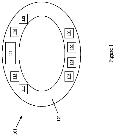
The spectrometer may further comprise a support shaped to couple to a head of the subject and support the light source, the reference light source, the optical delivery system, the detector and the reference detector with the head of the subject. The support and the light source, the reference light source, the optical delivery system, the detector and the reference detector may have a weight within a range from about 150 grams to about 250 grams. The support and the light source, the reference light source, the optical delivery system, the detector and the reference detector may weigh no more than about 125 grams. The support may comprise one or more of an eyeglass frame, a helmet, goggles, or a spiral extension to wrap around a head of the subject. The support may be configured to support the processor and a power supply and wireless communication circuitry. The processor and the power supply may be located away from the light source, the reference light source, the optical delivery system, the detector and the reference detector in order to distribute weight of the support when placed on the head of the subject. The support may comprise a cover to inhibit ambient light from illuminating the region.
The spectrometer may further comprise a support configured to support the light source, the reference light source, the optical delivery system, the detector and the reference detector, wherein the support is configured for placement on a table top.
The spectrometer may further comprise a support shaped to support a chin of the subject in order to align the light source, the reference light source, the optical delivery system, the detector and the reference detector.
The support may comprise a visual illuminator visible to the user to direct a gaze of the viewer in order to align the tissue with the optical system. The visible illuminator may be located on a first side of the eye on the support and the optical system and detector may be located on a second side of the eye on the support in order to measure a conjunctiva of the eye when the subject views the visible illuminator.
The blood vessels may be part of a conjunctiva of an eye of the subject or a tympanic membrane of an ear of the subject. The epithelium may comprise an epithelium of the one or more of the conjunctiva of the eye of the subject or the tympanic membrane of the ear of the subject.
One or more of the reference beam or an additional light beam and one or more of the reference detector or another detector may be configured to measure absorption of the region. One or more of the reference beam or an additional light beam and one or more of the reference detector or another detector may be configured to measure near infrared light energy of the region. One or more of the reference beam or an additional light beam and one or more of the reference detector or another detector may be configured to measure near infrared spectra of the region.
The processor may be configured to repeatedly measure the Raman peaks and the portion with a plurality of measurements to increase a signal to noise ratio. The processor may be configured with instructions to extract data from signals from the detector and the reference detector in response to the resonance Raman peaks and the portion of the reference beam using signal processing comprising one or more of filtering, subtraction of a baseline, a Fourier transform, a wavelet transform, a partial least square regression, a differentiation, or a ratio of a signal from the detector to a ratio of a signal from the reference detector.
The reference beam may comprise a calibration beam, and the amount may be determined in response to a ratio of a resonance Raman signal from the detector to a calibration signal from the portion of the beam received with the reference detector.
The monochromatic light beam and the reference beam may be arranged in a coaxial configuration to direct the monochromatic light beam and the reference beam along a common axis toward the region. The monochromatic light beam and the reference beam may be arranged in a confocal configuration to focus the monochromatic light beam and the reference beam together on a tissue volume of the region.
The spectrometer may further comprise a camera configured with an optic to view the blood vessels of the region and align the monochromatic beam and the measurement beam with the region.
The region may comprise a volume located below the epithelium.
The portion of the reference beam backscattered from the blood at the region may comprise one or more of light having wavelengths of the reference beam, secondary radiation from the reference beam, or fluorescence from the reference beam.
The spectrometer may further comprise instructions to determine the amount of the blood constituent from the resonance Raman peaks and the portion of the reference beam backscattered from the blood at the region with one or more of a processor electrically coupled to the detector and the reference detector, a processor of a mobile computing device, or a processor of a remote server. The processor electrically coupled to the detector and the reference detector may comprise the instructions to determine the amount of the blood constituent from the resonance Raman peaks and the portion of the reference beam backscattered from the blood at the region. The processor of the mobile computing device may comprise the instructions to determine the amount of the blood constituent from the resonance Raman peaks and the portion of the reference beam backscattered from the blood at the region. The remote server may comprise the instructions to determine the amount of the blood constituent from the resonance Raman peaks and the portion of the reference beam backscattered from the blood at the region. The mobile computing device may comprise instructions to display the amount to a user of the spectrometer.
The spectrometer may further comprise a plurality of structures configured for insertion and removal from the spectrometer by a user to measure a plurality of blood constituents each having different Raman peaks and different resonance Raman excitation wavelengths. The plurality of structures may comprise one or more of a plurality of cartridges or a plurality of arms.
In another aspect, disclosed herein is a head worn spectrometer to measure a blood constituent of a subject. The head worn spectrometer comprises a light source to generate a monochromatic light beam, the monochromatic beam comprising one or more wavelengths having a frequency at an electronic excitation of the blood constituent to generate resonance Raman peaks. The head worn spectrometer further comprises an optical system to deliver the monochromatic beam through the epithelium and vessel walls to the blood constituent at the region and receive light energy from the blood constituent through the epithelium and the vessel walls. The optical system may comprise an optical wavelength separator to selectively transmit resonance the Raman peaks. The head worn spectrometer further comprises a detector coupled to the optical delivery system to measure the resonance Raman peaks from the blood constituent at the region. The head worn spectrometer further comprises a reference light source to generate a reference beam to illuminate blood at the region, and a reference detector to measure a portion of the reference beam backscattered from the blood at the region. The head worn spectrometer further comprises a support to couple to the head of the wearer and support the light source, the optical system, the detector, the reference light source and the reference detector. The head worn spectrometer further comprises a processor coupled to the light source, the reference light source, the detector and the reference detector to measure the resonance Raman peaks and the portion of backscatter from the reference beam. The head worn spectrometer may comprise a weight within a range from about 150 grams to about 250 grams.
In another aspect, disclosed herein is a method of non-invasively measuring a blood constituent through an epithelium and vessel walls of blood vessels at a region of a subject with light with a spectrometer. The method comprises generating a monochromatic light beam with a light source, the monochromatic beam comprising one or more wavelengths having a frequency at an electronic excitation of the blood constituent to generate resonance Raman peaks. The method further comprises delivering the monochromatic light beam through the epithelium vessel walls to the blood constituent at the region with an optical delivery system. The method further comprises receiving with an optical separator light energy from the blood constituent through the epithelium and the vessel walls to selectively transmit the resonance Raman peaks. The method further comprises measuring the resonance Raman peaks from the blood constituent at the region with an optical detector. The method further comprises generating a reference beam to illuminate blood at the region with the reference light beam, and measuring a portion of the reference beam backscattered from the blood at the region with a reference detector in order to determine an amount of the blood constituent from the resonance Raman peaks and the portion of the reference beam backscattered from the blood at the region.
The method may further comprise determining the amount of the blood constituent from the resonance Raman peaks and the portion of the reference beam backscattered from the blood at the region with one or more of a processor electrically coupled to the detector and the reference detector, a processor of a mobile computing device, or a processor of a remote server.
The method may further comprise placing a spectral device anterior to an eye of the subject with the eye covered to inhibit ambient light from reaching the region.
In some embodiments of the method, gaze of the subject may be directed away from a spectrometer in order to measure a conjunctiva of an eye of the subject. In some embodiments of the method, a position of the eye may be verified by one or more of a measurement beam or an operator of the spectrometer.
The region may comprise a volume of blood and the reference beam may measure a second constituent of blood, the second constituent comprising a greater amount by volume than the constituent of interest.
The processor may be coupled to the light source and configured to transmit the monochromatic light beam to the region with an amount of energy greater than the reference beam. The processor may be further configured to detect a presence of the blood vessels with the portion of the reference beam and transmit the monochromatic beam in response to the presence of the blood vessel. The amount of energy of the monochromatic beam may exceed a retina safe threshold of the measurement beam and the reference beam may not exceed a retina safe threshold of the reference beam. The processor may be configured to transmit the reference beam and not to transmit the monochromatic light beam when the presence of the blood vessels has not been detected.
The optical system may be configured to focus the monochromatic light beam on a blood vessel beneath the epithelium and the optical system may be configured to receive the resonance Raman peaks from the blood constituent through the epithelium and direct the resonance Raman peaks to the detector. The optical system may comprise a focusing element to collect the backscattered light from a region of the tissue. The focusing element may comprise one or more of a lens, an objective lens, a reflector, a curved reflector, a spherical reflector, or a curved reflector with an aperture, a concave reflector with an aperture, a convex reflector, a convex reflector aligned with an aperture of a concave reflector or Schwarzschild optics. The focusing element may be configured to receive light from the region and transmit the resonance Raman peaks in a substantially collimated configuration toward the detector.
The monochromatic light source may be transmitted toward the region in a substantially collimated configuration. The optical system may comprise an optical path along which the resonance Raman peaks and the portion are transmitted coaxially toward the detector and the reference detector.
The processor may be coupled to the reference detector and configured to determine the amount of the constituent in response to an intensity of the portion of the beam received from the tissue and an intensity of the resonance Raman peaks. The processor may be coupled to the reference detector and configured to determine a concentration of the constituent in blood in response to an intensity of the portion of the beam received from the tissue and an intensity of the resonance Raman peaks. The processor may be coupled to the reference detector and configured to determine a concentration of the constituent in response to an intensity of the portion of the beam received from the tissue and an intensity of the resonance Raman peaks, the concentration comprising the amount of the constituent per unit volume of the blood.
The wavelength separator may be configured to separate non-resonance Raman light energy from the resonance Raman peaks, wherein the non-resonance Raman light energy may comprise one or more of fluorescent light, light having wavelengths of the monochromatic illumination beam, ambient light or other parasitic light.
An electronic excitation of the constituent may correspond to a precisely excited state of the biomarker. An electronic excitation of the constituent may be above a virtual state corresponding to Raman emission of the constituent.
The monochromatic light beam may comprise a bandwidth of no more than about ten nanometers. The monochromatic light beam may comprise a bandwidth of no more than about three nanometers.
The laser may comprise a tunable laser wherein a bandwidth of the tunable laser may comprise a bandwidth of no more than about a nanometer when tuned to a wavelength.
The wavelength separator may comprise one or more of a prism, a granting, a mirror, an etalon, an optical filter or a plurality of optical filters.
The detector may comprise one or more of a plurality of detectors or a plurality of detector elements.
The blood constituent may comprise a biomarker. The biomarker may comprise one or more of Glucose, Troponin Complex, Troponin T (TnT), Troponin I (TnI), Troponin C (TnC), a cardiac biomarker, a Troponin cardiac biomarker, a breast cancer biomarker, Breast Cancer 1 Biomarker (BRCA1), Breast Cancer 2 Biomarker (BRCA2), a biomarker related to coronary disease, B-type Natriuretic Peptide (BNP) and N-terminal proBNP (NT-proBNP), an infection specific biomarker, a biomarker related to dementia, Beta Amyloid, Low Density Lipoprotein (LDL), High Density Lipoprotein (HDL), Cholesterol, a Triglyceride, a Thyroid Stimulating Hormone (TSH), Creatine Kinase, Prostate Specific Antigen (PSA), Creatinine, Globulin, Adenovirus DNA, Alanine Aminotransferase (ALT/SGPT), Albumin, Alkaline Phosphatase (ALP), Alpha-1-Acid Glycoprotein, Alpha-1-Antitrypsin, Alpha-Fetoprotein (AFP), Amphetamines, Amylase, Androstenedione, RBC Antibody Detection, Anti-Mullerian Hormone (AMH), Antinuclear Antibodies, Apolipoprotein (apo A-1, apo B), Apolipoprotein A-1 (apo A-1), Apolipoprotein B (apo B), Aspartate Aminotransferase (AST/SGOT), B Cell, Barbiturates, Benzodiazepines, Beta-2 Microglobulin, Bilirubin, Blood Type (ABO/RhD), Blood Urea Nitrogen (BUN), Borrelia Antibody (Lyme Disease), Calcitonin, Calcium, Cancer Antigen 125 (CA 125), Cancer Antigen 15-3 (CA 15-3), Cancer Antigen 27.29 (CA 27.29), Cancer Antigen-GI (CA 19-9), Carbon Dioxide, Carcinoembryonic Antigen (CEA), Cardiolipin Antibody (ACA), CBC (Complete Blood Count), CD4, CD8, Celiac Panel, Chlamydia Trachomatis, Gonorrhea, Chloride, Cholinesterase, Cocaine, Complement Component 3 antigen, Complement Component 4 antigen, Cortisol, C-Peptide, C-Reactive Protein (CRP), Cyclic Citrullinated Peptide (CCP) Antibody, Cyclosporine A, Cystatin C, Cytomegalovirus (CMV) Antibody, Cytomegalovirus (CMV) Antibody, D-Dimer, Deamidated Gliadin Peptide (DGP) Antibody, Deamidated Gliadin Peptide (DGP) Antibody, Dehydroepiandrosterone Sulfate (DHEA-S), Deoxypyridinoline crosslinks (DPD) (Collagen crosslinks), Double-stranded DNA (dsDNA) Antibody, EBV Early D Antigen (EA-D), EBV Nuclear Antibody, EBV Viral Capsid Antigen (VCA), EBV Viral Capsid Antigen (VCA), Ecstasy (MDMA), Endomysial Antibody (EMA), Endomysial Antibody (EMA), Epstein-Barr (EBV) Antibody, Erythrocyte Sedimentation Rate (ESR/Sed Rate), Estradiol, Estriol, Estrone, Ethanol, Extractable, Ferritin, Fibrinogen, Folate (Folic acid), Follicle Stimulating Hormone (FSH), Gamma-Glutamyltransferase (GGT), Gastrin, Glucose, Growth Hormone (HGH), chronic Haptoglobin (hCG), Helicobacter Pylori ( H. Pylori ), Hematocrit (HCT), Hemoglobin (HGB), Hemoglobin Alc (HbAlc), Hemogram 2, Hepatitis A (HAV) Antibody, Hepatitis A (HAV) Antibody, Hepatitis B (HBV) Core Antibody, Hepatitis B (HBV) Core Antibody, Hepatitis B (HBV) Surface Antibody (HBsAb), Hepatitis B (HBV), Hepatitis C (HCV) Antibody, HER-2/neu, Herpes Simplex 1 (HSV1), Herpes Simplex 2 (HSV2), HIV-1, Homocysteine, IGF-1 (Insulin-like Growth Factor 1), Insulin, Iron, Lactate Dehydrogenase, Lead, Lipase, Lithium, Luteinizing Hormone (LH), Magnesium, Marijuana (THC), Measles, Mumps, and Rubella (MMR) Immunity, Methadone (dolophine), Methamphetamines, Microalbumin, Mumps Antibody, Myoglobin, N. Gonorrhea, Natural Killer Cells, Nuclear Antigen Antibody Jo-1, Nuclear Antigen Antibody RNP, Nuclear Antigen Antibody Scl-70, Nuclear Antigen Antibody Sm, Nuclear Antigen Antibody SSA, Nuclear Antigen Antibody SSB, Opiates, Parathyroid Hormone (PTH), Phencyclidine (PCP), Phosphorus, Platelets, Potassium, Prealbumin, Progesterone, Prolactin, Propoxyphene, Reticulocyte Count, Rheumatoid Factor, Rubella Antibody, Rubeola (Measles) Antibody, Sex Hormone-binding Globulin (SHBG), Sodium, Streptolysin O Antibody, Treponema Pallidum Antibody, T Cell, Triiodothyronine (T3), Testosterone, Thyroglobulin, Thyroglobulin Antibodies (TAA), Thyroid Peroxidase (TPO) Antibody, Thyroxine Binding Globulin (TBG), Thyroxine (T4), Tissue Transglutaminase (tTG) Antibody, Toxoplasma, Transferrin, Treponema Pallidum Antibody, Triiodothyronine (FT3), Uric Acid, Varicella-Zoster (VZV) Antibody, Vitamin B-12, Vitamin D 25-OH, or WBC.
The light source, the reference light source, the optical delivery system, the detector and the reference detector may be configured to be worn by the subject.
The method may further comprise providing a support shaped to couple to a head of the subject and support the light source, the reference light source, the optical delivery system, the detector and the reference detector with the head of the subject. The support and the light source, the reference light source, the optical delivery system, the detector and the reference detector may weigh no more than about 250 grams. The support and the light source, the reference light source, the optical delivery system, the detector and the reference detector may weigh no more than about 125 grams. The support may comprise one or more of an eyeglass frame, a helmet, goggles, a spiral extension to wrap around a head of the subject. The support may be configured to support the processor and a power supply and wireless communication circuitry and the processor and the power supply may be located away from the light source, the reference light source, the optical delivery system, the detector and the reference detector in order to distribute weight of the support when placed on the head of the subject. The support may comprise a cover to inhibit ambient light from illuminating the region.
The method may further comprise providing a support configured to support the light source, the reference light source, the optical delivery system, the detector and the reference detector, wherein the support is configured for placement on a table top.
The method may further comprise providing a support shaped to support a chin of the subject in order to align the light source, the reference light source, the optical delivery system, the detector and the reference detector.
The support may comprise a visual illuminator visible to the user to direct a gaze of the viewer in order to align the tissue with the optical system. The visible illuminator may be located on a first side of the eye on the support and the optical system and detector may be located one a second side of the eye on the support in order to measure a conjunctiva of the eye when the subject views the visible illuminator.
The blood vessels may be part of a conjunctiva of an eye of the subject or a tympanic membrane of an ear of the subject. The epithelium may comprise an epithelium of the one or more of the conjunctiva of the eye of the subject or the tympanic membrane of the ear of the subject.
One or more of the reference beam or an additional light beam and one or more of the reference detector or another detector may be configured to measure absorption of the region. One or more of the reference beam or an additional light beam and one or more of the reference detector or another detector may be configured to measure near infrared light energy of the region. One or more of the reference beam or an additional light beam and one or more of the reference detector or another detector may be configured to measure near infrared spectra of the region.
The processor may be configured to repeatedly measure the Raman peaks and the portion with a plurality of measurements to increase a signal to noise ratio.
The processor may be configured with instructions to extract data from signals from the detector and the reference detector in response to the resonance Raman peaks and the portion of the reference beam using signal processing comprising one or more of filtering, subtraction of a baseline, a Fourier transform, a wavelet transform, a partial least square regression, a differentiation, or a ratio of a signal from the detector to a ratio of a signal from the reference detector.
The reference beam may comprise a calibration beam wherein the amount may be determined in response to a ratio of a resonance Raman signal from the detector to a calibration signal from the portion of the beam received with the reference detector.
The monochromatic light beam and the reference beam may be arranged in a coaxial configuration to direct the monochromatic light beam and the reference beam along a common axis toward the region. The monochromatic light beam and the reference beam may be arranged in a confocal configuration to focus the monochromatic light beam and the reference beam together on a tissue volume of the region.
The method may further comprise providing a camera configured with an optic to view the blood vessels of the region and align the monochromatic beam and the measurement beam with the region.
The region may comprise a volume located below the epithelium.
The portion of the reference beam backscattered from the blood at the region may comprise one or more of light having wavelengths of the reference beam, secondary radiation from the reference beam, or fluorescence from the reference beam.
The method may further comprise providing instructions to determine the amount of the blood constituent from the resonance Raman peaks and the portion of the reference beam backscattered from the blood at the region with one or more of a processor electrically coupled to the detector and the reference detector, a processor of a mobile computing device, or a processor of a remote server. The processor electrically coupled to the detector and the reference detector may comprise the instructions to determine the amount of the blood constituent from the resonance Raman peaks and the portion of the reference beam backscattered from the blood at the region. The processor of the mobile computing device may comprise the instructions to determine the amount of the blood constituent from the resonance Raman peaks and the portion of the reference beam backscattered from the blood at the region. The remote server may comprise the instructions to determine the amount of the blood constituent from the resonance Raman peaks and the portion of the reference beam backscattered from the blood at the region. The mobile computing device may comprise instructions to display the amount to a user of the spectrometer.
The method may further comprise providing a plurality of structures configured for insertion and removal from the spectrometer by a user to measure a plurality of blood constituents each having different Raman peaks and different resonance Raman excitation wavelengths. The plurality of structures may comprise one or more of a plurality of cartridges or a plurality of arms.
In another aspect, disclosed herein is a spectrometer to non-invasively measure a blood constituent through an epithelium and vessel walls of blood vessels at a region of a subject with light. The spectrometer comprises a light source to generate a measurement light beam to measure the constituent, and an optical system to deliver the measurement light beam through the epithelium and vessel walls to the blood constituent at the region and receive light energy from the blood constituent through the epithelium and the vessel walls. The optical system may comprise an optical wavelength separator to selectively transmit light from the region. The spectrometer further comprises a detector coupled to the optical delivery system to measure the selectively transmitted light from the region, a reference light source to generate a reference beam to illuminate blood at the region, a reference detector to measure a portion of the reference beam backscattered from the region, and a processor coupled to the detector and the reference detector. The processor may be configured with instructions to measure the resonance Raman peaks and the portion in order to determine an amount of the blood constituent from the selectively transmitted light at the region and the portion of the reference beam backscattered from the region.
The description continues in the full USPTO document.
 Lapsed, fee not paid10 drawings