Field of the invention
The invention relates to a combination spinal disk herniation repositioning and radiofrequency ablation device, apparatus, system, and method for treating spinal disc herniation.
Background
Spinal Disk Herniation is a common disease wherein it has been reported that many patients who suffered from back or leg pain are diagnosed with spinal disk herniation. In recent years, the number of patients have increased significantly throughout the world. It is a disease that has affected almost all age groups, and thus it is important to develop new minimally invasive procedures that help treating millions of patients. The existing methods of treating Spinal Disk Herniation include, e.g., (a) Massage/Physical Therapy wherein the effectiveness of this method is often questioned and is not very reliable; (b) Surgeries, wherein while this is a proven procedure, its drawbacks include the obvious: high cost and risks as known; and (c) Existing minimally invasive procedures, including ozone injection, radiofrequency ablation, and laser disk decompression, which are typically less invasive and often carry less risks, resulting in quicker recovery, and thus can be favored by patients. However, such minimally invasive procedures are often not as effective as traditional surgeries. For both patients and clinical professionals, there is a need to provide a procedure that is not only minimum invasive, but also clinically effective.
Summary
Accordingly, the invention provides devices and methods to treat disc abnormalities such as disc degeneration or herniation without major surgical intervention or substantial destruction to the disc. The invention further optionally provides devices and methods for the treatment of disc abnormalities via controlled high-energy input available through radio frequency energy, optionally to the nucleus pulposus at the posterior, posterior lateral and the posterior medial regions of the inner wall of the annulus fibrosis; optionally further administering materials to, or remove materials from, a precise, selected location within the disc, optionally to the location of the annular fissure. The invention also optionally provides thermal energy into collagen in the area of the fissure to strengthen the annulus, optionally to fuse collagen to the sides of the fissure, optionally at the posterior, posterior lateral and the posterior medial regions of the inner wall of the annulus fibrosus.
The invention optionally provides or more or devices, components, apparatus, and/or methods for using and making a combination spinal disk herniation repositioning and radiofrequency apparatus (RFA) or device that uses one or more repositioning probes and one or more needles/electrodes, wherein the device's probe repositions, and the electrode or needle treats or ablates, an injured, torn, herniated, or displaced vertebral disc propulus and/or nucleus, and wherein one or more side probes or needles are used to reposition or treat the spinal disk herniation, optionally followed by medicine/ozone therapy of the disc being treated.
Non limiting embodiments of the invention can include one or more of devices, components, apparatus, and/or methods for using and making a combination spinal disk herniation repositioning and radiofrequency ablation (RFA) apparatus or device that uses two needles/electrodes, wherein the electrode's probe treats or ablates an injured, torn, herniated, or displaced vertebral disc propulus and/or nucleus, and optionally wherein one or more side needles/electrodes and/or probes are used to reposition the spinal disk herniation, followed by RFA treatment of the injured, torn, herniated, and/or displaced vertebral disc, optionally further comprising medicine/ozone therapy of the disc being treated.
Such a nonlimiting embodiment of the invention can provide minimally invasive procedure(s) with comparable clinical effect as compared to known or traditional surgeries. Such advantages can include one or more of, but not limited to: reduced or avoided need for more invasive surgeries; less pain for the patients; and/or quicker recovery, hospital stay, and/or follow up or treatment.
Non limiting optional embodiments of the invention can also optionally provide a new way to treat spinal disk herniation through a radiofrequency ablation electrode with spinal disk herniation repositioning features in the same device or shaft, e.g., using concentric shafts which can also provide administration or removal of compounds, fluids, or agents.
The method optionally overcomes the shortcomings of traditional minimally invasive techniques, allowing patients to have immediate recovery after the procedure. Under the guidance of imaging devices (e.g. CT, CAT, MRI, etc.), physicians or medical professionals would optionally use the electrode's probe to puncture the body. After the needle reaches the injured disk, the physician can optionally deploy the side needle in order to reposition the spinal disk herniation or other anatomical structures to expose or separate the damaged or diseased tissue in preparation for ablation. Later the physician can optionally further perform radiofrequency ablation treatment and/or medicine/ozone therapy with the device.
Non-limiting optional embodiments of the invention can use mechanical or other forces to separate the diseased tissue from the trapped spinal disk herniation tissues: termed “repositioning”. When those tissues are pushed back into their original positions, patients' back pain will be greatly relieved. Later radiofrequency ablation procedure will be performed in order to reduce the size of the diseased tissues, further reducing the chance of oppressing the nerves by those tissues. With the same radiofrequency ablation electrode, physician could also inject ozone or collagenase later to deal with infections and to further reduce pains. This comprehensive approach of treatment can be as effective as, or an improvement over, surgeries, but also offers the benefits of avoiding surgeries (quicker recovery, less pain, less damage to the body, etc.).
Another object of the invention is to provide a minimally invasive method and apparatus for treating morphological abnormalities of discs at selected locations within the disc via radio frequency ablation electrode(s) in combination in a single device with a second needle to reposition, retract or move adjacent or nearby diseased tissue.
Another object of the invention is to provide a device which has a distal end that is inserted into the disc and accesses the posterior, posterior lateral and the posterior medial regions of the inner wall of the annulus fibrosis for application of RF energy at such location in combination in a single device with a second needle to reposition, retract or move adjacent or nearby diseased tissue
Another object of the invention is to provide an apparatus which is advanceable and navigable at the inner wall of the annulus fibrosus to provide localized heating at the site of the annular fissure in combination in a single device with a second needle to reposition, retract or move adjacent or nearby diseased tissue.
Another object of the invention include providing apparatus and methods for diagnosing an abnormality and/or adding or removing a material at a preselected location of a disc via a functional element in combination in a single device with a second needle to reposition, retract or move adjacent or nearby diseased tissue.
Another object of the invention is to provide a device which has a distal end that is inserted into the disc and accesses the posterior, posterior lateral and the posterior medial regions of the inner wall of the annulus fibrosus in order to repair or shrink an annular fissure at such a location in combination in a single device with a second needle to reposition, retract or move adjacent or nearby diseased tissue.
Another object of the invention is to provide a non-destructive method and apparatus for treating morphologic abnormalities of discs in combination in a single device with a second needle to reposition, retract or move adjacent or nearby diseased tissue.
Another object of the invention is to provide a method and apparatus to treat degenerative intervertebral discs by delivering thermal energy to denervate selective nerves embedded in the walls of the disc in combination in a single device with a second needle to reposition, retract or move adjacent or nearby diseased tissue.
Another objective of the invention is to provide a method and apparatus to treatment of degenerative intervertebral discs by delivering thermal energy to cauterize granulation tissue that is ingrown in the wall of the disc in combination in a single device with a second needle to reposition, retract or move adjacent or nearby diseased tissue.
Another object of the invention is to provide a method and apparatus to treat degenerative intervertebral discs by delivering thermal energy to break down selected enzyme systems and neurotransmitters that generate pain within the disc in combination in a single device with a second needle to reposition, retract or move adjacent or nearby diseased tissue.
Another object of the invention is to provide a method and apparatus to treat degenerative intervertebral discs by shrinking a selected amount of collagen in the annulus fibrosis of the disc and remove a redundancy in the disc roll in combination in a single device with a second needle to reposition, retract or move adjacent or nearby diseased tissue.
Another object of the invention is to provide a method and apparatus to treat degenerative intervertebral discs by delivering thermal energy to at least a portion of the nucleus pulposus to reduce water content of the nucleus pulposus and shrink the nucleus pulposus without creating a contained herniated disc in combination in a single device with a second needle to reposition, retract or move adjacent or nearby diseased tissue.
Another object of the invention is to provide a method and apparatus to treat degenerative intervertebral discs by supplying sufficient thermal energy to shrink the nucleus pulposus and tighten the disc in combination in a single device with a second needle to reposition, retract or move adjacent or nearby diseased tissue.
Another object of the invention is to provide an apparatus to treat degenerative intervertebral discs which is advanceable and navigational adjacent to an inner wall of the annulus fibrosis in combination in a single device with a second needle to reposition, retract or move adjacent or nearby diseased tissue.
Another object of the invention is to provide a thermal energy delivery device which has a distal end that is inserted into the nucleus pulposus and accesses the posterior, posterior lateral and the posterior central regions of the inner wall of the nucleus fibrosis in combination in a single device with a second needle to reposition, retract or move adjacent or nearby diseased tissue.
The invention optionally provides in non-limiting embodiments an intervertebral disc apparatus that includes an introducer with an introducer lumen and a catheter. The catheter is at least partially positioned in the introducer lumen and includes a probe section including a nerve repositioner and an energy delivery device coupled to the intradiscal section. The intradiscal section is configured to be advanceable through a nucleus pulposus of the intervertebral disc and positionable adjacent to a selected site of an inner wall of an annulus fibrosis. The energy delivery device is configured to deliver sufficient energy to heat at least a portion of the intervertebral disc without substantially removing intervertebral disc material positioned adjacent to the energy delivery device.
The invention also includes providing in non limiting optional embodiments an externally guidable intervertebral disc apparatus for manipulation of disc tissue present at a preselected location of an intervertebral disc, the disc having a nucleus pulposus, an annulus fibrosis, and an inner wall of the annulus fibrosis, the nucleus pulposus having a first diameter and a disc playing between opposing sections of the inner wall, proximity to the nucleus being provided by an introducer comprising an internal introducer lumen with an opening at a terminus of the introducer, comprising a catheter having a distal end and a proximal end having a longitudinal access, the catheter being adapted to slidably advance through the introducer lumen, the catheter having an intradiscal section at the distal end of the catheter, the intradiscal section being extendable through the opening of the introducer and having sufficient rigidity to be advanceable through the nucleus pulposus of the disc and around the inner wall of the annulus fibrosis under a force applied longitudinally to the proximal end and having insufficient penetration ability to be advanceable through the inner wall of the annulus fibrosis under the force; and a heating element located at the intradiscal section selected from the group consisting of
RF heating elements, resistive heating elements, chemical heating elements, and ultrasound heating elements, in combination with at least one spinal disk herniation repositioning element and further comprising in combination in the same distal end a spinal disk herniation repositioning element, needle, tip, or the like.
A non limiting optional embodiment of the invention is based on a catheter for delivering energy to a surgical site in combination with the same device and concentric positioned spinal disk herniation repositioning element, needle or tip that is moveable longitudinally within the catheter, which can be stiff and made of at least one of metal, plastic, polymer or fiber. The catheter optionally includes at a proximal end a handle and at a distal end a probe, shaft and/or tip. The catheter optionally includes at least one energy delivery device and an activation element and a spinal disk herniation repositioning element. The at least one energy device is located at the distal end of the catheter to deliver energy to portions of the surgical site in combination with a spinal disk herniation repositioning element. The activation element is optionally located at the distal end of the catheter, to transition the probe from a linear to a multi-dimensional shape, within the surgical site. In another optional embodiment of the invention, the catheter includes a substrate and a heating element. The substrate is located at the distal end of the catheter.
In another optional embodiment of the invention the catheter includes a first probe section, at least one energy delivery element, a tip and a spinal disk herniation repositioning element or blade. The first probe section optionally defines along a length thereof at least one first lumen. The at least one energy delivery element is optionally located at the distal end of the catheter to deliver energy to portions of the intervertebral disc while also repositioning spinal disk herniation as needed. The tip is optionally coupled to the first probe section at a terminus thereof. The tip optionally defines on an exterior face a second lumen substantially concentric with said first lumen. The spinal disk herniation repositioning element or blade is optionally positioned within the first lumen and is extensible from a first position within said first probe section, to a second position extending through the second lumen and beyond the tip, to reposition spinal disk herniation and/or cut selected portions within the intervertebral disc.
In another optional embodiment of the invention a catheter optionally includes an energy delivery element, a material transfer element, and at least one interface on the handle thereof. The energy delivery element is optionally located at the distal end of the catheter to deliver energy to portions of the intervertebral disc. The optionally material transfer element is optionally located at the distal end of the catheter to transfer material to and from the intervertebral disc. The at least one interface on the handle optionally couples the energy delivery element and the spinal disk herniation repositioning and/or material transfer element to external devices for energy and spinal disk herniation repositioning and/or material transfer to and from the intervertebral disc.
In still another optional embodiment of the invention a method for deploying a probe portion of a catheter in a multi-dimensional shape within a surgical site is optionally disclosed. The method optionally includes the steps of: configuring the probe of the catheter in a substantially linear configuration; applying a sufficient force to advance the probe of the catheter through the nucleus pulposus, which force is insufficient to puncture the annulus fibrosus; deploying the probe in a substantially arcuate configuration within the inner wall of the annulus fibrosus, and repositioning spinal disk herniation and delivering energy from the same probe in an optional concentric configuration to portions of the intervertebral disc.
Description of figures
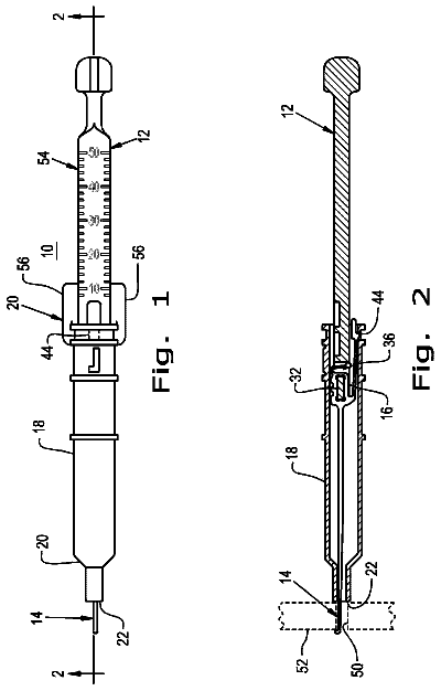
FIG. 1 is an exterior view of a schematic picture of a non-limiting example of a device of the invention.
FIG. 2 is a cut away view of a schematic picture of a non-limiting example of a device of the invention.
FIG. 3 : is a schematic picture of a non-limiting example of a device of the invention showing a probe's distal end and a side needle.
FIG. 4 : is a schematic picture of a non-limiting example of a device of the invention showing a distal end slope tip and side hole
FIG. 5 : is a schematic picture of a non-limiting example of a device of the invention showing a proximal end and sheath
FIG. 6 : is a schematic picture of a non-limiting example of a device of the invention showing a electrical connections
FIG. 7 : is a schematic picture of a non-limiting example of a device of the invention showing medicine injection channels.
FIG. 8 : is a schematic picture of a non-limiting example of a device of the invention showing Spinal Disk Herniation Treatment.
Description
The present invention provides in optional non-limiting embodiments a method and apparatus for treating intervertebral disc disorders by the application of controlled RF or heating in combination with spinal disk herniation repositioning in the same device or shaft to a localized region of an intervertebral disc. Such related disorders can include but are not limited to (i) degenerative discs which have tears or fissures in the annulus fibrosis, particularly fissures of the annulus fibrosis, which may or may not be accompanied with contained or escaped extrusions, (ii) contained disc herniation with focal protrusions, and/or (iii) bulging discs.
Degenerative discs with tears or fissures can be treated non-destructively, optionally without the significant removal of disc tissue other than torn or damaged or adjacent tissue by limited ablation and spinal disk herniation repositioning to the nucleus pulposus which can optionally change some of the water content, shape, or composition of the nucleus pulposus. Electromagnetic such as RF energy can optionally be delivered to a selected section of the disc in combination with spinal disk herniation repositioning in the same instrument wherein the energy can optionally be provided in an amount which does not create a destructive lesion to the disc. Sufficient electromechanical or RF energy is delivered to the disc to change its biochemical, neurophysiologic and/or biomechanical properties in order to optionally reduce at least one of pain, inflammation, tearing, displacement, swelling, necrosis, nerve impingement, and/or damage. Neurophysiologic modifications can optionally include denervation of nociceptors in a tear or fissure in the annulus fibrosis.
Degenerative intervertebral discs with fissures can optionally be treated by repositioning, cutting, and/or denervating selected or relevant nerves that are optionally embedded in the interior wall of the annulus fibrosis optionally as well as nerves outside of the interior wall including those on the surface of the wall. Electromagnetic or RF energy can optionally be used to cauterize granulation or other tissue which can be pain sensitive areas and formed in the annulus fibrosis wall. Electromagnetic or RF energy can also optionally be used to break down selected enzyme systems or neurotransmitters that generate pain within or related to the disc or nerves passing through the disc. Generally, these enzymes and neurotransmitters can work within a small bandwidth of both pH and temperature.
Electromagnetic such as RF energy is applied to shrink collagen in the annulus fibrosis and/or nucleus pulposus. This reduces the redundancy in the disc roll that is created in a degenerative disc. Delivery of electromagnetic energy to the nucleus pulposus removes some water and permits the nucleus pulposus to withdraw. This reduces a “pushing out” effect that created a contained herniation. Combinations of shrinking the disc, shrinking of the nucleus pulposus by reducing water content, as well as tightening up the annulus fibrosis wall, in combination with spinal disk herniation repositioning, optionally creates a rejuvenation or repair of the disc and/or reduction in pain or inflammation. Reducing the pressure in the disc and tightening the annulus fibrosis optionally produces a favorable biomechanical effect. Application of electromagnetic energy locally increases the stiffness of the disc.
The annulus fibrosis is comprised primarily of fibrosis-like material and the nucleus pulposus is comprised primarily of an amorphous colloidal gel. The distinction between the annulus fibrosis and the nucleus pulposus becomes more difficult to distinguish when a patient is 30 years old or greater. There is often a transition zone between the annulus fibrosis and the nucleus pulposus made of fibrosis-like material and amorphous colloidal gel. For purposes of this disclosure, the inner wall of the annulus fibrosis includes the young wall comprised primarily of fibrosis-like material as well as the transition zone which includes both fibrous-like material and amorphous colloidal gels (hereinafter collectively referred to as “inner wall of the annulus fibrosis”).
In general, an apparatus of the invention is optionally in the form of an externally guidable intervertebral disc apparatus for accessing and manipulating disc tissue present at a selected location of an intervertebral disc having a nucleus pulposus and an annulus fibrosus, the annulus having an inner wall. Optional use of a temperature-controlled energy delivery element, combined with spinal disk herniation positioning element and navigational control of the inventive catheter, provides preferential, localized heating and spinal disk herniation reposition to treat the fissure. For ease of reference to various manipulations and distances described below, the nucleus pulposus can be considered as having a given diameter in a disc plane between opposing sections of the inner wall. This nucleus pulposus diameter measurement allows instrument sizes (and parts of instruments) designed for one size disc to be readily converted to sizes suitable for an instrument designed for a different size of disc.
The operational portion of the apparatus of the invention is optionally brought to a location in or near the disc's fissure using techniques and apparatuses typical of percutaneous interventions as known in the art. For convenience and to indicate that the apparatus of the invention can be used with any insertional apparatus that provides proximity to the disc, including many such insertional apparatuses known in the art, the term “introducer” is used to describe this aid to the method. An introducer has at least one internal introducer lumen with a distal opening at a terminus of the introducer to allow insertion (and manipulation) of the operational parts of the apparatus into (and in) the interior of a disc.
The operational part of the apparatus optionally comprises at least one elongated element referred to as a catheter, various parts of which are located by reference to a distal end and a proximal end at opposite ends of its longitudinal axis. The proximal end is the end closest to the external environment surrounding the body being operated upon (which may still be inside the body in some embodiments if the catheter is attached to a handle insertable into the introducer). The distal end of the catheter is optionally intended to be located inside the disc under conditions of use. The catheter is optionally a traditional medical catheter (i.e., an elongate hollow tube for admission or removal of fluids from an internal body cavity) but is a defined term for the purposes of this specification. “Catheter” has been selected as the operant word to describe this part of the apparatus, as the inventive apparatus is optionally a long, flexible, partly flexible or rotatably, or at least partially stiff or rigid tube which transmits energy, spinal disk herniation repositioning, and/or material from or to a location external to the body to a location internal to the disc being accessed upon, such as optionally a collagen solution, spinal disk herniation repositioning, and./or heat to the annular fissure. Alternatively, material can be transported in the other direction to remove material from the disc, such as removing material by aspiration to decrease pressure which is keeping the fissure open and aggravating the symptoms due to the fissure.
The catheter is optionally adapted to slidably advance through the introducer lumen, the catheter optionally having a probe section at the distal end of the catheter, the probe section being extendible through the distal opening at the terminus of the introducer into the disc and can include an RF needle, electrode or tip and a spinal disk herniation repositioning needle or tip. Although the length of the probe portion can vary with the intended function as explained in detail below or as known in the art, a typical distance of extension is optionally at least one-half the diameter of the nucleus pulposus, preferably in the range of one-half to one and one-half times the circumference of the nucleus.
In order that the functional elements of the catheter (e.g., an electromagnetic probe, such as, an RF electrode or a resistance heater, and a spinal disk herniation repositioning element, e.g., probe, needle or tip) can be readily guided to the desired location within a disc, the probe portion of the catheter is manufactured with sufficient rigidity to avoid collapsing upon itself while being advanced through the nucleus pulposus and navigated around the inner wall of the annulus fibrosus. The probe portion, however, can have insufficient rigidity to puncture the annulus fibrosus under the same force used to advance the catheter through the nucleus pulposus and around the inner wall of the annulus fibrosus. Absolute penetration ability will vary with sharpness and stiffness of the tip of the catheter, but in all cases a catheter of the present invention will optionally advance more readily through the nucleus pulposus than through the annulus fibrosus.
In optional embodiments, the probe section of the catheter further has differential bending ability in two orthogonal directions at right angles to the longitudinal axis. This optionally causes the catheter to bend along a desired plane (instead of at random). Also when a torsional (twisting) force is applied to the proximal end of the catheter to optionally re-orient the distal end of the catheter, controlled advancement of the catheter in the desired plane can be provided.
A further component of the catheter optionally is a functional element located in the probe section for diagnosis or for adding energy and adding and/or removing material at the selected location of the disc where the annular tear is to be treated. The apparatus optionally allows the functional element to be controllably guided by manipulation of the proximal end of the catheter into a selected location for localized treatment of the annular fissure.
The optional method embodiment of the invention, which optionally involves manipulating disc tissue at the annular fissure, is easily carried out with an apparatus of the invention. An introducer is provided that is located in a patient's body so that its proximal end is external to the body and the distal opening of its lumen is internal to the body and
internal to the annulus fibrosus or
adjacent to an annular opening leading to the nucleus pulposus, such as an annular tear or trocar puncture that communicates with the nucleus pulposus. The catheter is optionally then slid into position in and through the introducer lumen so that the functional elements in the catheter are positioned at the selected location of the disc by advancing or retracting the catheter or probe in the introducer lumen and optionally twisting the proximal end of the catheter to precisely navigate the catheter. By selection of the rigidity of the catheter and by making it sufficiently blunt to not penetrate the annulus fibrosus, and by selection of the flexibility in one plane versus the orthogonal plane, the distal portion of the catheter optionally will curve along the inner wall of the annulus fibrosus as it is navigated and is selectively guided to an annular tear at selected location(s) in the disc. Energy and spinal disk herniation repositioning is optionally applied and/or material is added or removed at the selected location of the disc via the functional elements.
Each of the elements of the apparatus and method will now be described in more detail. However, a brief description of disc anatomy is provided first, as sizes and orientation of structural elements of the apparatus and operations of the method can be better understood in some cases by reference to disc anatomy.
A Non Limiting Exemplary Surgical Site
The annulus fibrosus is comprised primarily of tough fibrous material, while the nucleus pulposus is comprised primarily of an amorphous colloidal gel. There is a transition zone between the annulus fibrosus and the nucleus pulposus made of both fibrous-like material and amorphous colloidal gel. The border between the annulus fibrosus and the nucleus pulposus becomes more difficult to distinguish as a patient ages, due to degenerative changes. This process may begin as early as 30 years of age. For purposes of this specification, the inner wall of the annulus fibrosus can include the young wall comprised primarily of fibrous material as well as the transition zone which includes both fibrous material and amorphous colloidal gels (hereafter collectively referred to as the “inner wall of the annulus fibrosus”). Functionally, that location at which there is an increase in resistance to catheter penetration and which is sufficient to cause bending of the distal portion of the catheter into a radius less than that of the internal wall of the annulus fibrosus is considered to be the “inner wall of the annulus fibrosus.”
As with any medical instrument and method, not all patients can be treated, especially when their disease or injury is too severe. There is a medical gradation of degenerative disc disease (stages 1-5). See, for example, Adams et al., “The Stages of Disc Degeneration as Revealed by Discograms,” J. Bone and Joint Surgery, 68, 36-41 (1986), entirely incorporated herein by reference. As these grades are commonly understood, the methods of instrument navigation described herein can distinguish between the nucleus and the annulus in degenerative disease of grades, such as in discs in stages 3 and 4, optionally up to 5, and optionally as early as stages 1 and 2, but which are often asymptomatic in most patients, and stage 5 may require disc removal and fusion.
Some of the following discussion refers to motion of the catheter inside the disc by use of the terms “disc plane,” “oblique plane” and “cephalo-caudal plane.” These specific terms refer to orientations of the catheter within the intervertebral disc.
Referring to FIGS. 1-8 , when the side needle ( 2 ) of the electrode is deployed, the probe and the side needle would form an “r” shape, allowing physicians to use mechanical force to push annulus and diseased tissues away from the nerves.
The side needle ( 2 ) is also equipped with a thermocouple, allowing physicians to monitor and control the temperature when performing radiofrequency ablation procedures.
The electrode itself has the following major components: Probe ( 1 ); Side needle ( 2 ); Operation handle ( 3 ); Push button ( 4 ); Injection nozzle ( 5 ); Electric cable ( 8 ); Power plug ( 7 ).
In this electrode, the probe ( 1 ) and the side needle ( 2 ) are both made of metal tubes. The side needle ( 2 ) is connected to the proximal end (B) of the sheath ( 10 ) through the probe ( 1 ); the sheath ( 10 ) is connected to the push button ( 4 ) through a connection piece ( 12 ), thus when the physician moves the push button ( 4 ), the side needle ( 2 ) could be either deployed or retrieved.
The side needle ( 2 ) is also equipped with a thermocouple ( 13 ) at the tip position. The thermocouple ( 13 ) is connected to the inner chamber of the side needle ( 2 ), the electrical cable ( 8 ) and the power plug ( 7 ), allowing physicians to monitor and control the temperature when performing radiofrequency ablation procedure.
In addition, medicines could be injected through the injection nozzle ( 5 ), injection tube ( 14 ), injection sheath ( 6 ) and the inner chamber of the probe ( 1 ) to reach the distal end of the probe as well the side holes (D). Similarly, ozone or collagenase could be injected through the same mechanism. Figure Descriptions of Optional Non Limiting Embodiments
FIG. 1 : shows a schematic of an outside view of the electrode and two or more of the following major components: Probe ( 1 ); Side needle ( 2 ); Operation handle ( 3 ); Push button ( 4 ); Injection nozzle ( 5 ); Electric cable ( 8 ); and Power plug ( 7 ). The side needle ( 2 ) can optionally be located at the same side as the push button ( 4 ). The probe and the side needle can optionally form an “r” shape, making it easier for physicians to puncture through the tissues.
FIG. 2 : shows an optional inside view of the electrode, e.g., but not limited to, wherein the probe ( 1 ) and operation handle ( 3 ) can be attached or associated, by mechanical or other connection (e.g., but not limited to attached or glued together at point A). The sheath ( 10 ) can optionally be a tube outside the proximal end of the probe ( 1 ). The distal end of the sheath ( 10 ) can optionally have a seal pipe ( 11 ) to seal the space between the operation handle ( 3 ) and the probe ( 1 ). The proximal end of the sheath ( 10 ) and the proximal end of the side needle ( 2 ) can be attached or associated, by mechanical or other connection (e.g., but not limited to attached or glued together. With the protection of the seal pipe ( 11 ), the sheath ( 10 ) and the probe ( 1 ) can optionally slide smoothly without or substantially reduced leakage problems. The sheath ( 10 ) can optionally be connected to the push button ( 4 ) through a connection piece ( 12 ), thus when the physician moves the push button ( 4 ), the side needle ( 2 ) can optionally be either deployed or retrieved.
FIG. 3 : shows optional probe's distal end and side needle: The distal end of the probe ( 1 ) can optionally have a sloped shape. Underneath the slope optionally is a side hole: the hole is optionally either rectangular or oval shaped, allowing side needle ( 2 ) to be deployed or retrieved.
FIG. 4 shows the distal end slope and side hole. The tip of the probe ( 1 ) optionally has a sloped shape (point C). A rectangular-shaped hole optionally sits underneath the slope.
FIG. 5 shows an optional proximal end and sheath. The sheath ( 10 ) optionally is a tube outside the proximal end of the probe ( 1 ). The distal end of the sheath ( 10 ) optionally has a seal pipe ( 11 ) to seal the space between the operation handle ( 3 ) and the probe ( 1 ). The proximal end of the sheath ( 10 ) and the proximal end of the side needle ( 2 ) can be attached or associated, by mechanical or other connection (e.g., but not limited to attached or glued together, e.g., optionally using a sealant ( 9 ).
FIG. 6 shows optional electrical connections. The side needle ( 2 ) is optionally equipped with a thermocouple ( 13 ) at the tip position. The thermocouple ( 13 ) optionally is connected to the inner chamber of the side needle ( 2 ), optionally through a thermocouple cable ( 15 ). The thermocouple cable ( 15 ) is optionally also connected to the electrical cable ( 8 ) and the power plug ( 7 ). There is optionally a radiofrequency cable ( 16 ) between the proximal end of the side needle ( 2 ) and the power plug ( 7 ).
FIG. 7 shows optional medicine injection channels. Medicines (e.g. ozone and collagenase) optionally are injected through the injection nozzle ( 5 ), injection sheath ( 6 ) and the inner chamber of the probe ( 1 ) to reach the distal end of the probe as well the side holes, and to enter the diseased or other tissues for therapy or diagnosis.
FIG. 8 shows optional Spinal Disk Herniation Treatment. Optionally under the guidance of imaging devices (e.g. CT), physicians can optionally use the electrode's probe to puncture the body. After the needle reaches the injured disk, the physician optionally deploys the side needle in order to reposition the spinal disk herniation. Later the physician optionally performs radiofrequency ablation treatment as well as optional medicine/ozone therapy with the device.
In a non limiting optional embodiment of a catheter of the invention as it would appear inserted into the lumen of an introducer. The catheter includes handle, stem, probe section and a tip. The handle at the proximal end of the catheter is coupled via the stem to the probe section, which is located proximate the distal end of the device. At the terminus of the probe, i.e., the distal end of the device, is the tip. The tip may be axially displaced from the probe section. Functional elements for delivery or energy or material to or from the site in combination with a spinal disk herniation repositioning element can be placed within the probe. These can, via connections within the probe, stem and handle, be coupled to either an RF energy delivery device 1 and the repositioning needle 2 or a material transfer device. Therefore no limitation should be placed on the types of energy, spinal disk herniation repositioning, force, or material transporting elements present in the catheter. These are merely some of the possible alternative functional elements that can be included in the probe portion of the catheter. The flexible, movable catheter is at least partially positionable in the introducer lumen, to bring the probe section, which is designed to be the portion of the catheter that will be pushed out of the introducer lumen and into the nucleus pulposus and into the selected location(s) with regard to the annular tear.
The description continues in the full USPTO document.
 Lapsed, fee not paid4 drawings