 Lapsed, fee not paid14 drawings
Consumer Products · US 9,883,785 B2
Dispensing pad cleaner
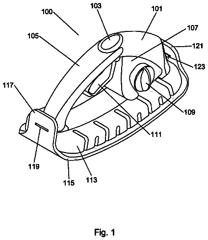
A dispensing pad cleaner having a novel integrated dispensing system for delivering cleaning solutions and similar liquids to a surface to be cleaned.
US 9,884,065 B2 · Assignee: Buck Institute for Research on Aging · Inventors: Campisi; Judith et al.
Sheet 1 of 38 from the published document. All sheets in the USPTO PDF
Methods are provided herein for enhancing the effectiveness of medical therapies by administering agents that suppress a biological damage response that is inducible by the medical therapy administered to a subject. In certain embodiments, a method is provided for administering an anti-senescent cell agent that suppresses a biological response comprising cellular senescence that is induced by the medical therapy.
Technical Field Methods for enhancing the effectiveness of various medical therapies used for treating diseases, such as cancer, HIV/AIDS, and autoimmune diseases, are provided herein. Agents used in these methods include agents that suppress a biological damage response. Description of the Related Art Cytotoxic and genotoxic therapies are administered to hundreds of thousands of patients each year for treatment of a variety of diseases, most notably, cancers. Cancer includes a broad range of diseases and affects approximately one in four individuals worldwide. In the United States, cancer is the second leading cause of death, accounting for 23% of all deaths. While the five-year relative survival rate for all cancers diagnosed is approximately 68%, treatments and their rates of success vary between cancer types. Even though chemotherapies and radiotherapies are designed to target cancer
1 of 38 drawing sheets so far from the published document, cropped to the drawing. Every sheet is in the USPTO PDF.
What the patent claimed, word for word. All of it is now free to use.
The Sequence Listing associated with this application is provided in text format in lieu of a paper copy, and is hereby incorporated by reference into the specification. The name of the text file containing the Sequence Listing is 200201_402WO_SEQUENCE_LISTING.txt. The text file is 30 KB, was created on Dec. 13, 2012 and is being submitted electronically via EFS-Web.
Technical Field
Methods for enhancing the effectiveness of various medical therapies used for treating diseases, such as cancer, HIV/AIDS, and autoimmune diseases, are provided herein. Agents used in these methods include agents that suppress a biological damage response.
Description of the Related Art
Cytotoxic and genotoxic therapies are administered to hundreds of thousands of patients each year for treatment of a variety of diseases, most notably, cancers. Cancer includes a broad range of diseases and affects approximately one in four individuals worldwide. In the United States, cancer is the second leading cause of death, accounting for 23% of all deaths. While the five-year relative survival rate for all cancers diagnosed is approximately 68%, treatments and their rates of success vary between cancer types. Even though chemotherapies and radiotherapies are designed to target cancer cells, the therapies can adversely affect normal cells and tissue to an extent that the beneficial effect of the cancer therapy can be significantly compromised.
Highly active anti-retroviral therapy administered to men and women who are HIV infected and have developed AIDS has contributed to extending the lifespan and improving the general health of those infected. However, this therapy can also adversely affect normal cell physiology as well.
Briefly, provided herein are methods for enhancing the effectiveness of a medical therapy by administering an agent that suppresses a biological damage response, including cellular senescence, which is inducible by the medical therapy. Provided herein are the following embodiments.
In one embodiment, a method is provided herein for enhancing the effectiveness of a medical therapy in a subject, the method comprising administering to the subject an agent that suppresses a biological damage response inducible by the medical therapy. In a specific embodiment, the method for enhancing the effectiveness of a medical therapy in a subject comprises administering to the subject an agent that suppresses a biological damage response inducible by the medical therapy, wherein the agent is administered prior to, subsequent to, or concurrent with administration of the medical therapy. In certain embodiments, the medical therapy increases the proportion of senescent cells in the subject. In another specific embodiment, a method is provided for enhancing the effectiveness of a medical therapy in a subject comprising administering to the subject an agent that suppresses a biological damage response inducible by the medical therapy, wherein the agent is administered prior to, subsequent to, or concurrent with administration of the medical therapy, wherein the agent is selected from (a) an agent that selectively destroys or facilitates selective destruction of one or more senescent cells; and (b) an agent that inhibits expression or secretion of one or more senescence cell-associated molecules produced by a senescent cell. In a specific embodiment, the agent is administered to the subject at least 3 days, at least 4 days, at least 5 days, at least 6 days, at least 7 days, at least 10 days, at least 30, at least 60, or at least 90 days subsequent to administration of the medical therapy. In a particular embodiment, the agent that suppresses the biological damage response selectively destroys or facilitates selective destruction of one or more senescent cells. In another embodiment, the agent is administered to the subject prior to administration of the medical therapy. In particular embodiments the agent is administered to the subject at least 1 day, at least 2-6 days, at least 1 week, at least 2 weeks, at least 3 weeks, at least 4-5 weeks, at least 6-8 weeks, or at least 10-12 weeks prior to administration of the medical therapy. In still another specific embodiment, the agent is administered concurrently with at least a portion of the administered medical therapy. In the embodiments described above and herein, the agent that suppresses the biological damage response inhibits expression or secretion of one or more senescence cell-associated molecules produced by a senescent cell. In a specific embodiment, the agent is a small molecule, polypeptide, peptide, antibody, antigen-binding fragment, peptibody, recombinant viral vector, or a nucleic acid. In certain embodiments, the medical therapy increases the proportion of senescent cells in a subject. In another specific embodiment, the medical therapy comprises radiation, a chemotherapy, an anti-viral therapy, or a hormone. In certain embodiments, the subject has a cancer, is in cancer remission, is at risk of developing a recurrence of a cancer, or is at risk of developing a cancer, and wherein the medical therapy comprises an anti-cancer therapy. In a more specific embodiment, the cancer comprises a solid tumor, and in other specific embodiments, the cancer comprises a liquid tumor. In other specific embodiments of the methods described above and herein, the cancer is metastatic cancer. In another particular embodiment, the methods for enhancing the effectiveness of a medical therapy described above and herein may be used when the subject has a cancer and has received or will receive a stem cell transplant, and wherein the medical therapy is high dose chemotherapy or high dose radiotherapy or a combination thereof. In particular embodiment, the stem cell transplant is an autologous or allogenic stem cell transplant.
In another particular embodiment of the methods described above and herein, the anti-viral therapy is an HIV/AIDS management therapy, wherein in one embodiment, the HIV/AIDS management therapy comprises a highly active antiretroviral therapy (HAART). In still another specific embodiment of the methods described above and herein, the subject has a cardiovascular disease or is at risk of developing a cardiovascular disease, and the medical therapy is angiotensin. In yet another specific embodiment of the methods described above and herein, the subject has diabetes, and the medical therapy is insulin.
In one embodiment, a method is provided for enhancing the effectiveness of a medical therapy in a subject comprising (a) administering to the subject the medical therapy, which medical therapy induces senescence in one or more cells of the subject; and then (b) administering to the subject an anti-senescent cell agent, which agent selectively destroys or facilitates the selective destruction of the one or more senescent cells. In a specific embodiment, the agent is administered to the subject at least 3 days, at least 4 days, at least 5 days, at least 6 days, at least 7 days, or at least 10 days, at least 30 days, at least 60 days, or at least 90 days subsequent to administration of the medical therapy. In particular embodiments, the agent is a small molecule, polypeptide, peptide, antibody, antigen-binding fragment, peptibody, recombinant viral vector, or a nucleic acid. In certain embodiments, the medical therapy increases the proportion of senescent cells in a subject. In certain embodiments, the medical therapy comprises radiation, a chemotherapy, an anti-viral therapy, or a hormone. In a specific embodiment, the subject has a cancer, is in cancer remission, is at risk of developing a recurrence of a cancer, or is at risk of developing a cancer, and wherein the medical therapy comprises an anti-cancer therapy. In another specific embodiment, the cancer comprises a solid tumor, and in yet another specific embodiment, the cancer is a liquid tumor. In other specific embodiments, the cancer is metastatic cancer. In another particular embodiment, the methods for enhancing the effectiveness of a medical therapy described above and herein may be used when the subject has a cancer and has received or will receive a stem cell transplant, and wherein the medical therapy is high dose chemotherapy or high dose radiotherapy or a combination thereof. In particular embodiment, the stem cell transplant is an autologous or allogenic stem cell transplant.
In another particular embodiment of the methods described above and herein, the anti-viral therapy is an HIV/AIDS management therapy, wherein in one embodiment, the HIV/AIDS management therapy comprises a highly active antiretroviral therapy (HAART). In still another specific embodiment of the methods described above and herein, the subject has a cardiovascular disease or is at risk of developing a cardiovascular disease, and the medical therapy is angiotensin. In yet another specific embodiment of the methods described above and herein, the subject has diabetes, and the medical therapy is insulin.
In another embodiment, is provided a use of an agent that suppresses a biological damage response inducible by a medical therapy for enhancing the effectiveness of the medical therapy, wherein the agent is suitable for administration prior to, subsequent to, or concurrent with administration of the medical therapy. In a specific embodiment, the agent is suitable for administration at least 3 days, at least 4 days, at least 5 days, at least 6 days, at least 7 days, at least 10 days, at least 30, at least 60, or at least 90 days subsequent to administration of the medical therapy. In one embodiment, the agent that suppresses the biological damage response selectively destroys or facilitates selective destruction of one or more senescent cells. In another specific embodiment, the agent is administered prior to administration of the medical therapy. In still another embodiment, the agent is administered at least 1 day, at least 2-6 days, at least 1 week, at least 2 weeks, at least 3 weeks, at least 4-5 weeks, at least 6-8 weeks, or at least 10-12 weeks prior to administration of the medical therapy. In another specific embodiment, the agent is administered concurrently with at least a portion of the administered medical therapy. In certain embodiments, the agent that suppresses the biological damage response inhibits expression or secretion of one or more senescence cell-associated molecules produced by a senescent cell. In certain embodiments, the agent is a small molecule, polypeptide, peptide, antibody, antigen-binding fragment, peptibody, recombinant viral vector, or a nucleic acid. In still another particular embodiment, the medical therapy increases the proportion of senescent cells in a subject. In one embodiment, the medical therapy comprises radiation, a chemotherapy, an anti-viral therapy, or a hormone. In another embodiment, the medical therapy is an anti-cancer therapy. In still another embodiment, the cancer comprises a solid tumor or a liquid tumor, which in certain embodiments, is metastatic cancer. In still other embodiments, the anti-viral therapy is an HIV/AIDS management therapy. In another embodiment, the HIV/AIDS management therapy comprises a highly active antiretroviral therapy (HAART). In as specific embodiment, the medical therapy is high dose chemotherapy or high dose radiotherapy or a combination thereof, which is administered prior to or subsequent to administration of a stem cell transplant. In another embodiment, the stem cell transplant is selected from (a) an autologous stem cell transplant, and (b) an allogenic stem cell transplant. In still another embodiment, the agent is useful for treating or preventing a cardiovascular disease wherein the medical therapy is angiotensin. In another embodiment, the agent is useful for treating or preventing diabetes, wherein the medical therapy is insulin.
Also provided herein is a use of an anti-senescent cell agent for enhancing the effectiveness of a medical therapy wherein the medical therapy induces senescence in one or more cells, and wherein the agent selectively destroys or facilitates the selective destruction of the one or more senescent cells.
In the following description, certain specific details are set forth in order to provide a thorough understanding of various embodiments. However, one skilled in the art will understand that the invention may be practiced without these details. In other instances, well-known structures have not been shown or described in detail to avoid unnecessarily obscuring descriptions of the embodiments. Unless the context requires otherwise, throughout the specification and claims which follow, the word “comprise” and variations thereof, such as, “comprises” and “comprising” are to be construed in an open, inclusive sense, that is, as “including, but not limited to.” In addition, the term “comprising” (and related terms such as “comprise” or “comprises” or “having” or “including”) is not intended to exclude that in other certain embodiments, for example, an embodiment of any composition of matter, composition, method, or process, or the like, described herein, may “consist of” or “consist essentially of” the described features. Headings provided herein are for convenience only and do not interpret the scope or meaning of the claimed embodiments.
Reference throughout this specification to “one embodiment” or “an embodiment” means that a particular feature, structure or characteristic described in connection with the embodiment is included in at least one embodiment. Thus, the appearances of the phrases “in one embodiment” or “in an embodiment” in various places throughout this specification are not necessarily all referring to the same embodiment. Furthermore, the particular features, structures, or characteristics may be combined in any suitable manner in one or more embodiments.
Also, as used in this specification and the appended claims, the singular forms “a,” “an,” and “the” include plural referents unless the content clearly dictates otherwise. Thus, for example, reference to “a non-human animal” may refer to one or more non-human animals, or a plurality of such animals, and reference to “a cell” or “the cell” includes reference to one or more cells and equivalents thereof (e.g., plurality of cells) known to those skilled in the art, and so forth. When steps of a method are described or claimed, and the steps are described as occurring in a particular order, the description of a first step occurring (or being performed) “prior to” (i.e., before) a second step has the same meaning if rewritten to state that the second step occurs (or is performed) “subsequent” to the first step. The term “about” when referring to a number or a numerical range means that the number or numerical range referred to is an approximation within experimental variability (or within statistical experimental error), and thus the number or numerical range may vary between 1% and 15% of the stated number or numerical range. It should also be noted that the term “or” is generally employed in its sense including “and/or” unless the content clearly dictates otherwise. The term, “at least one,” for example, when referring to at least one compound or to at least one composition, has the same meaning and understanding as the term, “one or more.” BRIEF DESCRIPTION OF THE DRAWINGS
FIGS. 1A and 1B show radiation induces persistent senescent cells in p16-3MR transgenic mice and that GCV treatment leads to depletion of senescent cells and reduction of the level of several SASP (senescence associated secretory phenotype) biomarkers. The transgenic p16-3MR mice were mock irradiated (Ctrl) or irradiated (IR) (7 Gy whole body X-ray), housed for 3 months, and then treated with vehicle or GCV as described herein. Various tissues were isolated (results here shown are for lung tissue) and measured for bioluminescence ( FIG. 1A ) and the abundance of mRNAs encoding the p16INK4a, mRFP, IL-6 and MMP-3 proteins. Results are shown in arbitrary units (AU) after setting Ctrl levels at 1.
FIGS. 2A-2C show senescent cells induced in p16-3MR transgenic mice by irradiation promote primary and metastatic tumor growth. The transgenic p16-3MR mice were mock-irradiated (Ctrl) or irradiated (IR). Three months later, the irradiated mice were treated with vehicle (IR) or GCV (IR+GCV), then injected with fLUC-expressing B16 melanoma cells into the tail veins. Fifteen days later, bioluminescence of the B16 melanoma cells was measured.
FIG. 3 shows full body luminescence measurements of the B16 melanoma cells from the mice in FIGS. 2A-2C . Irradiated mice were moribund at day 15-16 and were sacrificed.
FIGS. 4A-4C show that elimination of senescent cells suppresses the development of metastases. The p16-3MR transgenic mice of FIG. 2 were followed for an additional three days (i.e., day 18). The irradiated mice treated with GCV (in which senescent cells were eliminated) eventually developed primary tumors in the lungs ( FIG. 4A ). But, despite the presence of primary tumors in the lung, the fat and liver tissues remained relatively metastasis free ( FIG. 4C ). In contrast, irradiated mice not treated with GCV (which retain senescent cells) showed metastatic tumors in the liver and fat tissue ( FIG. 4B ).
FIGS. 5A-5B show that treatment with doxorubicin induces persistent senescent cells in p16-3MR transgenic mice. The transgenic p16-3MR mice were mock treated with vehicle (Ctrl) or treated with 10 mg/kg of doxorubicin (DOXO). Various tissues were isolated (liver, heart, lung, kidney, and spleen) and measured for abundance of mRNAs encoding mRFP ( FIG. 5A ) and p16INK4a ( FIG. 5B ) (normalized to actin).
FIG. 6 shows that doxorubicin induces persistent senescent cells in p16-3MR transgenic mice and that GCV treatment leads to depletion of senescent cells and reducing the level of SASP biomarkers, p16INK4 and mRFP. Skin biopsies were isolated and measured for abundance of p16INK4 and mRFP (normalized to actin). Results are shown in arbitrary units (AU) after setting Ctrl levels at 1.
FIG. 7 shows senescent cells induced in p16-3MR transgenic mice by doxorubicin treatment promoted primary tumor growth. The transgenic p16-3MR mice were vehicle-treated (Ctrl) or treated with doxorubicin (10 mg/kg). 7 days later, the doxorubicin treated mice were mock treated with vehicle (DOXO) or GCV (DOXO+GCV), then injected subcutaneously with fLUC-expressing B16 melanoma cells. Twelve days later, bioluminescence of the B16 melanoma cells was measured.
FIG. 8 shows that clearance of senescent cells in doxorubicin treated p16-3MR transgenic mice reduced tumor size. The transgenic p16-3MR mice were vehicle-treated (Ctrl) or treated with doxorubicin (10 mg/kg). 7 days later, the doxorubicin treated mice were mock treated with vehicle (DOXO) or GCV (DOXO+GCV), then injected subcutaneously with fLUC-expressing B16 melanoma cells. Twelve days later, primary tumor diameter was measured.
FIG. 9 shows that elimination of senescent cells suppresses the multiplicity of K-Ras induced lung tumors as compared to mice in which senescent cells were not cleared or reduced.
FIGS. 10A-10D provide a listing of an illustrative transgene selectively expressed in senescent cells, the nucleic acid sequence of a pBLUESCRIPT II KS vector containing a p16.sup.Ink4a promoter-FKBP-caspase-IRES-GFP nucleic acid construct (SEQ ID NO:1).
FIG. 11A-11F provide a listing of the nucleic acid sequences of FIG. 10 with the various vector components and construct components labeled.
FIGS. 12A-12E show that corticosterone and cortisol partially suppress the SASP. ( FIG. 12A ) Senescent X-irradiated with 10 Gy (Sen (XRA)) HCA2 fibroblasts were incubated in medium plus 10% serum containing the indicated concentrations of corticosterone or the highest concentration of DMSO (vehicle control). The cells were given corticosterone or DMSO immediately after irradiation and analyzed 6 days later. The cells were washed and incubated in serum-free medium without corticosterone to generate conditioned media. Conditioned media from pre-senescent (Pre) and control or corticosterone-treated Sen (XRA) cells were analyzed by ELISA for IL-6. ( FIG. 12B ) Cells were treated, and conditioned media were generated and analyzed as described in ( FIG. 12A ) except cortisol was used at the indicated concentrations. ( FIG. 12C ) Conditioned media were collected from presenescent (PRE) or senescent (XRA) cells that were treated with DMSO, corticosterone (50 nM), or cortisol (100 nM) as described in ( FIG. 12A ). The conditioned media were analyzed by antibody arrays. Average signal from PRE and XRA DMSO cells was used as the baseline. Signals higher than baseline are light gray (see +1 on scale to right); signals lower than baseline are dark gray as illustrated by PRE DMSO (see −1 on scale at right). Color intensities represent log.sub.2-fold changes from the average value. The hierarchical clustering relationship between samples is shown as a dendrogram (left). *Factors significantly (P<0.05) suppressed by cortisol. ‡Factors significantly suppressed by corticosterone (P<0.05). ( FIG. 12D ) Cells were infected with RAS- or MKK6-expressing lentiviruses. After selection, the cells were given DMSO-, 500 nM corticosterone (C1) and 100 nM cortisol (C2) for 6 days. Conditioned media were generated as previously described and analyzed by ELISA for IL-6. *Factors significantly different from DMSO treatment (P<0.05). ( FIG. 12E ) Cells were treated with 500 nM corticosterone for the indicated intervals (a-d, indicated by the thick lines in the lower panel) before or after X-irradiation (XRA, indicated by the arrow). Conditioned media were prepared and analyzed by ELISA for IL-6 (upper panel). *Factors significantly different from DMSO treatment (P<0.05).
FIGS. 13 A 1 - 13 G show the effect of glucocorticoids on the SASP depends on the glucocorticoid receptor (GR). (FIG. 13 A 1 ) mRNA was extracted from presenescent (Pre) or senescent (Sen (XRA)) HCA2 cells treated with DMSO, 500 nM corticosterone (C1) or 100 nM cortisol (C2) as described in the legend to FIG. 12 . (FIG. 13 A 2 ) mRNA was extracted from presenescent (Mock) or senescent X-irradiated HCA2 cells treated with DMSO, 500 nM corticosterone, or 100 nM cortisol as described in FIG. 12 . Transcripts for IL-5, IL-6, IL-8, MMP-3, IL-1α, MCP-2, MCP-3, and GM-CSF were quantified by quantitative PCR (normalized to tubulin). *Factors significantly different from DMSO treatment (P<0.05). ( FIG. 13B ) mRNA was extracted from Pre and Sen (XRA) cells treated with DMSO, 500 nM corticosterone (C1), or 100 nM cortisol (C2) as previously described, and transcripts for GR were quantified by PCR (normalized to tubulin). Although GR mRNA levels tended to be slightly elevated in senescent cells, the increase was not statistically significant. (FIG. 13 C) Pre and Sen (XRA) cells treated with DMSO, 500 nM corticosterone, or 100 nM cortisol as previously described were immunostained for GR 1, 4, and 7 days after X-irradiation. ( FIG. 13D ) Cells were infected with lentiviruses expressing shRNAs against GFP (control) or GR and selected. Seven days after selection, mRNA was extracted and transcripts for GR were quantified by PCR (normalized to tubulin). ( FIG. 13E ) Total cell clysates were prepared from the shGFP- and shGR-expressing cells described in ( FIG. 13D ) and analyzed by western blotting for GR and actin (control). ( FIG. 13F ) Cells infected with shGFP- or shGR-expressing lentiviruses were X-irradiated and treated immediately thereafter with DMSO, 500 nM corticosterone, or 100 nM cortisol. Conditioned media were collected 7 days later and analyzed by ELISA for IL-6. ( FIG. 13G ) Cells were treated as described in ( FIG. 13F ) except for the addition of RU-486 at the indicated doses. Conditioned media were collected and analyzed by ELISA for IL-6 secretion.
FIGS. 14A-14C show that glucocorticoids repress IL-1α expression. ( FIG. 14A ) Presenescent (Pre) HCA2 cells were treated with DMSO, 500 nM corticosterone, or 100 nM cortisol for 24 hours or were induced to senesce by X-irradiation (Sen (XRA)) and given DMSO, corticosterone, or cortisol immediately thereafter. mRNA was extracted after the indicated intervals, and transcripts for IL-1α were quantified by PCR (normalized to tubulin). ( FIG. 14B ) mRNA extracted from cells described in ( FIG. 14A ) was used to quantify transcripts for IL-6 (normalized to tubulin). ( FIG. 14C ) Pre and Sen (XRA) cells, prepared as described in ( FIG. 14A ), were immunostained for IL-1α. Sen (XRA) cells were immunostained 7 days after irradiation.
FIGS. 15A-15F show that glucocorticoids impair the IL-1α/NF-κB pathway and suppress the ability of the SASP to induce tumor cell invasion. ( FIG. 15A ) Total HCA2 cell lysates were prepared from presenescent (Pre) cells, or senescent cells (Sen (XRA)) treated with DMSO, 500 nM corticosterone (C1), or 100 nM cortisol (C2) in the absence (left panel) or presence (right panel) of recombinant IL-1α protein (rIL-1α). The lysates were analyzed by western blotting for IRAK1, IκBα, RelA, and actin (control). ( FIG. 15B ) After irradiation, Sen (XRA) cells were given DMSO, 50 nM corticosterone, or 100 nM cortisol. Six days later, the cells were given recombinant IL-la protein at the indicated doses in the presence of the glucorticoids in serum free media. Conditioned media were collected 24 hours later and analyzed by ELISA for IL-6. ( FIG. 15C ) Nuclear extracts were prepared from Pre cells and Sen (XRA) cells treated with DMSO, 500 nM corticosterone (C1) or 100 nM cortisol (C2) as described above, and analyzed for NF-κB DNA binding activity. ( FIG. 15D ) Cells were infected with a lentivirus carrying an NF-κB-luciferase reporter construct, irradiated, and allowed to senesce. Immediately after irradiation cells were treated with DMSO, 500 nM corticosterone, or 100 nM cortisol, plus 0.5 μM RU-486 or 2.5 ng mL.sup.−1 IL-1α, as indicated. Seven days after irradiation, cells were trypsinized, counted, lysed, and assayed for luciferase activity, which was normalized to cell number. ( FIG. 15E ) Conditioned media from presenescent (Pre) or senescent (Sen (XRA)) cells that had been treated with corticosterone (C1) or cortisol (C2) as described in FIG. 12 were prepared. The conditioned media were then assayed for ability to stimulate T47D human breast cancer cells to invade a basement membrane, as described in the Experimental Procedures. ( FIG. 15F ) Nuclear extracts were prepared from Pre cells, and Sen (XRA) treated cells treated with DMSO, 500 nM corticosterone, or 100 nM cortisol in the absence (left panel) or presence (right panel) of recombinant IL-1α protein (rIL-1α) and analyzed for NF-κB DNA binding activity.
FIG. 16A-16E — FIG. 16A shows IMR-90 fibroblasts that were induced to senesce by X-irradiation (10 Gy; Sen (XRA)) and treated immediately after irradiation with the indicated concentrations of corticosterone or the highest concentration of DMSO (vehicle control) for 7 days. Conditioned media from presenescent (Pre) and the control and glucocorticoid-treated Sen (XRA) cells were analyzed by ELISA for IL-6. FIG. 16B shows Sen (XRA) HCA2 cells that were treated with DMSO, 500 nM corticosterone (C1), or 100 nM cortisol (C2) for 7 days. The percentage of presenescent (Pre) and Sen (XRA) cells that express SA-Bgal were scored (upper panel). A representative field corresponding to each condition is also shown (bottom panels). FIG. 16C shows the Pre and Sen (XRA) HCA2 cells described in (B), given BrdU for 24 hours, fixed, and immunostained for nuclear BrdU staining, and then analyzed for the percentage of BrdU-positive cells. FIG. 16D shows Pre and Sen (XRA) cells described in (B) immunostained for 53BP1. The percentage of cells with >2 53BP1 nuclear foci was determined using C ELL P ROFILER software. At least 200 cells were analyzed per condition. FIG. 16E shows the average number of 53BP1 foci from (D), determined using the C ELL P ROFILER software.
FIG. 17A-17B — FIG. 17A shows presenescent (A) or Sen (Xra) HCA2 cells immunostained for the mineralocorticoid receptor. Sen (XRA) cells were given DMSO, 500 nM corticosterone, or 100 nM cortisol immediately after irradiation and immunostained 1 or 7 days thereafter. FIG. 17B shows Sen (XRA) HCA2 cells that were treated with DMSO, 500 nM corticosterone, or 100 nM cortisol in the presence or not (−) of RU486, and immunostained for the GR.
FIG. 18 mRNA extracted from Pre HCA2 cells treated with DMSO, 500 nM corticosterone, or 100 nM cortisol for 24 hours and Sen (XRA) HCA2 cells treated with these compounds for 7 days starting immediately after X-irradiation. mRNA extracts were analyzed for IκBα transcripts by quantitative PCR (normalized to tubulin). The level of IκBα mRNA in DMSO-treated Pre cells was arbitrarily assigned a value of 1.
FIG. 19 shows that apigenin treatment partially suppresses SASP. Conditioned media from control (Mock irradiated, DMSO-treated), DMSO-treated (DMSO) or apigenin-treated senescent (Api) cells were analyzed by multiplex ELISA for expression of SASP. Results are shown as fold difference over control (Mock irradiated, DMSO-treated) cells with the vertical axis in log scale.
Provided herein are methods for enhancing the effectiveness of a medical therapy by administering an agent that suppresses a biological damage response that is inducible by the medical therapy. Medical therapies, such as cancer chemotherapy, radiation treatment, hormone therapy, and various anti-viral therapies, are intended and designed to target aberrant or abnormal cells that cause the disease, which because of aberrant metabolism, proliferation, repair capacity and/or other physiological and biological properties are presumed to be more sensitive to these therapies. However, these medical therapies, particularly those that are administered systemically, act on normal cells resulting in cell damaging, cytotoxic, and/or genotoxic effects, including inducing cellular senescence. The biological response of the damaged normal cells and tissue to the medical therapy may result in a reduction in the effectiveness of the therapy to treat the underlying disease, for example, by promoting resistance to the medical therapy and/or by exacerbating the underlying disease. Accordingly, the disclosure herein contributes to the medical art by providing methods for enhancing the effectiveness of a medical therapy by administering agents that suppress a damaging biological response.
In one embodiment, agents that suppress a biological damage response and that are useful in the methods described herein include agents (called herein anti-senescent cell agents) that (a) selectively destroy (kill, clear, remove) one or more senescent cells or that facilitate selective destruction, killing, clearance, or removal of one or more senescent cells and/or (b) suppress production and secretion of one or more senescence cell-associated molecules (e.g., by way of non-limiting example, cytokines, chemokines, growth factors, and proteases) by senescent cells. As exemplified herein, even after cellular senescence has been established in animals due to exposure to a cancer therapy, removal of senescent cells by an anti-senescent cell agent enhanced the efficacy of the therapy to inhibit tumor progression, and significantly reduced metastatic disease. Also described herein, when a biological damage response comprises induction of cellular senescence, agents that inhibit expression or secretion of senescence cell-associated molecules by the senescent cell suppress tumor cell invasiveness.
Methods for Enhancing the Effectiveness of a Medical Therapy
By suppressing the biological damage response that is inducible by the medical therapy, the suppressive agents administered to a subject in need thereof provide enhancement (i.e., improvement) of the effectiveness (i.e., efficacy) of the medical therapy. Administration of an agent that suppresses a biological damage response inducible by the therapy results in an improvement or increase of the medical therapy's therapeutic and/or prophylactic benefit compared with the benefit observed in the absence of administering the agent. In certain embodiments, enhancing the effectiveness of the medical therapy comprises suppressing the deleterious biological and physiological effects of the medical therapy.
As described herein, methods are provided for enhancing the effectiveness of a medical therapy in a subject who is in need thereof, which method comprises administering to the subject an agent capable of suppressing (i.e., reducing, decreasing, preventing, inhibiting, attenuating) a biological damage response that is inducible by exposure to the medical therapy. Agents that suppress the biological damage response may be administered to the subject prior to or subsequent to administration of the medical therapy. In certain embodiments, agents may be administered concurrently with the medical therapy. These agents include, by way of example, a small molecule, polypeptide, peptide, antibody, antigen-binding fragment, peptibody, recombinant viral vector, or a nucleic acid.
A biological damage response that is inducible by a medical therapy includes a cellular, tissue-related, and/or systemic response of the subject, which response is induced upon exposure of the treated subject to the therapy. The biological damage response inducible by the medical therapies described herein includes, but is not limited to, cellular senescence, a DNA damage response (also called herein and in the art, DDR), a tumor-promoting response in a subject who has cancer, or combinations thereof. In certain instances, when medical therapies induce cellular senescence, the proportion of senescent cells in the subject is increased. Stated another way, the number of senescent cells in the subject is greater than would be present in the subject in the absence of receiving the medical therapy.
Agents useful for suppressing a biological damage response include agents that alter the activity or physiology of a senescent cell in a manner that blunts or reduces (suppresses) the biological damage response. Agents that suppress (i.e., reduce or inhibit in a statistically or clinically significant manner) the biological damage response include those that inhibit or reduce expression and/or secretion of a polypeptide (for example, a senescence cell-associated polypeptide) that is expressed and/or up-regulated by a cell in response to a medical therapy that is cell damaging. Accordingly, such agents include those that inhibit, prevent, or disrupt a cell signaling pathway or that inhibit or reduce, or in some manner interfere with, transcription or translation or transport of the polypeptide.
In instances when the induced biological damage response comprises induction and establishment of cellular senescence, useful agents include an anti-senescent cell agent that suppresses the damage response by destroying or facilitating destruction (or clearance, killing, removal) of senescence cells. Accordingly, in a specific embodiment, methods are provided that comprise administering to the subject the medical therapy, which medical therapy induces senescence in one or more cells of the subject; and then administering to the subject an anti-senescent cell agent, which agent selectively destroys or facilitates the selective destruction of the one or more senescent cells. Also useful are agents that alter (e.g., block or inhibit) a biological activity or in some manner alter the physiology of a senescent cell such that the agent has suppressed a biological damage response. In certain embodiments, the agent inhibits the expression or secretion of one or more senescence cell-associated molecules produced by the cell.
As discussed in greater detail herein, reducing production and secretion of senescence cell-associated polypeptides may suppress a biological damage response, which with respect to a cancer, thereby reduces tumor progression and/or metastasis. Senescence cell-associated polypeptides include those described in greater detail herein and in the art that are components or molecules of a senescence associated secretory phenotype (SASP) of the senescent cell. In certain embodiments, an agent useful for suppressing a biological damage response include an agent (also called herein anti-senescent cell agents) that selectively destroys (or kills, removes, clears) one or more senescent cells or that facilitate selective destruction, killing, clearance, or removal of one or more senescent cells. In vitro cell studies indicate that senescence is established between approximately 3 to 10 days after exposure to irradiation as evidenced by the time before a SASP was established (see, e.g., Coppe et al., PLoS Biol. 6:2853-68 (2008); Rodier et al., Nature Cell Biol. 11:973-70 (2009); Laberge et al., Aging Cell 11(4):569-578 (2012). doi: 10.1111/j.1474-9726.2012.00818.x. Epub 2012 Apr. 17)). In particular embodiments, agents useful in the methods described herein include those capable of suppressing, inhibiting, eliminating, or reducing the biological damage response (e.g., cellular senescence) once it has been induced by exposure of cells and tissues to a medical therapy. Accordingly, in one embodiment, such an agent that suppresses this biological damage response is administered subsequent to administration of the medical therapy.
Agents useful in the methods described herein also include agents that specifically bind to and interact with a senescent cell-associated molecule to inhibit one or more biological activities of the senescent cell-associated molecule. Alternatively, an agent may bind to a cognate ligand (e.g., a cell receptor or other cell surface polypeptide, signaling molecule, secreted molecule) of a senescent cell-associated molecule thereby blocking or inhibiting binding of the senescent cell-associated molecule to its cognate ligand. This inhibition or blocking may then suppress a biologically damaging activity that would have otherwise occurred in the absence of the agent.
As discussed in greater detail herein, surprisingly, reducing production and/or secretion of polypeptides of senescent cell associated molecules characteristic of a SASP and/or reducing populations of senescent cells (characterized by a senescence associated secretory phenotype) can improve therapeutic (or prophylactic) outcome (e.g., a reduction in tumor progression and/or metastasis). In one embodiment, methods are provided for enhancing the effectiveness of a medical therapy that is a cancer therapy (e.g., irradiation, chemotherapy). As described herein, a biological damage response induced by cancer therapies, such as radiation and chemotherapy drugs, comprises cellular senescence. The presence of senescent cells promotes tumor progression, which may include promoting tumor growth and increasing size, promoting metastasis, and altering differentiation. As exemplified herein, when senescent cells are destroyed, tumor progression is significantly inhibited, resulting in tumors of small size and with little or no observed metastatic growth.
The description continues in the full USPTO document.
About 5,992 words. The USPTO PDF has it with every drawing.
Fees are due 3.5, 7.5 and 11.5 years after grant. This patent expired on February 6, 2026, so the fee marked "not paid" was the one that went unpaid.
METHODS FOR IMPROVING MEDICAL THERAPIES
Filed Dec 2012 · published Nov 2014METHODS FOR IMPROVING MEDICAL THERAPIES
Filed Mar 2016 · published May 2017Inhibiting activity of senescent cells using a glucocorticoid
Filed Mar 2016 · granted Feb 2018Earlier publications, parents and continuations. None of them can still be enforced, or this patent would not be listed.
Prior art cited by the examiner or applicant. Useful when you check your own idea for novelty.
Everything on this page comes from the documents linked above.


