Cross-reference to related applications
Not applicable.
Statement regarding federally sponsored research or development
Not applicable.
The names of parties to a joint research agreement
Not applicable. INCORPORATION-BY-REFERENCE OF MATERIAL SUBMITTED ON A COMPACT DISC OR AS A TEXT FILE VIA THE OFFICE ELECTRONIC FILING SYSTEM (EFS-WEB)
Not applicable.
Statement regarding prior disclosures by the inventor or a joint inventor
Not applicable.
Background of the invention
1. Field of the invention
The present invention relates to a device for viewing the inside of a mouth, namely a device permitting to see an inner portion located under an outer surface of an organ arranged in the mouth.
2. Description of related art
Including Information Disclosed under 37 CFR 1.97 and 37 CFR 1.98
The known devices aimed at viewing in the field of dental treatments of the invisible sub-gingival, coronary, radicular or osseous parts include the following devices: a penetrating ray emitter such as an X-ray device operating in 2D (conventional radiography or radiovisiography), in 2D½ or 3D (scanner, cone beam, panoramic scanner or orthopantomograph) or an MRI device, an ultrasound device, a device operating with terahertz radiation or a device operating with the techniques derived from the holographic interferometry (OCT); eventually an intraoral camera for taking an impression by optical means (radiation ranging from deep blue to X-ray, even ultrasound), whether using a structured projected light or not; and a remote display screen permitting to see, on the one hand, the view of the modeling from the device emitting penetrating rays and, on the other hand, the modeling obtained as a result of the scanning performed using the intra-oral camera displayed after the clinician has performed the optical impression.
Radiology devices used in dental treatments can be divided into two large categories, those located close to the dental unit and the remote ones.
In the first category we find devices using silver-based, phosphor-based or digital supports (radiovisiography or RVG).
Though the silver-based devices are increasingly less used, this is not the case for the other two, since they permit to scan indirectly (phosphor-based supports) or directly (RVG) the pixelated radiological image obtained from osseous tissue by transparency to X-rays. In both cases, the image obtained is scanned in grayscale and displayed in 2D on a screen near the practitioner, in black and white or in reconstructed virtual colors. This image allows him to know the sub-gingival condition of the osseous tissue, but also of the crowns and roots of the teeth.
The clinician carries over and intuitively matches the viewed shapes seen on the 2D screen onto the parts visible in the mouth of his patient. This allows him to have a very rough idea of knowing the shape and length of a root, of knowing whether there are pathological pictures and to imagine the position of the nerves and the big blood vessels. If he also wants to monitor over time whether his treatment is effective or not, he will have to make several successive pictures.
With the emergence of a more demanding dentistry, in particular addressing the treatments in periodontology and implantology, more complex devices, which represent a second category, have been used. These devices are rarely present in the dental office, but they allow the dentist to have a general view of the entire mouth in 2D, 2D½, even 3D if he uses magnetic resonance (MRI).
In this category we have found over the last thirty years the oral scanners (pantographs, panoramic scanners) providing 2D images of the entire arch in one picture, the CT scanner providing 2D½ images that permit, thanks to the different voxel planes, to reconstruct a false 3D image (scanner) and more recently the cone beams combining the advantages of the traditional scanner and the CT scanner providing a very fast and much more accurate 2D½ picture of the osseous tissue.
The latter images are widely used in implantology where the practitioner should exactly know the position of the underlying organs such as the sinuses and the various osseous structures when preparing the site for receiving his future implant.
In all cases, these spatial 2D½ (or false 3D) images are shown on a remote 2D screen permitting to move them in three planes in space and to know where the interesting areas or risk areas are located.
Finally, some practitioners use real 3D images in MRI, but this is still seldom and very expensive. In this case too, the display will occur on a remote monitor.
Recently, and in view of the inaccuracy of the radiological image, some clinicians have decided to associate with the inaccurate X-ray image (100 to 200 microns) a much more accurate (10 to 30 microns) image of the outer portion obtained using an intraoral camera for optical impression. By blending the first and the second image, they get on the remote 2D screen a combined view of the tissues and the underlying organs and the optical impression of the teeth and the gums.
Unfortunately, though the knowledge of the proximity of an underlying organ is acceptable to within about one hundred microns, this is not true for the accuracy of a crown or the cylinder of an implant, which must be known to within about ten microns.
If they use the systems described above for the sub-gingival view, they need in addition an optical impression camera in order to have a sufficiently accurate external view.
Nowadays, as a direct result of the works by the inventor François Duret, there exist different kinds of methods for taking an intraoral optical impression in the dental practice, which can be combined in a radiological image. We find: those projecting onto the tooth a structured light, which may be a dot, a line or a complete grid. They have been widely known for several decades and are very well described in the article by G. Hausler and Col “light sectioning with large depth and high resolution” in Appl. Opt. 27 (1988). They can use, for example, projections of grids with variable pitch (“numerical stereo camera” SPIE Vol 283 3-D, 1981), the principle of the profilometric phase (Duret U.S. Pat. No. 5,092,022 and U.S. Pat. No. 4,952,149), the best known of which is the CEREC (Sirona GmbH), the one that combines the projection of the fringe and phase variations of the Hint-Els Company (USA) or the parallel confocal principle such as the Itero (US.0109559) from Cadent (USA). those that do not use the projection of active or structured light, but the stereoscopic interferometry. This is the case of the Lava AWS camera from 3M (Rohaly and Co, U.S. Pat. No. 7,372,642) or the Condor camera from Duret and V & O Querbes (U.S. Pat. No. 8,520,925).
Though we can say that all these works and inventions have led to many embodiments and to more than twenty commercially available systems (F. Duret, dental floss No. 63, May 2011, “the great adventure of the CADCAM at the IDS in Cologne” 14-26), none of them has provided an original solution permitting to display the impression of the visible and invisible parts directly in the mouth during and after their taking.
All these described methods, implemented in dental offices or in another room for large radiology devices, use the same display system: a remote screen close to or far away from the operator. Irrespective of the complexity of these devices, with all the cameras or radiology devices that we have described above is associated a screen. It can be placed on a kart, be connected to or depending (all-in-one) on a computer or be part of or the whole laptop or tablet.
In the case of a data-processing monitor (video, plasma, LCD or LED). The screen is specific to the application, radiological or display of the optical impression being taken. Sometimes it combines the two methods (Planmeca, Carestream) by displaying in two different windows the video picture from the camera view and the modeled picture resulting from the radiological and/or intraoral digital processing.
On this same screen can be displayed the practitioner's interactive view that permits him to complete the information relating to the patient: the medical characteristics and the care to be brought or already brought. This is referred to as the patient card. In this case, it is no problem to display this information on a remote screen, since the elements contained in this card are rarely completed during the actions or need not be displayed during same. Although this has already led to making an augmented-reality application, for us it is of little interest to the patient's health. This is not case as regards the displaying of his physiological data during the intervention, as we will see in the accessory applications of our invention.
The digital central processing unit (CPU) collects and processes the information proceeding from the intraoral camera and the radiology devices, then displays them on the display screens.
We immediately understand that the first problem faced by the operator is to have to look on one or more remote screens at the radiological view and the one proceeding from his intraoral camera. If he uses a silver-base support, he has no option but to use a light box. This obliges him to look away and to never have any accurate match between his clinical space, which is what he sees in his patient's mouth, and the sub-gingival area, which is radiologically known and displayed on the monitor.
We understand why the clinician must constantly take his eyes away from his operating field to the remote image.
In addition, though he is provided with augmented-reality indications on the remote screen, he must not only make the effort of moving his eyes from his operating field to the monitor, but also of transposing with his brain and virtually these indications and information displayed on the remote 2D screen to the operating field, with the risk of being inaccurate or of doing it wrong.
This is extremely uncertain, especially since the only region corresponding to a common volume between the visible part and the sub-gingival part permitting a correlation by the mind is in the radiological view displayed in 2D on the screen, while in the mouth his vision is three-dimensional. The operation is so inaccurate in implantology that the clinicians must use guides, which are secured to the teeth, so that their drill bits do not injure the underlying tissue.
We easily understand that seeing indirectly the course and the result of his work is dangerous for the patient, inaccurate, incomplete and extremely damaging in daily practice. We can summarize the issues arising from this way of displaying on a remote screen as follows: this obliges the latter to permanently move his eyes between the body part on which he is working and the remote screen. Indeed, if the practitioner wishes to follow the evolution of his endodontic or surgery work, he must move his eyes away from the body area on which he is working and watch his video or digital screen (monitor) in order to guess where his work is located, this movement can lead to adverse, inaccurate and uncontrolled movements of his hands during his work, which issue is especially important when he works for a long period (fatigue), this movement is dangerous because his eyes regularly leave the operating field at the risk of causing an injury in the patient's mouth or body or of breaking his instruments. this is also very tiring because the existence of a remote display requires eye gymnastics at a very high pace. It is thus possible to have more than 20 to-and-fro movements of his eyes per minute.
This excludes any additional directly correlated information about the viewed field as is now possible with the augmented reality. Having no correlation between the actual view and the information proceeding for example from the augmented reality on a remote screen excludes any real time and any accurate information in the operating field. Even though this information appears on the remote screen, the display will never be in real time and the clinician's gesture will not be positioned accurately in the working field.
This action is inaccurate: we see that though it is possible to see the underlying tissues on a remote screen, the direct viewing of his work is never secure, because moving his eyes and changing the clinical action area during his work makes difficult the correlation between the two observations. There exists no real correlation between the RX representation and the working field, due to the use of the remote screen. This also applies to any information from the augmented-reality software transferred onto the remote screen.
This operation is insufficient: the RX radiation produces a 2D or 2D½ display transferred onto a 2D screen, which makes it especially difficult, even impossible, to estimate what has been x-rayed with respect to what is actually present in front of the operator in 3D eye vision.
This medical procedure is not secure: we can say that no simple and especially secure solution has been found to meet the needs of the clinician. For his action to be secure, he must see the area that has been X-rayed and the area on which he is working combined in real time in one and the same repository. This is the essential condition for being able to work safely, quickly, with total comfort and with the accuracy required for this type of intervention.
Brief summary of the invention
The present invention aims at coping with these aforementioned drawbacks by providing a new viewing device.
The invention relates to a device for viewing the inside of a patient's mouth, the viewing device comprising a penetrating ray emitter adapted to take a picture of an inner portion located under an outer surface of an organ arranged in the mouth, wherein it comprises a pair of augmented-reality glasses having, on the one hand, an optical glass through which a user of the pair of glasses can see the inside of the mouth, and, on the other hand, a viewing camera adapted for taking an image of what the user sees through the optical glass, a central unit being adapted to correlate first images corresponding to those taken by the viewing camera with second images corresponding to those taken by the viewing camera with second images corresponding to those taken by the penetrating ray emitter.
According to a first embodiment, the central unit is adapted to orient the second images depending on the orientation of the pair of augmented-reality glasses.
According to a second embodiment, the central unit is adapted to project onto the optical glass the correlation of the first images with the second images.
According to a peculiarity of the second embodiment, the central unit is adapted to project onto the optical glass, at request by the user, images from a selection of anatomical components of the organ taken by the penetrating ray emitter.
According to a third embodiment, the viewing device includes a medical treatment instrument comprising, on the one hand, a tool that is adapted to process anatomical components of an organ, which it is into contact with, and, on the other hand, a reference mark, which is adapted to be spatially identified during the processing of the anatomical components, and wherein the central unit is adapted to know the dimensions of the tool and the distance separating the tool from the reference mark, and to determine the position of the tool in the organ during the treatment.
According to a first peculiarity of the third embodiment, the central unit is adapted to make third images that represent the tool used for the treatment, to correlate them with the second images, and to project the correlation so as to permit viewing the tool in the organ being treated.
According to a second peculiarity of the third embodiment, since the length of displacement of the tool is equal to the length of displacement of the reference mark, the central unit is adapted to determine the direction and the direction of movement of the tool relative to the anatomical components, which it is into contact with, the direction and the direction of movement of the tool being either equal to the direction and the direction of movement of the reference mark, when the tool is not deformable relative to these anatomical components, or determined by the relief of these anatomical components, when the tool is deformable relative to the latter.
According to a third peculiarity of the third embodiment, the central unit is adapted to determine the ideal movement of the tool used to carry out a treatment. According to an advantageous embodiment of the third peculiarity of the third embodiment, the central unit is adapted to guide the user for the tool being used to follow the ideal movement.
According to a first preferred embodiment of the advantageous embodiment of the third peculiarity of the third embodiment, the guidance of the user is carried out by displaying the ideal movement on the optical glass correlated with the second images.
According to a second preferred embodiment of the advantageous embodiment of the third peculiarity of the third embodiment, the guiding of the user is carried out by emitting a sound signal depending on the position of the tool being used.
According to a fourth peculiarity of the third embodiment, the tool being used is identified by an identifier and in that the central unit is adapted to receive the identifier and to determine the corresponding tool.
According to an advantageous embodiment of the fourth peculiarity of the third embodiment, the central unit comprises a library of identifiers, each identifier corresponding to a tool that is part of the viewing device.
According to a fourth embodiment, the viewing device comprises a camera for taking an optical impression adapted to take an optical impression of an outer surface of an organ arranged in the mouth, the central unit being adapted to correlate fourth images corresponding to those taken by the camera for taking an optical impression with the first images.
According to a fifth embodiment, the correlation of the images performed by the central unit is a superposition and/or a replacement of the images on the optical glass.
According to a sixth embodiment, the central unit is adapted, at request by the user, to change the contrast and the transparency of the images it processes.
According to a seventh embodiment, the penetrating ray emitter is adapted to transmit digitally to the central unit the images it takes.
According to an eighth embodiment, the viewing device comprises a scanning device adapted to scan the non-digital images emitted by the penetrating ray emitter and to transmit the scanned images to the central unit.
According to a ninth embodiment, the central unit is adapted to project onto the optical glass additional information relating to the patient.
According to a first peculiarity of the ninth embodiment, the additional information relating to the patient comprises data to be respected for making a dental prosthesis.
According to a second peculiarity of the ninth embodiment, the viewing device comprises at least one peripheral instrument connected to the central unit and adapted to capture additional information relating to the patient.
According to an advantageous embodiment of the second peculiarity of the ninth embodiment, one of the peripheral instruments permits either to capture the static occlusion and jaw movements or to capture the color of teeth, or to capture the shape of the face, or to capture of the patient's physiological data.
According to a tenth embodiment, the viewing device comprises a microphone adapted to capture control commands proceeding from the user and to transmit them to the central unit.
According to an eleventh embodiment, the pair of augmented-reality glasses comprises a spatial marking instrument.
According to a twelfth embodiment, the viewing device comprises a lighting system adapted to illuminate the organ arranged in the mouth.
According to a peculiarity of the twelfth embodiment, the lighting system comprises light-emitting diodes the wavelength of which is adapted to permit the identification of diseases.
According to a thirteenth embodiment, the central unit is adapted to project on a remote screen images relating to the organ arranged in the mouth.
According to a fourteenth embodiment, the central unit is adapted to control a numerical-control machine for making a prosthesis relating to the organ arranged in the mouth.
Thus, the device according to the invention combines in one and the same field, perfectly correlated or very close, the direct viewing through augmented-reality glasses of the operating area the practitioner sees in the mouth or on the face of the patient through his glasses, the modeling obtained by radiography (RX, ultrasound, MRI or holographic interferometry—OCT), eventually supplemented with the modeling proceeding from the processing resulting from the reading of the optical impression of a very accurate intraoral camera and all the additional information that may help the surgical procedure, which is in turn correlated in the same repository.
By additional information, we understand, and this is just one example, the path followed by a canal-treatment, a surgical treatment instrument or by drill bits normally invisible in implantology when we do not use X-rays. This point is extremely important, because it should permit to follow, without increasing the exposures to RX, real-time procedures in the mouth without these being visible through normal glasses.
This invention thus fully solves the problems set forth by providing an adaptable inexpensive solution usable in all dental practices in a simplified and patient friendly form.
In particular, it solves the many above-mentioned problems: thanks to this new and original organization the practitioner can see through his augmented-reality glasses, in the same field, i.e. in the mouth of his patient, (a) the body part he is analyzing and on which he is working, (b) the sub-gingival and osseous view obtained from the radiology, ultrasound, MRI or holographic interferometry (OCT . . . ) devices, (c) eventually, if he wants accuracy, the modeling he obtains by optical impression with his three-dimensional reading intraoral camera, the three views being totally combined without using the remote screen. Indeed, if the practitioner wants to monitor the evolution of his surgery (implants, extractions . . . ) or endodontics work, he will see through superimposition or any other form viewable as a change in intensity, color or contrast, and this is given only as an example, the supra-gingival surface (teeth and gums . . . ) and the sub-gingival part (bones, nerves, vessels, sinus . . . ) without moving his eyes away from the body area on which he is working and is making his diagnosis. He can therefore monitor in real time or with delay the environment and the result of his supra- and sub-gingival action without taking his eyes away from his operating field. thanks to the matching of this information, he is no longer likely to make harmful and uncontrolled moves of his hands during his work, which advantage is especially important if he wants to permanently monitor his actions in areas inaccessible for the eyes, without using penetrating radiations (RX . . . ). thanks to the elimination of the taking away his eyes from his operating field he will no longer risk causing an injury in the mouth or on the body of his patient, because his actions and the information attached to the result of his action or helping him to achieve them will permanently be visible in his working area. by choosing to make a correlation between the actual view and the sub-gingival and osseous invisible view after processing the information, it is possible to use any kind of method for taking an accurate optical impression, irrespective of it being or not an impression resulting from a method using a structured active light. It is also possible to use any kind of penetrating radiation like X-rays, ultrasound, MRI or holographic interferometry (OCT . . . ). This method of superimposition and/or augmented-reality substitution is fully independent from the type of reading being adopted, as is the additional information from the augmented reality. by using a central unit, he will be able to store the follow-up of all these actions, which is very important during examinations (implantology, temporal or post-operative semiotics . . . ). due to the absence of any eye movements likely to involve strong eye gymnastics at a very high pace, the operation will become very relaxing for the clinician. thanks to the use of glasses having the possibility of displaying an augmented reality it will be possible to provide information in real time or with delay, at the discretion of the clinician, in the operating field. This includes any directly related additional information on the viewed field, like the augmented reality nowadays permits, but also information from additional information sources like that from telemedicine. thanks to the optional additional information from the augmented reality, it also permits: to guide the operator on the site by telemedicine, but also by a personalized expert or learning system when important areas are not treated properly. to show specifically and on site sub-gingival information from a fragile or important environment. to warn the clinician during the surgical procedure when it is not performed perfectly. It is possible, for example, to indicate incomplete root canal treatments, drilling of insufficiently or incorrectly positioned implant cylinders, incomplete extractions or curettage. to reveal and to permit to view on site the dynamic movements of the instruments being used or parts of the body being treated during the performing of difficult extractions, the fitting of implants or the drilling of root canals. to highlight in the mouth the distribution of the dental tissues, for example the proximity of the pulp, during the preparation of cavities for receiving a filling and a crown. to follow in the mouth and in real time the path followed by any instrument the clinician uses, in order to increase his efficiency and to avoid accidents on the environment (veins, nerves . . . ). thanks to the means being implemented, the device is simple to be manufactured, which makes it particularly strong. It also permits: to significantly reduce the manufacturing cost, hence the sales price since the democratization of the electronic elements being used, such as the new generation Condor cameras, the virtual-reality glasses or the LEDs. to choose a wired or a wireless connection, including at the level of the camera, which permits fully free movements of the clinician. to have the stereoscopic natural 3D restitution without being obliged to use 3D screens, which are always expensive and often inefficient.
Further aims and advantages of the present invention will become clear from the following description relating to an embodiment given by way of an indicative and non-restrictive example. The understanding of this description will be facilitated when referring to the attached drawings.
Brief description of the several views of the drawings
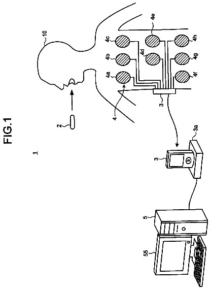
FIG. 1 is a schematic view of a representation of the whole of the device comprising all the main elements necessary for its proper operation, but also the additional peripheral, but not compulsory elements;
FIG. 2 is a schematic view of an overall representation of the partially made prototype including the camera, the connectors, the computer (here a laptop) and eventually a box containing the processing card.
FIG. 3 represents a complete diagram of the essential elements of the device specific for the invention.
FIGS. 4A, 4B, 4C, 4D, 4E, 4F and 4G show schematic views of the various steps of correlation between the visible and invisible part permitting to create the complemented object based on their common areas, here the crowns of the teeth.
FIGS. 5A, 5B, 5C and 5D show various schematic views of a complemented object observed by the clinician in the mouth of his patient through augmented-reality glasses when he moves his eyes.
FIGS. 6A, 6B, 6C and 6D show schematic views of the various planes that can be observed by the clinician in the mouth of the complemented object when he uses the transparency function of the present invention.
FIG. 7 represents the schematic view of the complemented object in the application of the present invention during the making of prosthetic preparations.
FIGS. 8A, 8B, and 8C show schematic views of the complemented object observed in the mouth by the practitioner when using a stored or recognizable instrument deformable for a root canal treatment or non-deformable for drilling an implant or a surgery operation.
FIG. 9 is a diagram representing the various steps of the clinical manipulation permitting the implementation of the present invention.
Detailed description of the invention
The present invention relates to a novel device in the dental field for intraoral viewing and/or measuring directly on the working site, i.e. in the patient's mouth, bringing together in one and the same three-dimensional or slightly shifted repository (a) the direct view of the teeth and gums in the patient's mouth through the augmented-reality glasses, (b) one or more modelings from taking radiological impressions—OCT and/or MRI, (c) one or more references or modelings from the cameras present on the augmented-reality glasses through which the practitioner views the patient's mouth, (d) eventually one or more references or modeling from an optical impression using structured light or not made using an intraoral camera, so that they complement and/or replace each other in order to enrich themselves using the principle of the augmented reality, and (e) eventually additional information associated herewith and provided by other peripheral devices, in order to permit the clinician to never take his eyes away from his intraoral working site, namely during gingival, coronary, root canal, surgical or bone treatment, in order to secure, facilitate and optimize his clinical action, this application being however non-restrictive in that the device is also applicable in the follow-up of all the clinical activities of the dental office.
To this end, this device according to the invention permits to view directly in the patient's mouth, through augmented-reality glasses, and in a way perfectly correlated, both the visible and the invisible part of the mouth in the form of one single object, which is here referred to as the complemented object.
The visible part is the surface of the teeth and the gums, the tongue and inner side of the cheeks. It can be seen directly through the glasses, but it can also be seen in the form of a scanned view resulting from the reading of a stereo-dynamic camera, or of several cameras located on the glasses or, more accurately, thanks to the digitized picture obtained by the scanning performed using the intraoral camera. The scanning can be substituted, without this being compulsory, by a direct vision in the scanned parts from the less accurate to the more accurate one, i.e. in the order of the most accurate domination (the intraoral camera) replaces the less accurate (camera of the glasses), which can in turn be substituted by the non-digitized direct view.
The invisible part results from a reading made separately before the therapeutic action through peripheral devices capable of providing RX, MRI, terahertz or ultrasonic images of the invisible parts located under the teeth, under the gums or under the skin, as are the bone, the epithelial and conjunctive tissues, the blood vessels and the nerves. These devices permit to statically or dynamically know, store and scan the invisible underlying anatomy.
For these two volume units, the visible and invisible part then forms one single unit, the central unit seeks the common parts and combines the two objects based on these common parts. These common parts can be anatomical objects, such as the crowns of the teeth or applied objects such as the locating wedges fixed in the mouth, for example on the teeth, if one wishes to avoid any abnormal mobility. These wedges or these anatomical reference marks are also used as an identification for following up the movement of the instruments made for this purpose.
Preferably and in some cases, it is possible to scan simultaneously invisible and visible parts. This is the case if we use an ultrasonic or terahertz device.
To this end, the invention is comprised of 1 ) a viewing device operating in real time or with a delay using augmented-reality glasses, which a three-dimensional spatial locating system (at least accelerometers/gyroscope and cameras) can be associated to and the function of which is to permit the practitioner not only to see his operating field in direct vision, but also to have punctual indications or external views, as all the glasses of this kind permit for assisting surgery, which permits him to normally follow the normally visible progression of his work (for example root canal treatment or surgical actions), i.e. the external part of the teeth and gums. It also permits him to add in addition correlated images, and this is the essential feature of the invention, resulting from 2 ) a second peripheral device. This second peripheral device is capable of providing RX, MRI, or terahertz ultrasonic images of the invisible parts that are located under the teeth and under the gums, such as the bone, the epithelial and conjunctive tissues, the blood vessels and the nerves, and to permit to know, store and scan the invisible underlying anatomy. The two devices 1 ) and 2 ) are dependent 3 ) on a central unit the function of which is to scan the views from the cameras located on the glasses and on the peripheral device in order to correlate them to combine them in one and the same repository, so that the clinician sees in the mouth of his patient, through his augmented-reality glasses, one single object from the combination of the view he naturally gets through his augmented-reality glasses, with which is combined permanently, dynamically and in real or almost real time, the various information from both external elements, the teeth and the gums, but also of the invisible elements, which permits the latter to have in its field of view in the mouth his patient the visible part, but also the invisible part located under the gums and under the teeth.
This thus permits the user to follow his action without taking his eyes away and to know the consequence of his action in a portion normally inaccessible to his eyes in the mouth. It should be noted that the present device, in order to avoid a permanent irradiation of the patient, needs only at least an initial 3D image and that he will correlate it in real time or almost real time depending on the viewing field, varying depending on the direction of the eyes of the practitioner, the cameras on augmented-reality glasses are filming.
To this device will preferably be added 4 ) an intraoral camera for taking an accurate optical impression using coherent radiation or not, with or without projection of active/or structured light the function of which is to perform a very accurate record of the shapes and colors of the anatomy present in the visible part of the patient's mouth, such as his teeth and/or his gums, this taking of an impression being correlated and combined by the central unit 3 ) with the previous views and more particularly with the less accurate view from the cameras carried by the augmented-reality glasses 1 ), but also and therefore with the sub-epithelial images from the external peripheral device 2 ). This permits the clinician to have in his working field an extremely accurate view of the portion resulting from the augmented-reality treatment.
Eventually and preferably, there will be added 5 ) a lighting system on the intraoral camera or on the glasses, the function of which is to optimize the diagnostic analysis such as for example the highlighting by special radiation of the carious areas on the hard tissues or tumors on the soft tissues.
In order to follow the movements in endodontics or surgery, it is enough to correlate the displayed instruments, known or calibrated on the double view—visible and invisible.
This process permits to optimize the therapeutic action of the clinician by significantly increasing the safety necessary for his actions while ensuring the structural integrity of the human body and providing accuracy within one micron. It permits especially to completely release the practitioner from determined constraints obliging him to watch a remote screen, to correlate different images of the visible and invisible area and to stay close to his working unit.
The invention comprises a hardware device and a software system.
The hardware device associates 1 ) a system for direct dental viewing of the visible tissues associated with the specific and miniaturized augmented reality, 2 ) a system for scanning the underlying tissues invisible to the naked eye, 3 ) a central unit for analog/digital conversion, management and correlation of the data, 4 ) eventually an accurate intraoral three-dimensional reading camera with or without structured light, 5 ) eventually an intraoral illumination specific for diagnosis, and 6 ) calibrated and known instruments used in the visible and invisible fields.
To this end, the object of the present invention is a device comprising specifically a viewing/capturing system with augmented-reality glasses, a device permitting to scan the parts invisible to the naked eye, a central unit, an intraoral accurate scanning camera, a lighting system and auxiliary devices.
The display/capturing system with augmented-reality glasses 1 permits to see the area of therapeutic action in direct viewing, while being able to correlate with same, then to add to same, when they have common connecting areas, additional information invisible to the eye directly from independent peripheral devices such as the images from the RX, MRI, terahertz or ultrasonic reading systems.
This viewing, displaying and capturing system may consist, for example, and this is only a non-restrictive example for the invention, of “Google Glass”, “Vuzix Smart Glass”, “Sony”, “K-Glass” or “HoloLens” glasses.
To these glasses are added one or more cameras permitting to readjust permanently and in real time by successive tracking the modeling resulting from the reading of the sub-gingival peripheral devices using common markers such as, and this is only an example, the crowns of the teeth or markers voluntarily placed on their surfaces or on the gum on what the clinician sees in the patient's mouth.
Eventually, advantageously according to an additional feature of the device according to the invention, for financial reasons, the device can be based on a 2D viewing, the essential function of the glasses is to show additional information with inaccurate adjustment on the working area in relief. The central unit is nowadays capable of correlating the 2D views on a 3D pattern.
It can also create a 3D image using two or more 2D radiologic images by applying equations that are now well known.
In the case of a 2D½ or 3D viewing, i.e. since these glasses have spatial vision, generally using stereoscopy, without this being systematic, the correlation is very accurate and the indications occur on parts of the body read in 3 dimensions. This is made possible by the presence of dedicated specific screens existing on this kind of glasses.
The description continues in the full USPTO document.
 Lapsed, fee not paid12 drawings