 Lapsed, fee not paid5 drawings
Biotech & Lab · US 9,863,941 B2
Microchip and method for detecting molecules and molecular interactions
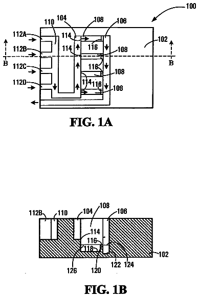
A microchip with flow-through inlet ( 104 ) and outlet ( 106 ) channels and test channels ( 108 ).
US 9,863,944 B2 · Assignee: ASAHI KASEI FIBERS CORPORATION · Inventors: Horii; Atsushi et al.
Sheet 1 of 2 from the published document. All sheets in the USPTO PDF
Provided is an immunochromatographic diagnostic kit which renders prompt diagnosis possible, has high analysis sensitivity, and gives test results with excellent reproducibility. The immunochromatographic diagnostic kit includes: a conjugate pad that contains a conjugate containing chromogenic particles which have an average particle diameter of 100-1,000 nm and attain a color intensity of 1.0-10.0, 10-90 wt % of the particles being derived from cellulose and 90-10 wt % of the particles being derived from a colorant; and a sample pad for an in vitro diagnostic reagent, the sample pad being constituted of nonwoven fabric that has a basis weight of 10-150 g/m.sup.2 and a thickness of 0.07-1.00 mm and comprises regenerated cellulosic fibers.
In recent years, convenient examination reagents, diagnostic reagents and diagnostic kits have been developed for conducting various examinations for the presence of pathogenic infection by viruses, bacteria or the like, the presence or absence of pregnancy, the presence or absence of cancer markers, and the presence or absence of specific raw materials or hazardous substances such as residual pesticides in foods, in short periods of time. These utilize specific reactions between the different substances to be examined and substances that specifically react with the substances to be examined. In particular, a large number of assay methods have been developed as immunoassays using antigen-antibody reaction between antigens and antibodies, including immunochromatographic assays, turbidimetric immunoassays, enzyme immunoassays, chemiluminescent assays, radioimmunoassays, assay methods using
All 2 drawing sheets from the published document, cropped to the drawing.
What the patent claimed, word for word. All of it is now free to use.
This application is a national phase application based on PCT/JP2014/065243, filed Jun. 9, 2014, which claims the priority of Japanese Patent Application No. 2013-121898, filed Jun. 10, 2013, the contents of both of which are incorporated herein by reference.
The present invention relates to an immunochromatographic diagnostic kit containing a sample pad for an in vitro diagnostic reagent, and more specifically it relates to a lateral flow-type immunochromatographic diagnostic kit.
In recent years, convenient examination reagents, diagnostic reagents and diagnostic kits have been developed for conducting various examinations for the presence of pathogenic infection by viruses, bacteria or the like, the presence or absence of pregnancy, the presence or absence of cancer markers, and the presence or absence of specific raw materials or hazardous substances such as residual pesticides in foods, in short periods of time. These utilize specific reactions between the different substances to be examined and substances that specifically react with the substances to be examined. In particular, a large number of assay methods have been developed as immunoassays using antigen-antibody reaction between antigens and antibodies, including immunochromatographic assays, turbidimetric immunoassays, enzyme immunoassays, chemiluminescent assays, radioimmunoassays, assay methods using surface plasmon resonance, and the like. These assay methods are also utilized for disease examination at hospitals, clinics and the like, and for food examination at food companies, for example. Among these, immunochromatographic assays do not require special equipment, devices or knowledge and their operation is convenient and economical, while they also allow rapid diagnosis, and therefore they are commonly implemented in a large range of examinations. Pregnancy examination agents, HIV examination agents and the like have come to be sold at ordinary pharmacies in recent years, allowing measurement by ordinary consumers, and it has also become possible to perform not only qualitative examinations for examining the presence or absence of substances to be examined, but also quantitative examinations for measurement of their amounts.
The principle of measurement in an immunochromatographic assay may be that of the “sandwich method” or of the “competitive method”. Moreover, the measurement system may be based on a flow-through method or a lateral flow method. It is possible to detect a variety of different substances as substances to be examined in a specimen, and a typical example is measurement by detection of antigen by the sandwich method, in which the following procedure is carried out in order.
A antibody that specifically binds with an antigen as the substance to be examined is immobilized at a prescribed section of a chromatographic medium such as a nitrocellulose membrane, and a reaction site known as the “test line” (hereunder referred to as “TL”) is formed at a desired location in the chromatographic medium.
A detection reagent is prepared by supporting an antibody that specifically binds with the substance to be examined, on a labeling substance such as an enzyme, chromogenic particles, fluorescent particles, magnetic particles or the like, and the detection reagent is coated and dried onto a conjugate pad or the like, a detection reagent-containing section is formed, and this is combined with the chromatographic medium to form an immunochromatographic diagnostic kit.
The actual specimen containing the antigen, or a solution containing it diluted with a desired liquid, is dropped onto a prescribed location of the immunochromatographic diagnostic kit, such as onto the sample pad, and the antigen and the detection reagent are developed on the chromatographic medium.
Using this series of procedures, the labeling substance is captured via the antigen on the antibody immobilized on the chromatographic medium at the reaction site, and the labeling substance signal is detected for diagnosis by the immunochromatographic diagnostic kit. Ordinarily, diagnosis is qualitative diagnosis that detects only the presence or absence of the antigen, but in recent years the strength of the signal can be visually or mechanically detected for quantitative diagnosis.
A rapid diagnosis time is a requirement for an immunochromatographic assay. This is in order to shorten the waiting time for the examination. A common method for meeting this requirement is a method of adjusting the pore sizes of the chromatographic medium to increase the traveling speed of the specimen.
Also, PTL 1 reports that a more rapid diagnosis time is possible by using a specific cellulosic fiber nonwoven fabric as the sample pad, at the section where the specimen sample containing the substance to be examined is to be dropped. PTL 1 mentions the liquid absorption speed and developability, but contains no concrete data for the diagnosis time and nowhere mentions a combination with specific chromogenic particles, as according to the present invention. In addition, it does not mention modification to the placement of the sample pad and conjugate pad.
Furthermore, PTL 2 reports that it is possible to achieve a more rapid diagnosis time by modifying the structure of the diagnostic kit and the like, but the shortest assessment in a hCG assay, for example, is 2 minutes and 35 seconds.
Increased analysis sensitivity is another requirement for an immunochromatographic assay. This means the ability to accomplish detection with an even lower amount of substance to be examined. In PTL 3, the present inventors have reported that analysis sensitivity can be increased by using cellulose particles having dense color and large particle diameters as the chromogenic particles. However, while it is mentioned that rapid diagnosis is possible by using chromogenic particles having dense color and large particle diameters, there is no concrete data for the diagnosis time and nothing is mentioned regarding combination with a specific sample pad, as according to the present invention.
Thus, there is generally a trade-off between speed of diagnosis time and increased analysis sensitivity, and a very great need exists for simultaneously achieving both.
In addition, PTL 4 discloses a semiquantitative test method using immunochromatography, and states that the antigen concentration and examination result signal strength can be made proportional for antigen concentrations in certain ranges. However, for quantitation in immunochromatography it is ideal to not only have proportionality between antigen concentration and examination result signal strength at certain antigen concentrations, but also to have reproducibility of test results, i.e. to obtain a signal of the same intensity when measuring antigen of the same concentration, but it is the general consensus that such reproducibility is still inadequate.
Also, PTL 5 discloses an immunochromatographic diagnostic kit using fluorescent particles as chromogenic particles, mentioning cellulose particles as an example for the material for the chromogenic particles and a cellulose nonwoven fabric as an example of the material for the sample pad, but neither are described in specific detail. In addition, it does not mention modification to the placement of the sample pad and conjugate pad. Naturally, nothing is mentioned regarding an effect by combination of the sample pad and the chromogenic particles, as according to the present invention. CITATION LIST Patent Literature
[PTL 1] Japanese Unexamined Patent Publication No. 2012-108031 [PTL 2] Japanese Unexamined Patent Publication HEI No. 7-55809 [PTL 3] International Patent Publication No. WO2011/062157 [PTL 4] International Patent Publication No. WO2006/080438 [PTL 5] International Patent Publication No. WO2013/151066 DISCLOSURE Of THE INVENTION Problems to be Solved by the Invention
In light of the aforementioned prior art, the problem to be solved by the present invention is to provide an immunochromatographic diagnostic kit that allows more rapid diagnosis, has high analysis sensitivity and also has excellent reproducibility of examination results. Means for Solving the Problems
The present inventors have conducted diligent research and much experimentation with the aim of solving this problem, and as a result have completed this invention upon finding, unexpectedly and surprisingly, that if particles with dense color and a large particle diameter are used as the chromogenic particles and a nonwoven fabric made of specific regenerated cellulosic fibers is used as the sample pad, by using these in combination, it is possible to increase the diagnosis speed and analysis sensitivity and to also achieve excellent reproducibility of examination results.
Specifically, the present invention is as follows.
[1] An immunochromatographic diagnostic kit including a conjugate pad containing a conjugate that includes chromogenic particles with an average particle diameter of 100 to 1000 nm and a color intensity of 1.0 to 10.0, the particle weight being 10 to 90 wt % derived from cellulose and 90 to 10 wt % derived from a colorant, and a sample pad for an in vitro diagnostic reagent composed of a nonwoven fabric made of regenerated cellulosic fibers, with a basis weight of 10 to 150 g/m.sup.2 and a thickness of 0.07 to 1.00 mm.
[2] An immunochromatographic diagnostic kit according to [1] above, wherein the regenerated cellulosic fibers are composed mainly of cuprammonium rayon fibers.
[3] An immunochromatographic diagnostic kit according to [1] or [2] above, wherein the regenerated cellulosic fibers are continuous filaments.
[4] An immunochromatographic diagnostic kit according to any one of [1] to [3] above, wherein the fiber loss number of the nonwoven fabric composed of the regenerated cellulosic fibers is less than 5000/m.sup.2.
[5] An immunochromatographic diagnostic kit according to any one of [1] to [4] above, wherein the colorant is a reactive dye.
[6] An immunochromatographic diagnostic kit according to any one of claims 1 to 5 , wherein the coverage factor on the sample pad for the in vitro diagnostic reagent is 50 to 100% with respect to the conjugate pad. Effect of the Invention
The immunochromatographic diagnostic kit of the invention includes prescribed chromogenic particles as the labeling substance, and employs a nonwoven fabric made of prescribed regenerated cellulosic fibers as the sample pad, whereby diagnosis is more rapid and the analysis sensitivity is increased, while the reproducibility of examination results is also excellent.
The present invention is an invention based on the finding by the present inventors that by combining prescribed chromogenic particles and a prescribed sample pad, it is possible to unexpectedly and surprisingly achieve a more rapid diagnosis time, increase the analysis sensitivity and increase the reproducibility of examination. As disclosed in PTL 3, it has been shown that chromogenic particles with a large particle diameter and high color intensity have higher analysis sensitivity even when used alone. In addition, such high analysis sensitivity also allows the diagnosis time to be shortened to some extent. However, because an immunochromatographic diagnostic kit comprises different porous structures such as the chromatographic medium, chromogenic particles with large particle diameters sometimes encounter interference with the pores of the porous structures, inhibiting their flow. When particles have been generated that have reduced flow, the diagnosis time can potentially be delayed. The present inventors considered that it may be possible to further shorten the diagnosis time by allowing chromogenic particles with large particle diameters to flow more easily. Upon repeated research based on this hypothesis, it was found that by using a nonwoven fabric made of regenerated cellulosic fibers and having low bulk density and a small thickness as the sample pad, it is possible to increase the flow rate per unit time for supply to the conjugate pad including the chromogenic particles, and to facilitate flow of the chromogenic particles in the conjugate pad, thereby shortening the diagnosis time. Chemical agents such as surfactants are sometimes added to facilitate the flow of chromogenic particles in sample pads, and since the sample pad of the present invention instantly absorbs a dropped specimen, it allows a fixed concentration of chemical agent to be constantly provided to facilitate flow of the chromogenic particles in the conjugate pad or chromatographic medium. Thus, the chromogenic particles easily flow without encountering interference in the pores of the porous structures. In an ordinary sample pad, the concentration of chemical agents that serve to facilitate flow of the chromogenic particles is not constant and clogging of the particles and the like can occur, but with the aforementioned effects it is possible to successfully allow all of the chromogenic particles to contribute to the examination, resulting in increased analysis sensitivity and also increased reproducibility of examination, while it was also found that it is possible to augment these effects by further modifying the placement of the sample pad and the conjugate pad.
FIG. 1 is a cross-sectional view of an immunochromatographic diagnostic kit as one embodiment of the invention.
FIG. 2 is a cross-sectional view of an immunochromatographic diagnostic kit as an embodiment of the invention, showing the furthest downstream section (h) of the sample pad, where the coverage factor of the sample pad on the conjugate pad is 100%.
FIG. 3 is a cross-sectional view of an immunochromatographic diagnostic kit as an embodiment of the invention, showing the furthest downstream section (i) of the sample pad, where the coverage factor of the sample pad on the conjugate pad is 50%.
FIG. 4 is a cross-sectional view of an immunochromatographic diagnostic kit as an embodiment of the invention, with the coverage factor of the sample pad on the conjugate pad at 100%, and showing a state where it is set inside an enclosure (j).
FIG. 5 is a graph showing the relationship between signal strength of examination results (TL color intensity) for examination of a positive specimen with constant concentration using an immunochromatographic diagnostic kit of the invention, and its change over time (min).
Embodiments of the invention will now be explained in detail.
The term “chromogenic particles”, for the purpose of the invention, refers to particulate matter that is insoluble in water, buffering solutions and the like, and supports a pigment, dye or the like. The material composing the particles is not particularly restricted, and examples of such chromogenic particles include metal colloid particles such as gold colloid, platinum colloid, silver colloid, selenium colloid and the like, colored latex particles including colored styrene-based latexes such as polystyrene latex, or acrylic acid-based latexes, colored silica particles which are colored silica with a 3-dimensional structure made of silicon atoms and oxygen atoms, colored cellulose particles which are colored cellulose, and chromogenic particles or magnetic particles that are direct particles of a colorant such as carbon black. The chromogenic particles may also be fluorescent particles. Colored cellulose particles are preferred from the viewpoint of ease of adjustment of the particle characteristics, such as adjustment of the particle diameter, adjustment of the color intensity, adjustment of the color type, and adjustment of the particle surface condition. Because cellulose contains numerous hydroxyl groups, it has high hydrophilicity and excellent dispersion stability, and can contain large amounts of colorant.
There are no particular restrictions on the “method for producing the chromogenic particles”. There may be mentioned a method in which the particles are first formed and then the colorant such as the pigment or dye is supported on them, a method in which particles are formed, and smaller chromogenic particles of a metal colloid or pigment are supported on them, or a method in which a colorant such as a coloring substance, dye, pigment, metal colloid or the like is added and formed together with formation of the particles. Preferred among these is a method in which particles are formed first and a colorant such as a pigment or dye is supported on them, from the viewpoint of ease of adjustment of the particle characteristics, including adjustment of the particle diameter, adjustment of the color intensity, adjustment of the color type and adjustment of the particle surface condition. A dye is preferred as the supported colorant, from the viewpoint of easier supporting.
When a dye is used as the colorant, the “type of dye” is not particularly restricted. There may be used dyes such as reactive dyes, direct dyes, auriferous dyes, acidic dyes, basic dyes, disperse dyes, sulfurized dyes, plant dyes, naphthol dyes, fluorescent dyes or the like. Any desired dyes may of course be used in combination. When cellulose is used as the particles, a reactive dye that binds to the hydroxyl groups of the cellulose by covalent bonding is most preferred from the viewpoint of allowing a large amount of dye to be held and from the viewpoint of stability.
When the cellulose particles are formed first and the colorant is supported afterwards, the “method of forming the cellulose particles” is not particularly restricted. There may be mentioned a method in which natural cellulose is physically micronized using a ball mill or a high-pressure homogenizer, a method in which it is chemically treated with an acid or alkali for micronization, or a method in which cellulose is dissolved in a good solvent and particles are formed. Alternatively, derivatized cellulose may be dissolved and particulates formed, and the derivatized substituents reconverted to hydroxyl groups to prepare cellulose particles. These forming methods may also be combined. The “type of cellulose” is also not particularly restricted, and there may be used regenerated cellulose, refined cellulose, natural cellulose, the aforementioned derivatized cellulose, or cellulose having derivatized substituents reconverted to hydroxyl groups. The preferred method among these is a method of dissolution in a good solvent and forming particulates, from the viewpoint of adjustment of the particle diameter, adjustment of the particle shapes, etc., while the preferred type of cellulose is regenerated cellulose.
When the cellulose is dissolved in a good solvent and particulates are formed, the “type of good solvent that dissolves cellulose” is not particularly restricted, and there may be used various good solvents that can dissolve cellulose, such as copper ammonia solution, viscose solution, N-methylmorpholine, and different ionic liquids and the like. Copper ammonia solution is preferred from the viewpoint of adjustment of the particle diameter, adjustment of the particle shapes, and the like. The method of forming the dissolved cellulose into particles is also not particularly restricted. A phase separation method was selected for the present invention.
The “average particle diameter” of the chromogenic particles is the volume-average median diameter as measured by the dynamic light scattering method, the volume-average median diameter being in the range of 100 to 1000 nm. If the average particle diameter is within this range, the particle surface area will be large, such that the TL will be darker, i.e. the analysis sensitivity will be higher, for use as an immunochromatographic diagnostic kit. If the average particle diameter is too small the surface area will be reduced and the analysis sensitivity may fall, or particle aggregation may occur. From this standpoint, the particle diameter is preferably 200 nm or greater and more preferably 300 nm or greater. If the particle diameter is too large, clogging of the pores of the chromatographic medium such as nitrocellulose will cause coloration of the sections that would otherwise be white after the examination, producing an adverse effect on judgment of the examination results and often impairing the detection limit. From this standpoint, the particle diameter is preferably no greater than 800 nm and more preferably no greater than 600 nm. The average particle diameter referred to here is only an average value, and a portion of the particle size distribution may be outside of the range.
The reason for using the volume-average for evaluation of the particle diameter is that very large particles in the immunochromatographic diagnostic kit clog the chromatographic medium, such as nitrocellulose, but larger particles have a greater effect based on volume-average, and therefore the effect of even a trace presence of large particles will be reflected. Other various methods of expression exist for evaluating the particle diameter in addition to volume-average, such as number-average and area-average. Naturally, a different method of expression will give a different particle diameter value, but volume-average is employed for the present invention.
The term “color intensity” is the value defining the color intensity of particles. As the method for measuring this value, a purified water dispersion of chromogenic particles with known concentration is prepared and subjected to visible spectrophotometry using an integrating sphere in a range of 400 to 800 nm with an optical path length of 10 mm, the peak value of the obtained absorbance curve (ABS) is measured, the obtained value is divided by the weight percent of the chromogenic particles, and the result is defined as the value in terms of absorbance per 0.01 wt % of chromogenic particles. For example, if the concentration of the prepared chromogenic particles is 0.0045% and the peak value of the absorbance curve is 1.0, the color intensity is (1×0.01)+0.0045=2.2.
The reason for conducting visible spectrophotometry using an integrating sphere for measurement of the color intensity of the particles is to allow the most accurate measurement of the color intensity of the particles in a state dispersed in liquid. Another method for measuring the color intensity of the particles is a method of measurement of a solid obtained by drying the particles, using a colorimeter or the like, but accurate measurement of the color intensity of the particles is not possible by such a method. For example, metal colloids and the like differ in their color tone and maximum wavelength depending on the particle diameter, and the dried aggregated state cannot accurately reflect the color intensity exhibited in a liquid-dispersed state. Furthermore, the color intensity is reduced when aggregation occurs in a liquid, even if dispersion is at the same particle concentration. In addition, the reason for using an integrating sphere for the visible spectrophotometry is in order to eliminate the effect of scattering of the particles themselves. Ordinary visible spectrophotometry is a method of measuring transmitted light, and it will reflect not only absorption of an incident beam by the colorant but also the influence of scattering of the particles themselves. For example, gold colloids commonly used for immunochromatography have particle diameters of 40 nm to 60 nm, and sometimes up to even 100 nm, and since these are all small particle diameters, there is virtually no effect on scattered light. In contrast, polystyrene latex particles have large particle diameters and clearly the effect on scattered light is considerable. For this reason, in order to more accurately reflect the color intensity of the particles themselves when particle diameters or particle materials differ, the present invention employs visible spectrophotometry using an integrating sphere.
The “color intensity” according to the invention is 1.0 to 10.0. If the value is too large, the color intensity becomes darker and the analysis sensitivity increases when used as an immunochromatographic diagnostic kit. A larger value is better of course, and there may be employed a method such as utilizing a dark-colored dye, increasing the number of dyeings, linking through some compound used as a spacer, increasing the amorphous regions of the particles to facilitate infiltration of the dye, or creating porosity in the particles to facilitate infiltration of the dye. In consideration of economy, however, the upper limit is preferably no greater than 7.0 and more preferably no greater than 5.0. Also, since a smaller value lowers the analysis sensitivity for use as an immunochromatographic diagnostic kit, the lower limit is preferably 1.5 or greater and more preferably 2.0 or greater.
The “proportion of the colorant in the chromogenic particles” is the proportion of colorant of the total weight of the chromogenic particles. For example, when 1.0 g of chromogenic particles consists of 0.2 g of cellulose and 0.8 g of colorant, the proportion of colorant is 80 wt %. The proportion of the colorant in the chromogenic particles is preferably 10 to 90 wt %. Within this range, the analysis sensitivity will be high for use as an immunochromatographic diagnostic kit. Also, when dyed particles that are dyed cellulose are used as the chromogenic particles, having the dye held in this range in the cellulose allows a suitable degree of hydrophobicity to be imparted to the cellulose, and allows substances that specifically bind with detection targets such as antibodies to be held by adsorption. Naturally, rather than holding by adsorption, it is possible to introduce carboxyl groups or amino groups into the colored cellulose particles to allow holding of substances that specifically bind to detection targets by covalent bonding. If the proportion of the colorant is low it will not be possible to obtain sufficient color intensity, and the analysis sensitivity will be reduced when used as an immunochromatographic diagnostic kit. Furthermore, when particles that are cellulose and dyed with a dye are used as the chromogenic particles, it is sometimes possible to even increase substances that specifically bind with the detection target by increasing the proportion of colorant. From this viewpoint, the lower limit is preferably 20 wt % or greater and more preferably 30 wt % or greater. While there is no particular problem even if the proportion of colorant exceeds 90 wt %, it is preferably no greater than 85 wt % and more preferably no greater than 80 wt %, when economic considerations are taken into account. Also, if the hydrophobicity is too strong, aggregation or nonspecific reactions may occur.
The “method of calculating the proportion of the colorant in the chromogenic particles” may be calculation from the weight change before and after coloration. When calculation from the weight change is difficult, a procedure of separating the colorant from the particles may be carried out and the colorant or particles isolated to perform the calculation. For example, when the cellulose particles have been dyed with a reactive dye, the covalent bonds between the cellulose and the dye may be broken with an acid or alkali, and the cellulose particles collected by centrifugal separation for calculation. Also, cellulase may be used for decomposition of the cellulose alone, and calculation performed.
The “method of calculating the cellulose-derived component of the chromogenic particles” may be calculation from the aforementioned proportion of the colorant in the chromogenic particles, specifically, the calculation may be performed by the formula: “Proportion of cellulose-derived component of the chromogenic particles”=100%−(proportion of the colorant in the chromogenic particles). The proportion of the cellulose-derived component of the chromogenic particles is preferably 90 to 10 wt %. Within this range it will be possible to maintain dispersion stability for the cellulose particles. Also, for the same reason mentioned for the proportion of the colorant, the lower limit for the cellulose-derived component of the chromogenic particles is more preferably 15 wt % or greater and most preferably 20 wt % or greater, and the upper limit is more preferably no greater than 80 wt % and most preferably no greater than 70 wt %.
The term “particles” refers to structures having similar lengths for the long diameter (L) and the short diameter (D), and having nearly spherical shapes. Specifically, it refers to structures having an L/D ratio represented by L+D of 1.0 to 3.0. If the L/D ratio is within this range, clogging will be inhibited when used as an immunochromatographic diagnostic kit, the ratio being more preferably 1.0 to 2.0, even more preferably 1.0 to 1.5 and most preferably 1.0 to 1.3. As the measuring method, an electron microscope image of the particles is taken, the long diameters (L) and short diameters (D) of 100 particles are measured, and the average value for the 100 particles is calculated.
The term “regenerated cellulosic fibers” refers to fibers composed mainly of regenerated cellulose. The regenerated cellulose may be cupra, lyocell, rayon or the like. Regenerated cellulosic fibers have very high hydrophilicity due to the orientation and state of the surface molecules during formation of the fibers, as well as excellent liquid absorption properties, and are therefore preferred as the material for composing the sample pad, with cupra and lyocell being more preferred and cupra being most preferred. It may be composed of 100% of specific regenerated cellulosic fibers, or they may be used in admixture.
The phrase “nonwoven fabric made of regenerated cellulosic fibers” refers to a nonwoven fabric made of the aforementioned regenerated cellulosic fibers. The structure can be easily formed with a nonwoven fabric, and handling is also excellent, allowing winding into a roll or the like. The type of nonwoven fabric may be a staple fiber nonwoven fabric or long filament nonwoven fabric, but a continuous filament nonwoven fabric is preferred for its excellent water absorbing properties and low fiber loss. Also, for control of the form stability and liquid absorption speed, synthetic fibers may be mixed therewith in a range that does not impair the hydrophilicity, in which case the content of the regenerated cellulosic fibers is preferably at least 50 wt % of the area ratio. Synthetic fibers are not particularly restricted so long as they satisfy the desired physical properties of the sample pad, and may be, for example, polyolefin-based, polyester-based, polyamide-based or acrylic-based synthetic fibers, any one or more of which may be used. These may also be used with a binder if necessary, or subjected to tangling treatment or the like.
The “basis weight of the nonwoven fabric made of regenerated cellulosic fibers” is 10 to 150 g/m.sup.2. The basis weight may vary appropriately depending on the size of the sample pad, the liquid volume of the specimen, the type of examination, etc., but if the basis weight is within the aforementioned range, a satisfactory balance will be obtained for absorption of specimen fluid and development on the conjugate pad, and after the specimen has been rapidly absorbed it will be possible to rapidly supply the specimen to the conjugate pad. A nonwoven fabric with an excessively low basis weight will have a small thickness or a high void percentage, and poor migration of specimen fluid into the sample pad and developability on the conjugate pad. From this viewpoint, the lower limit for the basis weight is preferably 15 g/m.sup.2 or greater, more preferably 20 g/m.sup.2 or greater and most preferably 25 g/m.sup.2 or greater. Also, a nonwoven fabric with an excessively high basis weight will have a large thickness or low void percentage, and specimen fluid will not be able to easily penetrate into the sample pad, causing the fluid to slide over the sample pad. From this viewpoint, the upper limit for the basis weight is preferably no greater than 120 g/m.sup.2 and more preferably no greater than 100 g/m.sup.2.
The “thickness of the nonwoven fabric made of regenerated cellulosic fibers” is 0.07 to 1.00 mm. The thickness may vary appropriately depending on the size of the sample pad, the liquid volume of the specimen, the type of examination, etc., but if the thickness is within the aforementioned range, a satisfactory balance will be obtained for absorption of specimen fluid and development on the conjugate pad, and after the specimen has been rapidly absorbed it will be possible to rapidly supply the specimen to the conjugate pad. If the thickness is too small, it will not be possible to adequately receive specimen fluid. Handling of the diagnostic kit will also become more difficult during the production steps. From this viewpoint, the lower limit for the thickness is preferably 0.10 mm and more preferably 0.20 mm. Also, if the thickness is too large, too much specimen fluid will be held, which is undesirable. From this viewpoint, the upper limit for the thickness is preferably no greater than 0.80 mm and more preferably no greater than 0.70 mm.
The bulk density of the nonwoven fabric made of regenerated cellulosic fibers” is preferably 0.06 to 1.00 g/cm.sup.3. The bulk density may vary appropriately depending on the size of the sample pad, the liquid volume of the specimen, the type of examination, etc., but if the bulk density is within the aforementioned range, a satisfactory balance will be obtained for absorption of specimen fluid and development on the conjugate pad, and after the specimen has been rapidly absorbed it will be possible to rapidly supply the specimen to the conjugate pad. If the bulk density is too low, the voids in the fibers will increase, leading to poor migration of specimen fluid into the sample pad and developability on the conjugate pad. From this viewpoint, the lower limit for the bulk density is preferably 0.07 g/cm.sup.3 and more preferably 0.10 g/cm.sup.3. Also, if the bulk density is too high, the fibers will become too compact and specimen fluid will not be able to easily penetrate into the sample pad, causing the fluid to slide over the sample pad. From this viewpoint, the upper limit for the bulk density is preferably no greater than 0.70 g/cm.sup.3 and more preferably no greater than 0.50 g/cm.sup.3.
The “fiber loss number” is the number of fibers with sizes of 100 μm or greater that are lost per unit square meter when a 25 cm×25 cm sample is placed in 300 ml of purified water and allowed to stand for 2 minutes, and then the sample is removed and the remaining fluid is filtered with black filter paper (ADVANTEC NO131), the filter paper after filtration is set in a thermostatic chamber (20° C., 65% RH) for 12 hours and dried and the count is determined using a video microscope. According to the invention, the fiber loss number is preferably less than 5000/m.sup.2, more preferably less than 4000/m.sup.2, and even more preferably less than 3000/m.sup.2. If the fiber loss number is greater than 10,000/m.sup.2 this is undesirable because the fibers will easily drop off, and depending on the type of examination fluid, the fallen fibers will accumulate at the interface between the sample pad and the conjugate pad, impairing the developability and also contaminating the area around the production line during production, tending to result in poor yield. Since a lower fiber loss number is preferred, the lower limit is as low as 1/m.sup.2.
As shown in FIG. 1( a ) , the “sample pad” according to the invention is the section that first receives a specimen that is to be measured by immunochromatography. Common sample pads include cellulose filter paper, paper, glass fibers, glass fibers, acrylic fibers nylon fibers and various woven fabrics, but for the invention the sample pad used is a nonwoven fabric made of regenerated cellulosic fibers, as mentioned above. The nonwoven fabric made of regenerated cellulosic fibers has excellent liquid absorption properties, and therefore the specimen is rapidly absorbed and the specimen can migrate into the conjugate pad. Furthermore, if the structure of the nonwoven fabric made of regenerated cellulosic fibers is the structure described above, it is possible to increase the flow rate per unit time for supply to the conjugate pad, and to better facilitate initial release of the chromogenic particles in the conjugate pad, thereby shortening the diagnosis time. Also, a chemical agent for facilitating flow of the chromogenic particles can be supplied at a constant fixed concentration, to allow easier flow without clogging of the chromogenic particles. Since, as a result of this, almost all of the particles can contribute to the examination, the analysis sensitivity is increased and the reproducibility of examination can also be improved. Conversely, when the chromogenic particles in the conjugate pad are not initially released, the diagnosis time is lengthened. Moreover, when virtually all of the chromogenic particles are not released from the conjugate pad, the analysis sensitivity is reduced and the reproducibility of examination is poor.
According to the invention, the nonwoven fabric made of regenerated cellulosic fibers may contain different chemical agents or powders, or a portion of the cellulose may be derivatized, for control of the hydrophilicity/water-repellency of the sample pad or the water absorption factor, so long as the aforementioned physical properties are not adversely affected and the antigen-antibody reaction and antibody stability are not affected. Examples of chemical agents to be impregnated include surfactants, proteins, antibodies, resins, water-soluble polymers, antimicrobial agents, antiseptic agents, antioxidants and the like. Also, examples of derivatized cellulose forms include carboxymethylated, carboxyethylated, primary aminated, secondary aminated, tertiary aminated, quaternary aminated and hydroxylated forms.
According to the invention, the sample pad may be subjected to pretreatment if necessary. For example, it may be subjected to treatment for pre-addition of a buffering solution, a surfactant, a protein, a reagent to trap contaminants in the specimen sample, an antiseptic agent, an antimicrobial agent, an antioxidant, a humectant or the like. Also, there is no particular restriction on the form of the sample pad, and for example, the size of the sample pad is such that the length (length flowed by the fluid) is preferably about 10 to 25 mm in consideration of the associating property from the specimen fluid, and the diagnosis time, while there is no problem if the width (perpendicular to the flow of the fluid) is larger than the width of the conjugate pad. If the width is too narrow, the examination fluid can potentially seep around the edges of the sample pad.
The description continues in the full USPTO document.
About 6,083 words. The USPTO PDF has it with every drawing.
Fees are due 3.5, 7.5 and 11.5 years after grant. This patent expired on January 9, 2026, so the fee marked "not paid" was the one that went unpaid.
Immunochromatographic Diagnosis Kit
Filed Jun 2014 · published May 2016Immunochromatographic diagnosis kit
Filed Jun 2014 · granted Jan 2018Earlier publications, parents and continuations. None of them can still be enforced, or this patent would not be listed.
Prior art cited by the examiner or applicant. Useful when you check your own idea for novelty.
Everything on this page comes from the documents linked above.