 Lapsed, fee not paid10 drawings
Medical Devices · US 9,801,593 B2
Intraoral X-ray imaging sensor and readout
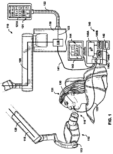
Devices, methods, and systems including an intraoral x-ray imaging sensor.
US 9,801,601 B2 · Assignee: LABORATOIRES BODYCAD INC. · Inventors: Rivet-Sabourin; Geoffroy et al.
Sheet 1 of 13 from the published document. All sheets in the USPTO PDF
A computer implemented method for performing bone segmentation in imaging data of a section of a body structure is provided. The method includes: Obtaining the imaging data including a plurality of 2D images of the section of the body structure; and performing a multiphase local-based hybrid level set segmentation on at least a subset of the plurality of 2D images by minimizing an energy functional including a local-based edge term and a local-based region term computed locally inside a local neighborhood centered at each pixel of each one of the 2D images on which the multiphase local-based hybrid level set segmentation is performed, the local neighborhood being defined by a Gaussian kernel whose size is determined by a scale parameter (σ).
There are several advantages that may occur from patient-specific orthopedic implants and surgeries including exact sizing of the implant according to the anatomy, such as reducing operating time, improving performance, etc. When designing and conceiving patient-specific orthopedic implants and planning surgeries, including alignment guides, all relevant components of an anatomical structure, e.g. an articulation, are to be modeled and segmented with high precision. Precise modelling and segmentation in turn ensures that the resulting prostheses and alignment guides accurately fit the unique shape and size of the anatomical structure. Furthermore, segmentation of bones from 3D images is important to many clinical applications such as visualization, enhancement, disease diagnosis, implant design, cutting guide design, and surgical planning. In the field of bone imaging, many imaging techn
1 of 13 drawing sheets so far from the published document, cropped to the drawing. Every sheet is in the USPTO PDF.
What the patent claimed, word for word. All of it is now free to use.
The present invention relates to the field of bone structure imaging for skeletal modelling. More particularly, it relates to a method for performing multi-bone segmentation in closely matching joints of a patient, as part of a bone imaging process.
There are several advantages that may occur from patient-specific orthopedic implants and surgeries including exact sizing of the implant according to the anatomy, such as reducing operating time, improving performance, etc.
When designing and conceiving patient-specific orthopedic implants and planning surgeries, including alignment guides, all relevant components of an anatomical structure, e.g. an articulation, are to be modeled and segmented with high precision. Precise modelling and segmentation in turn ensures that the resulting prostheses and alignment guides accurately fit the unique shape and size of the anatomical structure. Furthermore, segmentation of bones from 3D images is important to many clinical applications such as visualization, enhancement, disease diagnosis, implant design, cutting guide design, and surgical planning.
In the field of bone imaging, many imaging techniques and methods are known in the art in order to produce a skeletal model, such as a 3D skeletal model, of at least a portion of a body structure of a patient, such as a bone or series of bones on/between which an orthopedic implant is to be implanted.
For example and without being limitative, common imaging techniques, including magnetic resonance imaging (MRI), computed axial tomography (CAT scan), ultrasound, or the like are combined with three-dimensional image reconstruction tools, such as CAD software or the like, for the three-dimensional image reconstruction. In the case of bones with well-defined joints, known imaging techniques and three-dimensional image reconstruction tools are usually able to produce satisfactory models.
However, in the case of small and/or multiple adjacent bones such as, for example, bones of the hands and foot, where the distance between the bones is relatively small, thereby forming closely matching joints therebetween, and larger bones with outer edges closely matching one another, thereby also defining closely matching joints, such as hip bones or the like, known imaging techniques and three-dimensional image reconstruction tools often prove inadequate to perform the required individual multi-bone segmentation, i.e. the partitioning of a digital image into multiple segments in order to provide data that can be used for generating the three-dimensional image which clearly define the shape of each bone of the joints and is therefore more meaningful or easier to analyze. In fact, the failure of those well-known imaging techniques to segment these images is due to the challenging nature of the acquired images. For example, in CT imagery, when the boundaries of two bones are too close to each other, as described earlier, they tend to be diffused, which lower the contrast of the boundaries of the neighboring bones with respect to the background. Moreover, the bone structures have inhomogeneous intensities which involve an overlap between the distributions of the intensities within the regions.
In view of the above, there is a need for an improved method and system for performing bone segmentation which would be able to overcome or at least minimize some of the above-discussed prior art concerns.
According to a general aspect, there is provided a computer implemented method for performing bone segmentation in imaging data of a section of a body structure. The method comprises: Obtaining the imaging data including a plurality of 2D images of the section of the body structure; and Performing a multiphase local-based hybrid level set segmentation on at least a subset of the plurality of 2D images by minimizing an energy functional including a local-based edge term and a local-based region term computed locally inside a local neighborhood centered at each pixel of each one of the 2D images on which the multiphase local-based hybrid level set segmentation is performed, the local neighborhood being defined by a Gaussian kernel whose size is determined by a scale parameter (σ).
The minimization of the energy functional generates segmented blobs delimitating the regions of the 2D images, each one of the blobs substantially contouring one or more bones contained in the respective one of the 2D images. For instance, the segmented blobs are used to discriminate the background of the 2D images from the region(s) corresponding to the bones. The segmented blobs in each of the 2D images can be stacked to generate a plurality of 3D subvolumes, each one of the 3D subvolumes resulting from the combination of a plurality of corresponding segmented blobs. Each one of the 3D subvolumes corresponds to one or more bones. The plurality of 3D subvolumes can be combined to generate a 3D volume including a plurality of bones of the section of the body structure. The 3D volume is thus a 3D model of the bones of the section of the body structure.
In an embodiment, the local neighborhood is circular and performing the multiphase local-based hybrid level set segmentation further comprises: for each pixel of the 2D images, dynamically changing region descriptors based on a position of a center of the local neighborhood.
In an embodiment, the computer implemented method further comprises: selecting a value of λ to adjust a performance of the multiphase local-based hybrid level set segmentation with λ being greater than 0 and smaller than 1, wherein λ multiplies the local-based edge term and (1−λ) multiplies local-based region term.
In an embodiment, the computer implemented method further comprises: initializing the multiphase local-based hybrid level set segmentation with a contour obtained from a 3D adaptive thresholding, the contour being close to boundaries of the bones to be segmented.
In an embodiment, the 2D images include two phases and the energy functional is: .sub.2-phase(φ, c,b )=(1−λ) .sub.region(φ, c,b )+λ .sub.edge(φ)+μ .sub.p(φ) where λ is greater than or equal to 0 and smaller than or equal to 1, μ is a positive constant, b is a bias field accounting for intensity inhomogeneity, and c is a vector representing intensity-based constant values in disjoint regions, .sub.edge(φ)=ν .sub.g(φ)+α .sub.g(φ)
wherein ν and α are normalization constants,
ℒ g ( ϕ ) = Δ ∫ g σ , τ δ .Math. ( ϕ ) .Math. ∇ ϕ .Math. d x , �� g ( ϕ ) = Δ ∫ g σ , τ ℋ .Math. ( - ϕ ) d x , g σ , τ = Δ 1 1 + �� σ , τ , �� σ , τ ( x ) = ∫ K σ ( y - x ) u τ ( y ) d y , u τ = Δ .Math. ∇ G τ * I .Math. 2 with G.sub.τ being a Gaussian kernel with a standard definition τ and I being the image, K.sub.σ, a kernel function computed by means of a truncated Gaussian function of the scale parameter (σ), ρ being the radius of the local circular neighborhood:
K σ ( u ) = { 1 a e - .Math. u .Math. 2 / 2 σ 2 , .Math. u .Math. ≤ p 0 , otherwise and H .Math. ( ϕ ) = 1 2 [ 1 + 2 π arctan ( ϕ .Math. ) ] with ε being a parameter; and .sub.region(φ, c,b )=∫(Σ.sub.i=1.sup.N(∫ K .sub.σ( y−x )| I ( x )− b ( y ) c .sub.i|.sup.2 dy ) .sub.i(φ( x )) dx .sub.i is a membership function of each region Ω.sub.i, and is defined as: .sub.2(φ)= .sub.ε(φ) .sub.2(φ)=1− .sub.ε(φ) wherein .sub.p is a regularisation term: .sub.p(φ)=∫ p (|∇φ|) dx and the minimization of the hybrid energy functional is carried out by gradient descent method:
∂ ϕ ∂ t = - ∂ F 2 _ phase ∂ ϕ
In another embodiment, the energy functional is: .sub.multiphase(Φ, c,b )=(1−λ) .sub.region(Φ, c,b )+λ .sub.edge(Φ)+μ .sub.p(Φ) where λ is greater than or equal to 0 and smaller than or equal to 1, μ is a positive constant, b is a bias field accounting for intensity inhomogeneity, c is a vector representing intensity-based constant values in disjoint regions, and φ is a vector formed by k level set functions φi, i=1 . . . k for k regions or phases; Φ=(φ.sub.1( y ), . . . ,φ.sub.k( y )) and a number of the level set functions to be used is at least equal to: k =log.sub.2( ) where log.sub.2 is the logarithm to the base 2 and N is the number of the regions to be segmented in the image. .sub.region(Φ, c,b )=∫Σ.sub.i=1.sup.N e .sub.i( x ) M .sub.i(Φ) x )) dx With: e .sub.i( x )=∫ K .sub.σ |I ( x )− b ( y ) c .sub.i|.sup.2 dy, i=I, . . . ,k with K.sub.σ, a kernel function computed by means of a truncated Gaussian function of standard deviation σ, referred to as the scale parameter, .sub.i is a membership function of each region Ω.sub.i, and is defined as:
M i ( Φ ) = M i ( ϕ 1 ( y ) , .Math. , ϕ k ( y ) ) = { 1 , y ∈ Ω i 0 , else E edge ( Φ ) = v ℒ g ( Φ ) + α �� g ( Φ ) Where: .sub.g(Φ)=Σ.sub.j=1.sup.k .sub.g(φ.sub.j) .sub.g(Φ)=Σ.sub.j=1.sup.k .sub.g(φ.sub.j) wherein ν and α are normalization constants, wherein .sub.p is a regularisation term: .sub.p(φ)=∫ p (|∇φ|) dx and the minimization of the multiphase hybrid energy functional .sub.multiphase by gradient descent method:
∂ ϕ 1 ∂ t = - ∂ Fmult iphase ( Φ ) ∂ ϕ 1 , .Math. , ∂ ϕ k ∂ t = - ∂ Fmu ltiphase ( Φ ) ∂ ϕ k .
In an embodiment, the computer implemented method further comprises: identifying regions of interest (ROIs) on at least the subset of the plurality of 2D images of the section of the body structure; and performing the multiphase local-based hybrid level set segmentation on the regions of interest (ROIs).
According to another general aspect, there is provided a computer implemented method for performing bone segmentation in imaging data of at least a section of a body structure including a plurality of bones using anatomical knowledge data relative to the section of the body structure of the imaging data, the method comprising: Obtaining the imaging data including a plurality of 2D images of the section of the body structure; Generating primary image data from the imaging data using an image preprocessing including identifying regions of interest (ROIs) in the 2D images; Generating secondary segmented image data including a plurality of 2D binary images with segmented blobs by performing a multiphase local-based hybrid level set segmentation on the regions of interest (ROIs) by minimizing an energy functional including a local-based edge term and a local-based region term computed locally inside a local neighborhood centered at each point of a respective one of the regions of interest (ROIs), the local neighborhood being defined by a Gaussian kernel; Generating a 3D volume including a plurality of 3D subvolumes from the segmented blobs of the secondary segmented image data; and Associating an anatomical component to each one of the 3D subvolumes using the anatomical knowledge data relative to the section of the body structure of the imaging data.
In an embodiment, the image preprocessing further comprises performing a 3D adaptive thresholding processing to define thresholded blobs in the 2D images and generating binary masks from the thresholded blobs obtained by the 3D adaptive thresholding processing. The plurality of 2D images are greyscale images and the 3D adaptive thresholding processing can include the steps of: For at least a sample of the plurality of 2D greyscale images: Dividing each one of the 2D greyscale images of at least the sample in a plurality of sections; Computing a local pixel intensity section threshold for each one of the sections; Computing a global image pixel intensity threshold for each one of the 2D greyscale images of at least the sample using the local pixel intensity section thresholds computed for each one of the sections; Computing a global volume pixel intensity threshold using the global image pixel intensity thresholds; and Applying the global volume pixel intensity threshold to each one of the 2D greyscale images of the plurality of 2D greyscale images.
The global image pixel intensity threshold for each one of the 2D greyscale images of at least the sample can be computed as a maximum of the local pixel intensity thresholds for the corresponding image. The global volume pixel intensity threshold from the global image pixel intensity thresholds can be computed as a mean of the global image pixel intensity thresholds minus 1.5 times a standard deviation of the global image pixel intensity thresholds [mean(global image pixel intensity thresholds)−1.5std(global image pixel intensity thresholds)]. The image preprocessing can comprise computing thresholded blobs in the images following the 3D adaptive thresholding processing and creating binary masks from the thresholded blobs. Identifying regions of interest (ROIs) in the 2D images can comprise selecting regions in the 2D greyscale images of the imaging data including at least one of a respective one of the thresholded blobs and a respective one of the binary masks generated from the thresholded blobs.
In an embodiment, generating secondary segmented image data can comprise performing a blob masking validation following the multiphase local-based hybrid level set segmentation, the multiphase local-based hybrid level set segmentation generating a plurality of unmasked blobs, and wherein the blob masking validation comprises: Applying the binary masks to the unmasked blobs to obtain masked blobs; Determining at least one perceptual grouping property of each one of the masked blobs and the unmasked blobs; For each corresponding pair of masked blobs and unmasked blobs, Comparing the at least one perceptual grouping property of the masked blob to the at least one perceptual grouping property of the corresponding one of unmasked blobs; and Selecting the one of the masked blob and the corresponding one of unmasked blobs having the highest perceptual grouping property as the segmented blob of the secondary segmented image data.
The computer implemented method can further comprise initializing the multiphase local-based hybrid level set segmentation with the binary masks.
Performing the multiphase local-based hybrid level set segmentation on the regions of interest (ROIs) can comprise generating binary subimages including the segmented blobs and the method can further comprise merging the binary subimages to generate a respective one of the 2D binary images including the segmented blobs.
In an embodiment, generating secondary segmented image data comprises stacking the 2D binary images.
In an embodiment, the image preprocessing further comprises: Determining an initial image including at least one region of interest and determining a final image including at least one region of interest; and Selecting a subset of 2D images including the initial image, the final image, and the images extending therebetween, wherein the primary image data consists of the subset of 2D images including the regions of interest (ROIs). The computer implemented method of any one of claims 6 to 13 , wherein identifying anatomical components in the 3D volume comprises: Computing at least one subvolume feature for each one of the 3D subvolumes; For each one of the 3D subvolumes, carrying out a bone identification processing comprising: Identifying a closest one of the bones and comparing the at least one subvolume feature to features of the anatomical knowledge data corresponding to the closest one of the bones; If the at least one subvolume feature for the respective one of the 3D subvolumes substantially corresponds to the features of the anatomical knowledge data for the closest one of the bones, associating the respective one of the 3D subvolumes to the closest one of the bones; Otherwise, applying a selective 3D bone separation to the respective one of the 3D subvolumes and generating new 3D subvolumes.
Identifying anatomical components in the 3D volume can further comprise: Identifying a 3D anatomical point of interest within the 3D volume; Identifying a 3D subvolume closest to the 3D anatomical point of interest; and Performing sequentially the bone identification processing by proximity to a last one of associated 3D subvolumes, starting from the 3D subvolume closest to the 3D anatomical point of interest. The computer implemented method of one of claims 14 and 15 , wherein the 3D volume generated from the 2D binary images of the secondary segmented image data is a first 3D volume and the method further comprises: Carrying out a 2D blob separation on the secondary segmented image data and generating a second 3D volume by stacking binary images obtained following the 2D blob separation; and wherein identifying anatomical components is performed on the second 3D volume.
Carrying out a 2D blob separation can comprise: For each one of the segmented blobs of the secondary segmented image data: Creating straight segments from the contours of the respective one of the segmented blobs; Identifying points of interest using the straight segments; If there is at least one point of interest, identifying at least one bone attachment location close to the at least one point of interest; and separating the respective one of the segmented blobs by local morphological erosion along the at least one bone attachment location.
Identifying points of interest using the straight segments can comprise: Determining a length of the straight segments and an angle between consecutive ones of the straight segments, the consecutive one of the straight segments sharing a common point; For each pair of consecutive straight segments (s.sub.1, s.sub.2), computing a relevance measure (K.sub.relevance):
K relevance = β ( s 1 , s 2 ) l ( s 1 ) l ( s 2 ) l ( s 1 ) + l ( s 2 ) wherein β(s.sub.1, s.sub.2) is the angle between the two consecutive straight segments s.sub.1 and s.sub.2; I(s.sub.1) and I(s.sub.2) are lengths of the two consecutive straight segments s.sub.1 and s.sub.2 respectively; Comparing the computed relevance measure to a predetermined threshold; and If the computed relevance measure meets the predetermined relevance threshold, identifying the common point as being a point of interest.
Identifying at least one bone attachment location close to the at least one point of interest can comprise: Identifying if a respective one of the points of interest belongs to a linear bone attachment location defined by a pair of points of interest; and, for each identified linear bone attachment location, separating the respective one of the segmented blobs comprises performing a linear local morphological erosion along a line extending between the points of interest defining the linear bone attachment location; otherwise, identifying the respective one of the points of interest as a punctual bone attachment location and separating the respective one of the segmented blobs comprises performing local morphological erosion around the punctual bone attachment location.
Identifying a pair of points of interest can comprise: for each potential pair of points of interest, grouping the points of interest in a pair and computing a distance separating two grouped points of the pair, comparing the computed distance to a predetermined distance threshold; and if the computed distance meets the predetermined distance threshold, associating the potential pair of interest points as being one linear bone attachment location.
In an embodiment, the local neighborhood is circular and performing the multiphase local-based hybrid level set segmentation further comprises: for each pixel of the regions of interest (ROIs), dynamically changing region descriptors based on a position of a center of the local neighborhood.
In an embodiment, the computer implemented method further comprises: selecting a value of λ to adjust the performance of the multiphase local-based hybrid level set segmentation with λ being greater than 0 and smaller than 1, wherein λ multiplies the local-based edge term and (1−λ) multiplies local-based region term.
In an embodiment, the regions of interest (ROIs) include two phases and the energy functional is: .sub.2-phase(φ, c,b )=(1−λ) .sub.region(φ, c,b )+λ .sub.edge(φ)+μ .sub.p(φ) where λ is greater than or equal to 0 and smaller than or equal to 1, μ is a positive constant, b is a bias field accounting for intensity inhomogeneity, and c is a vector representing intensity-based constant values in disjoint regions, .sub.edge(φ)=ν .sub.g(φ)+α .sub.g(φ)
wherein ν and α are normalization constants,
ℒ g ( ϕ ) = Δ ∫ g σ , τ δ .Math. ( ϕ ) .Math. ∇ ϕ .Math. d x , �� g ( ϕ ) = Δ ∫ g σ , τ ℋ .Math. ( - δ ) d x , g σ , τ = Δ 1 1 + f σ , τ , �� σ , τ ( x ) = ∫ K σ ( y - x ) u τ ( y ) d y , u τ = Δ .Math. ∇ G τ * I .Math. 2 with G.sub.τ being a Gaussian kernel with a standard definition τ and I being the image, K.sub.σ, a kernel function computed by means of a truncated Gaussian function of the scale parameter (σ), ρ being the radius of the local circular neighborhood:
K σ ( u ) = { 1 a e - .Math. u .Math. 2 / 2 σ 2 , .Math. u .Math. ≤ p 0 , otherwise and H .Math. ( ϕ ) = 1 2 [ 1 + 2 π arctan ( ϕ .Math. ) ] with ε being a parameter; and .sub.region(φ, c,b )=∫(Σ.sub.i=1.sup.N(∫ K .sub.σ( y−x )| I ( x )− b ( y ) c .sub.i|.sup.2 dy ) .sub.i(φ( x )) dx .sub.i is a membership function of each region δ.sub.i, and is defined as: .sub.1(φ)= .sub.ε(φ) .sub.2(φ)=1− .sub.ε(φ) wherein .sub.p is a regularisation term: .sub.p(φ)=∫ p (|∇φ|) dx and the minimization of the hybrid energy functional is carried out by gradient descent method:
∂ ϕ ∂ t = - ∂ F 2 _ phase ∂ ϕ .
In another embodiment, the energy functional is: .sub.multiphase(Φ, c,b )=(1−λ) .sub.region(Φ, c,b )+λ .sub.edge(Φ)+μ .sub.p(Φ) where λ is greater than or equal to 0 and smaller than or equal to 1, μ is a positive constant, b is a bias field accounting for intensity inhomogeneity, c is a vector representing intensity-based constant values in disjoint regions, and φ is a vector formed by k level set functions φi, i=1 . . . k for k regions or phases; Φ=(φ.sub.1( y ), . . . ,φ.sub.k( y )) and a number of the level set functions to be used is at least equal to: k =log.sub.2( ) where log.sub.2 is the logarithm to the base 2 and N is the number of the regions to be segmented in the image. .sub.region(Φ, c,b )=∫Σ.sub.i=1.sup.N e .sub.i( x ) M .sub.i(Φ) x )) dx With: e .sub.i( x )=∫ K .sub.σ |I ( x )− b ( y ) c .sub.i|.sup.2 dy, i= 1, . . . , k with K.sub.σ, a kernel function computed by means of a truncated Gaussian function of standard deviation σ, referred to as the scale parameter, .sub.i is a membership function of each region Ω.sub.i, and is defined as:
0 M i ( Φ ) = M i ( ϕ 1 ( y ) , .Math. , ϕ k ( y ) ) = { 1 , y ∈ Ω i 0 , else E edge ( Φ ) = v ℒ g ( Φ ) + α �� g ( Φ ) Where: .sub.g(Φ)=Σ.sub.j=1.sup.k( .sub.g(φ.sub.j) .sub.g(Φ)=Σ.sub.j=1.sup.k .sub.g(φ.sub.j) wherein ν and α are normalization constants, wherein .sub.p is a regularisation term: .sub.p(φ)=∫ p (|∇φ|) dx and the minimization of the multiphase hybrid energy functional .sub.multiphase by gradient descent method:
∂ ϕ 1 ∂ t = - ∂ Fmult iphase ( Φ ) ∂ ϕ 1 , .Math. , ∂ ϕ k ∂ t = - ∂ Fmu ltiphase ( Φ ) ∂ ϕ k .
According to a general aspect, there is provided a computer implemented method for performing bone segmentation in imaging data of at least a section of a body structure including a plurality of bones using anatomical knowledge data relative to the section of the body structure of the imaging data. The method comprises: Obtaining the imaging data including a plurality of 2D greyscale images of the section of the body structure; Generating primary image data from the imaging data using an image preprocessing including: Performing a 3D adaptive thresholding processing to define thresholded blobs in each of the 2D greyscale images; Generating binary masks from the thresholded blobs; and Identifying regions of interest (ROIs) in the 2D greyscale images of the imaging data using the binary masks generated from the thresholded blobs; Generating secondary segmented image data including a plurality of 2D binary images with segmented blobs by: Carrying out a segmentation on the regions of interest (ROIs) to obtain a plurality of unmasked blobs; Applying the binary masks to the unmasked blobs to obtain masked blobs; Determining at least one perceptual grouping property of each one of the masked blobs and the unmasked blobs; For each corresponding pair of masked blobs and unmasked blobs, Comparing the at least one perceptual grouping property of the masked blob to the at least one perceptual grouping property of the corresponding one of the unmasked blobs; and Selecting the one of the masked blob and the corresponding one of unmasked blobs having the highest perceptual grouping property as the segmented blob of the secondary segmented image data; Generating a 3D volume including a plurality of 3D subvolumes from the segmented blobs of the secondary segmented image data; and Associating an anatomical component to each one of the 3D subvolumes using the anatomical knowledge data relative to the section of the body structure of the imaging data.
In an embodiment, the 3D adaptive thresholding processing includes the steps of: For at least a sample of the plurality of 2D greyscale images: Dividing each one of the 2D greyscale images of at least the sample in a plurality of sections; Computing a local pixel intensity section threshold for each one of the sections; Computing a global image pixel intensity threshold for each one of the 2D greyscale images of at least the sample using the local pixel intensity section thresholds computed for each one of the sections; Computing a global volume pixel intensity threshold using the global image pixel intensity thresholds; and Applying the global volume pixel intensity threshold to each one of the 2D greyscale images of the plurality of 2D greyscale images.
The global image pixel intensity threshold for each one of the 2D greyscale images of at least the sample can be computed as a maximum of the local pixel intensity thresholds for the corresponding image.
The global volume pixel intensity threshold from the global image pixel intensity thresholds can be computed as a mean of the global image pixel intensity thresholds minus 1.5 times a standard deviation of the global image pixel intensity thresholds [mean(global image pixel intensity thresholds)−1.5std(global image pixel intensity thresholds)].
Identifying regions of interest (ROIs) in the 2D greyscale images can comprise selecting regions in the 2D greyscale images of the imaging data including at least one of a respective one of the thresholded blobs and a respective one of the binary masks generated from the thresholded blobs.
The segmentation can be a multiphase local-based hybrid level set segmentation performed by minimizing an energy functional including a local-based edge term and a local-based region term computed locally inside a local neighborhood centered at each point of a respective one of the regions of interest (ROIs), the local neighborhood being defined by a Gaussian kernel and the method further comprises initializing the multiphase local-based hybrid level set segmentation with the binary masks.
In an embodiment, performing the segmentation on the regions of interest (ROIs) comprises generating binary subimages including the segmented blobs and the method further comprises merging the binary subimages to generate a respective one of the 2D binary images including the segmented blobs.
In an embodiment, generating secondary segmented image data comprises stacking the 2D binary images.
In an embodiment, the image preprocessing further comprises: Determining an initial image including at least one region of interest and determining a final image including at least one region of interest; and Selecting a subset of 2D images including the initial image, the final image, and the images extending therebetween, wherein the primary image data consists of the subset of 2D images including the regions of interest (ROIs).
In an embodiment, identifying anatomical components in the 3D volume comprises: Computing at least one subvolume feature for each one of the 3D subvolumes; For each one of the 3D subvolumes, carrying out a bone identification processing comprising: Identifying a closest one of the bones and comparing the at least one subvolume feature to features of the anatomical knowledge data corresponding to the closest one of the bones; If the at least one subvolume feature for the respective one of the 3D subvolumes substantially corresponds to the features of the anatomical knowledge data for the closest one of the bones, associating the respective one of the 3D subvolumes to the closest one of the bones; Otherwise, applying a selective 3D bone separation to the respective one of the 3D subvolumes and generating new 3D subvolumes.
Identifying anatomical components in the 3D volume can further comprise: Identifying a 3D anatomical point of interest within the 3D volume; Identifying a 3D subvolume closest to the 3D anatomical point of interest; and Performing sequentially the bone identification processing by proximity to a last one of associated 3D subvolumes, starting from the 3D subvolume closest to the 3D anatomical point of interest.
The 3D volume generated from the 2D binary images of the secondary segmented image data can be a first 3D volume and the method can further comprise: Carrying out a 2D blob separation on the secondary segmented image data and generating a second 3D volume by stacking binary images obtained following the 2D blob separation; and wherein identifying anatomical components is performed on the second 3D volume.
Carrying out a 2D blob separation can comprise: For each one of the segmented blobs of the secondary segmented image data: Creating straight segments from the contours of a respective one of the segmented blobs; Identifying points of interest using the straight segments; If there is at least one point of interest, identifying at least one bone attachment location close to the at least one point of interest; and separating the respective one of the segmented blobs by local morphological erosion along the at least one bone attachment location.
Identifying points of interest using the straight segments can comprise: Determining a length of the straight segments and an angle between consecutive ones of the straight segments, the consecutive one of the straight segments sharing a common point; For each pair of consecutive straight segments (s.sub.1, s.sub.2), computing a relevance measure (K.sub.relevance):
K relevance = β ( s 1 , s 2 ) l ( s 1 ) l ( s 2 ) l ( s 1 ) + l ( s 2 ) wherein β(s.sub.1, s.sub.2) is the angle between the two consecutive straight segments s.sub.1 and s.sub.2; I(s.sub.1) and I(s.sub.2) are lengths of the two consecutive straight segments s.sub.1 and s.sub.2 respectively; Comparing the computed relevance measure to a predetermined threshold; and If the computed relevance measure meets the predetermined relevance threshold, identifying the common point as being a point of interest.
Identifying at least one bone attachment location close to the at least one point of interest can comprise: Identifying if a respective one of the points of interest belongs to a linear bone attachment location defined by a pair of points of interest; and, for each identified linear bone attachment location, separating the respective one of the segmented blobs comprises performing a linear local morphological erosion along a line extending between the points of interest defining the linear bone attachment location; otherwise, identifying the respective one of the points of interest as a punctual bone attachment location and separating the respective one of the segmented blobs comprises performing local morphological erosion around the punctual bone attachment location.
Identifying a pair of points of interest can comprise: for each potential pair of points of interest, grouping the points of interest in a pair and computing a distance separating two grouped points of the pair, comparing the computed distance to a predetermined distance threshold; and if the computed distance meets the predetermined distance threshold, associating the potential pair of interest points as being one linear bone attachment location.
In an embodiment, the local neighborhood is circular and performing the multiphase local-based hybrid level set segmentation further comprises: for each pixel of the regions of interest (ROIs), dynamically changing region descriptors based on a position of a center of the local neighborhood.
In an embodiment, the computer implemented method further comprises: selecting a value of λ to adjust a performance of the multiphase local-based hybrid level set segmentation with λ being greater than 0 and smaller than 1, wherein λ multiplies the local-based edge term and (1−λ) multiplies local-based region term.
In an embodiment, the regions of interest (ROIs) include two phases and the energy functional is: .sub.2-phase(φ, c,b )=(1−λ) .sub.region(φ, c,b )+λ .sub.edge(φ)+μ .sub.p(φ) where λ is greater than or equal to 0 and smaller than or equal to 1, μ is a positive constant, b is a bias field accounting for intensity inhomogeneity, and c is a vector representing intensity-based constant values in disjoint regions, .sub.edge(φ)−ν .sub.g(φ)+α .sub.g(φ)
wherein ν and α are normalization constants, .sub.g(φ) ∫ g .sub.σ,τδ(φ)|∇φ| dx ,
�� ℊ ( ϕ ) = Δ ∫ ℊ σ , τ ℋ ( - ϕ ) d x , ℊ σ , τ = Δ 1 1 + �� σ , τ , σ , τ ( x ) = ∫ K σ ( y - x ) u τ ( y ) d y , u τ = Δ .Math. ∇ G τ * I .Math. 2 with G.sub.τ being a Gaussian kernel with a standard definition τ and I being the image, K.sub.σ, a kernel function computed by means of a truncated Gaussian function of the scale parameter (σ), ρ being the radius of the local circular neighborhood:
K σ ( u ) = { 1 a e - .Math. u .Math. 2 / 2 σ 2 , .Math. u .Math. ≤ ρ 0 , otherwise and H .Math. ( ϕ ) = 1 2 [ 1 + 2 π arctan ( ϕ .Math. ) ] with ⊖ being a parameter; and .sub.region(φ, c,b )=∫(Σ.sub.i=1.sup.N(∫ K .sub.σ( y−x )| I ( x )− b ( y ) c .sub.i|.sup.2 dy ) .sub.i(φ( x )) dx .sub.i is a membership function of each region Ω.sub.i, and is defined as: .sub.1(φ)= .sub.ε(φ) .sub.2(φ)=1− .sub.ε(φ) wherein .sub.p is a regularisation term: .sub.p(φ)=∫ p (|∇φ|) dx and the minimization of the hybrid energy functional is carried out by gradient descent method:
∂ ϕ ∂ t = - ∂ F 2 phase ∂ ϕ .
In another embodiment, the energy functional is: .sub.multiphase(Φ, c,b )=(1−λ) .sub.region(Φ, c,b )+λ .sub.edge(Φ)+μ .sub.p(Φ) where λ is greater than or equal to 0 and smaller than or equal to 1, μ is a positive constant, b is a bias field accounting for intensity inhomogeneity, c is a vector representing intensity-based constant values in disjoint regions, and φ is a vector formed by k level set functions φi, i=1 . . . k for k regions or phases; Φ=(φ.sub.1( y ), . . . ,φ.sub.k( y )) and a number of the level set functions to be used is at least equal to: k =log.sub.2( ) where log.sub.2 is the logarithm to the base 2 and N is the number of the regions to be segmented in the image. .sub.region(Φ, c,b )=∫Σ.sub.i=1.sup.N e .sub.i( x ) M .sub.i(Φ) x )) dx With: e .sub.j( x )=∫ K .sub.σ |I ( x )− b ( y ) c .sub.i|.sup.2 dy, i= 1, . . . , k with K.sub.σ, a kernel function computed by means of a truncated Gaussian function of standard deviation σ, referred to as the scale parameter, .sub.i is a membership function of each region Ω.sub.i, and is defined as:
M i ( Φ ) = M i ( ϕ 1 ( y ) , .Math. , ϕ k ( y ) ) = { 1 , y ∈ Ω i 0 , else edge ( Φ ) = v ℒ ℊ ( Φ ) + α �� ℊ ( Φ ) Where: .sub.g(Φ)=Σ.sub.j=1.sup.k .sub.g(φ.sub.j) .sub.g(Φ)=Σ.sub.j=1.sup.k .sub.g(φ.sub.j) wherein ν and σ are normalization constants, wherein .sub.p is a regularisation term: .sub.p(φ)=∫ p (|∇φ|) dx and the minimization of the multiphase hybrid energy functional .sub.multiphase by gradient descent method:
∂ ϕ 1 ∂ t = - ∂ Fmult iphase ( Φ ) ∂ ϕ 1 , .Math. , ∂ ϕ k ∂ t = - ∂ Fmu ltiphase ( Φ ) ∂ ϕ k .
According to still another general aspect, there is provided a system for generating segmentation data segmentation from imaging data of at least a section of a body structure including a plurality of bones using anatomical knowledge data relative to the section of the body structure of the imaging data. The system comprises: a processing unit having a processor and a memory; an image preprocessing module stored on the memory and executable by the processor, the image preprocessing module having program code that when executed, generates primary image data from the imaging data using an image preprocessing process, the primary image data including regions of interest in images of the imaging data; a multi-bone segmentation module stored on the memory and executable by the processor, the multi-bone segmentation module having a program code that when executed, generates a 3D volume by performing a multiphase local-based hybrid level set segmentation to obtain a plurality of segmented blobs and combining the segmented blobs to obtain the 3D volume including a plurality of 3D subvolumes, the multiphase local-based hybrid level set segmentation being carried out on each one of on the regions of interest (ROIs) by minimizing an energy functional including a local-based edge term and a local-based region term computed locally inside a local neighborhood centered at each point of a respective one of the regions of interest (ROIs), the local neighborhood being defined by a Gaussian kernel and generating a 3D volume following the multiphase local-based hybrid level set segmentation; and an anatomical component identification module stored on the memory and executable by the processor, the anatomical component identification module having a program code that, when executed, generates tertiary segmented imaging data through identification of the subvolumes defined in the 3D volume and identification of bones defined by the subvolumes.
In an embodiment, the program code of the anatomical component identification module, when executed, performs further 3D bone separation of at least one of the subvolumes.
According to a further general aspect, there is provided a computer implemented method for 3D adaptive thresholding of a 3D grayscale volume image including a plurality of 2D greyscale images. The method comprises: Selecting a subset of “N” 2D greyscale images from the plurality of 2D greyscale images, wherein “N” is smaller or equal to a number of images of the plurality of 2D greyscale images; Dividing each one of the “N” 2D greyscale images in “M” sections; Computing a set of “M” local pixel intensity thresholds for each one of the “N” 2D greyscale images divided into “M” sections; Computing a global image pixel intensity threshold for each one of the “N” 2D greyscale images to obtain “N” global image pixel intensity thresholds; Computing a global volume pixel intensity threshold from the “N” global image pixel intensity thresholds; and Applying the global volume pixel intensity threshold to threshold each one of the plurality of 2D greyscale images of the 3D grayscale volume image.
In an embodiment, the global image pixel intensity threshold for each one of the “N” 2D greyscale images is computed as a maximum of the “M” local pixel intensity thresholds for the corresponding one of the “N” 2D greyscale images.
The description continues in the full USPTO document.
About 6,870 words. The USPTO PDF has it with every drawing.
Fees are due 3.5, 7.5 and 11.5 years after grant. This patent expired on October 31, 2025, so the fee marked "not paid" was the one that went unpaid.
METHOD AND SYSTEM FOR PERFORMING MULTI-BONE SEGMENTATION IN IMAGING DATA
Filed Dec 2015 · published Sep 2016Method and system for performing multi-bone segmentation in imaging data
Filed Dec 2015 · granted Oct 2017Earlier publications, parents and continuations. None of them can still be enforced, or this patent would not be listed.
Prior art cited by the examiner or applicant. Useful when you check your own idea for novelty.
Everything on this page comes from the documents linked above.


