 Lapsed, fee not paid15 drawings
Biotech & Lab · US 9,771,620 B2
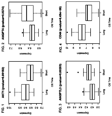
Biomarkers of high-grade serous ovarian carcinomas
The present disclosure provides biomarkers useful for determining the prognosis of conditions such as ovarian cancer.
US 9,772,334 B2 · Assignee: THE REGENTS OF THE UNIVERSITY OF CALIFORNIA · Inventors: Koon; Hon Wai et al.
Sheet 1 of 43 from the published document. All sheets in the USPTO PDF
Method of diagnosing and treating inflammatory bowel disease are disclosed herein. Inflammatory bowel disease can be treated and diagnosed using cathelicidin peptides and detection agents thereof. Specifically, method of treating and diagnosing Crohn's disease and ulcerative colitis are disclosed herein.
Chronic inflammatory disease is characterized by chronic, or persistent, inflammation. Chronic inflammatory disease encompasses a large number of diseases, many of which comprise a genetic component. Chronic inflammatory disease can develop as a result of a patient's exposure to harmful stimuli. For example, exposure to certain foods and environmental factors may trigger the development of chronic inflammatory disease. Chronic inflammatory disease can result in pain, fatigue, and digestive problems. Furthermore, the chronic nature of the inflammation may lead to tissue damage which can lead to a variety of additional problems. For example, chronic inflammation in the liver and digestive tract can lead to neurological changes such as fatigue and changes in personality. Chronic inflammation can also alter normal function of organs which can cause systemic disease and disorders in afflicted
1 of 43 drawing sheets so far from the published document, cropped to the drawing. Every sheet is in the USPTO PDF.
What the patent claimed, word for word. All of it is now free to use.
This invention relates to the use of a class of anti-microbial peptides for the detection and treatment of inflammatory disorders. Specifically, the anti-microbial peptide cathelicidin is useful for the detection and treatment of inflammatory bowel disease.
Chronic inflammatory disease is characterized by chronic, or persistent, inflammation. Chronic inflammatory disease encompasses a large number of diseases, many of which comprise a genetic component. Chronic inflammatory disease can develop as a result of a patient's exposure to harmful stimuli. For example, exposure to certain foods and environmental factors may trigger the development of chronic inflammatory disease. Chronic inflammatory disease can result in pain, fatigue, and digestive problems. Furthermore, the chronic nature of the inflammation may lead to tissue damage which can lead to a variety of additional problems. For example, chronic inflammation in the liver and digestive tract can lead to neurological changes such as fatigue and changes in personality. Chronic inflammation can also alter normal function of organs which can cause systemic disease and disorders in afflicted patients.
Examples of chronic inflammatory disease include celiac disease, vasculitis, lupus, chronic obstructive pulmonary disease (COPD), inflammatory bowel disease, atherosclerosis, arthritis, and psoriasis. Specifically, inflammatory bowel disease is a broad class of chronic inflammatory diseases. Examples of inflammatory bowel diseases are Crohn's disease, ulcerative colitis, collagenous colitis, lymphocytic colitis, ischaemic colitis, diversion colitis, Behcet's disease, indeterminate colitis. While many of these diseases have genetic components, the specific triggers and underlying biochemical causes for the onset of the diseases remain unknown. Furthermore, because the triggers and underlying biochemical causes of the diseases remain largely unknown, treatment regimes merely target reducing the symptoms without eliminating the disease or the chronic inflammation entirely.
Because the inflammation is not easily controlled, constant medication is often administered to lessen the symptoms and side effects of the inflammation. The medications often include antibiotics, aminosalicylates, corticosteroids, immune modifiers, and biologic therapies. However, the medication will often lead additional pain such as aching joints and headache, fatigue, digestive problems, fever, skin irritation and sensitivity, stomach pain and irritation, dizziness, increased blood pressure, fluid retention, cataracts, glaucoma, high blood sugar, increased risk of infection, osteoporosis, weak bones, suppressed adrenal gland hormone production, and increased risk of bruising and bleeding. Accordingly, even though the inflammation may lessen and worsen throughout the course of the chronic disease with the aid of medical therapies, it is often very difficult to treat and persists with most known treatment regimes.
Inflammatory bowel disease is a debilitating disease that is difficult to treat and incur high treatment cost to patients (Park K T, Bass D. Inflammatory bowel disease-attributable costs and cost-effective strategies in the United States: a review. Inflamm Bowel Dis. 2010; 17(7):1603-9). Inflamed colonic tissues express elevated levels of tumor necrosis factor alpha (TNFα) and other proinflammatory mediators, leading to tissue damage including apoptosis and loss of gut function (Ngo B, Farrell C P, Barr M, Wolov K, Bailey R, Mullin J M, et al. Tumor necrosis factor blockade for treatment of inflammatory bowel disease: efficacy and safety. Curr Mol Pharmacol. 2010; 3(3):145-52). Despite availability of medication like anti-TNFα antibodies, alternative therapeutic solutions are still being actively studied for better efficacy and safety (Rutgeerts P, Vermeire S, Van Assche G. Biological therapies for inflammatory bowel diseases. Gastroenterology. 2009; 136(4):1182-97).
The two major forms of Inflammatory Bowel Disease (IBD) are ulcerative colitis (UC) and Crohn's disease (CD). IBD is a chronic and remitting disease causing inflammation of the intestinal diseases. UC and CD have symptoms and pathologies in common, but they differ in the severity and location of the inflammation along the intestinal tract. Inflammation in UC patients is limited to the mucosal layer, and involves only the rectum and colon, while inflammation in CD patients penetrates the entire wall of the intestine and can occur anywhere along the intestinal tract. A clear diagnosis of the type of IBD is crucial to treatment decisions.
UC typically is characterized by ulcers in the colon and chronic diarrhea mixed with blood, weight loss, blood on rectal examination, and occasionally abdominal pain. UC patients may also present with a variety of other symptoms and extraintestinal manifestations including but not limited to anemia, weight loss, iritis, seronegative arthritis, ankylosing spondylitis, sacroiliitis, erythema nodosum, and pyoderma gangrenosum. Toxic megacolon is a life threatening complication of UC and requires urgent surgical intervention. UC usually requires treatment to go into remission. UC therapy includes anti-inflammatories, immunosuppressants, steroids, and colectomy (partial or total removal of the large bowel, which is considered curative). There is a significantly increased risk of colorectal cancer in UC patients several years after diagnosis, if involvement is beyond the splenic flexure, and a significant risk of primary sclerosing cholangitis, a progressive inflammatory disorder of the bile ducts.
Crohn's disease (CD) is also an IBD feat can affect the colon with symptoms similar to UC. Unlike UC, CD may affect any part of the gastrointestinal tract, and the inflammation penetrates deeper into the layers of the intestinal tact. Patients with CD may have symptoms and intestinal complications including abdominal pain, diarrhea, occult blood, vomiting, weight loss, anemia, fecal incontinence, intestinal obstructions, perianal disease, fistulae, and strictures, and apthous ulcers of the mouth. Extraintestinal complications include skin rashes, arthritis, uveitis, seronegative arthritis, peripheral neuropathy, episcleritis, fatigue, depression, erythema nodosum, pyoderma gangrenosum, growth failure in children, headache, seizures, and lack of concentration. The risk of small intestine malignancy is increased in CD patients. CD is believed to be an autoimmune disease, while it is uncertain whether there is an autoimmune component to UC. There is no known drug or surgical cure for CD; treatment focuses on controlling symptoms and maintaining remission to prevent relapse. Surgery is used for complications of Crohn's (e.g. strictures, fistulae, bleeding), and to remove segments of the intestine with active disease, but there is a high risk of recurrence; thus surgery is not considered curative.
Crohn's disease (CD) is a member of the broad class of inflammatory bowel diseases. One complication of CD is intestinal fibrosis. The intestine of CD patients develops strictures with overexpression of collagen (fibrogenic mediator) due to increased levels of transforming growth factor beta 1 (TGF-b1) and insulin like growth factor-1 (IGF-1), leading to obstruction of intestine and reduced gut motility. Intestines with strictures are unable to move, digest food or absorb nutrients. There is no satisfactory treatment of Crohn's disease associated fibrosis and stricture and surgery is often the only option for these patients. Recurrence of fibrosis or stricture formation is common in CD patients and leads to surgery that can be repeated several times and the disease progresses. Accordingly, intestinal fibrosis or stricture formation is a serious complication of CD and affects the daily live of these patients.
As described herein, cathelicidins are effective at significantly reducing intestinal fibrosis and treat inflammatory bowel diseases. Cathelicidins are a family of endogenous antimicrobial peptides which form a part of the innate immunity that protects the host from infection (Eckmann L. Defence molecules in intestinal innate immunity against bacterial infections. Curr Opin Gastroenterol. 2005; 21(2):147-51). Cathelicidin exists in human as LL-37 and in mice as mCRAMP (Gudmundsson G H, Agerberth B, Odeberg J, Bergman T, Olsson B, Salcedo R. The human gene FALL39 and processing of the cathelin precursor to the antibacterial peptide LL-37 in granulocytes. Eur J Biochem. 1996; 238(2):325-32; Gallo R L, Kim K J, Bernfield M, Kozak C A, Zanetti M, Merluzzi L, et al. Identification of CRAMP, a cathelin-related antimicrobial peptide expressed in the embryonic and adult mouse. J Biol Chem. 1997; 272(20):13088-93). Cathelicidin is secreted from the apical surface that is facing exterior environment such as intestine (Schauber J, Rieger D, Weiler F, Wehkamp J, Eck M, Fellermann K, et al. Heterogeneous expression of human cathelicidin hCAP18/LL-37 in inflammatory bowel diseases. Eur J Gastroenterol Hepatol. 2006; 18(6):615-21) and salivary gland (Murakami M, Ohtake T, Dorschner R A, Gallo R L. Cathelicidin antimicrobial peptides are expressed in salivary glands and saliva. J Dent Res. 2002; 81(12):845-50) by epithelial cells (Schauber J, Rieger D, Weiler F, Wehkamp J, Eck M, Fellermann K, et al. Heterogeneous expression of human cathelicidin hCAP18/LL-37 in inflammatory bowel diseases. Eur J Gastroenterol Hepatol. 2006; 18(6):615-21) and immune cells such as macrophages (Koon H W, Shih D Q, Chen J, Bakirtzi K, Hing T C, Law I, et al. Cathelicidin signaling via the Toll-like receptor protects against colitis in mice. Gastroenterology. 2011; 141(5):1852-63 e1-3).
Cathelicidins possess antimicrobial effects (Ho S, Pothoulakis C, Koon H W. Antimicrobial peptides and colitis. Curr Pharm Des. 2012; 19(1):40-7). Cathelicidin deficient mice have increased chance of infection, have reduced angiogenesis and wound healing (Ramos R, Silva J P, Rodrigues A C, Costa R, Guardao L, Schmitt F, et al. Wound healing activity of the human antimicrobial peptide LL37. Peptides. 2011; 32(7):1469-76). A previous study showed that low plasma level of cathelicidin is associated with increased infectious disease mortality in patients undergoing hemodialysis (Gombart A F, Bhan I, Borregaard N, Tamez H, Camargo C A, Jr., Koeffler H P, et al. Low plasma level of cathelicidin antimicrobial peptide (hCAP18) predicts increased infectious disease mortality in patients undergoing hemodialysis. Clin Infect Dis. 2009; 48(4):418-24. LL-37 mRNA expression is increased in colon biopsies from ulcerative colitis (UC), but not Crohn's disease (CD) patients (Schauber J, Rieger D, Weiler F, Wehkamp J, Eck M, Fellermann K, et al. Heterogeneous expression of human cathelicidin hCAP18/LL-37 in inflammatory bowel diseases. Eur J Gastroenterol Hepatol. 2006; 18(6):615-21). However, it was unknown whether cathelicidin expression in inflammatory bowel disease patients is associated with disease activity.
Currently, IBD (such as UC and CD) can only be definitively diagnosed by colonoscopy, a rather invasive procedure; even this invasive procedure is incapable of diagnosing approximately 10% of patients undergoing colonoscopy (Burczynski, J. Mol. Diag. 8 (1): 51 (2006)). Furthermore, there is currently no satisfactory treatment or prevention of intestinal fibrosis and a limited number of effective treatments for the broader class of inflammatory bowel diseases.
Provided herein are methods of diagnosis and treatment of inflammatory bowel disease using cathelicidin. As described herein, intra-colonic and intravenous administration of cathelicidin significantly reduces intestinal fibrosis in an experimental Crohn's disease model. It is also shown herein that cathelicidin administration also significantly reduced TGF-b1- and IGF-1-induced collagen expression in human colonic fibroblasts. Furthermore, cathelicidin has been shown by others to possess anti-inflammatory effects in a chemical (dextran sulfate) acute colitis mouse model, a Clostridium difficile infection colitis model, but never in Crohn's disease chronic colitis model associated with fibrosis. As described herein, two in vivo models (chronic trinitrobenzene sulfonic acid (TNBS) with intracolonic cathelicidin peptide and intravenous cathelicidin expressing lentivirus) and one in vitro model (human colonic fibroblasts) were used to demonstrate that cathelicidins have anti-fibrogenic effects. This demonstrates that cathelicidin has direct anti-fibrogenic effects apart from its anti-microbial or anti-inflammatory effects. Thus, exogenous administration of cathelicidin represents a new therapeutic approach against Crohn's disease-associated intestinal fibrosis.
In a first aspect, provided herein is a method of diagnosing inflammatory bowel disease. In certain embodiments, the method includes detecting cathelicidin protein expression with a probe that specifically binds cathelicidin in a biological sample from a patient. In specific embodiments, the biological sample is a colonic sample. In specific embodiments, the sample is selected from a group consisting of a blood sample, fecal sample, and intestinal sample. In specific embodiments, the sample is a colonic biopsy. In specific embodiments, the cathelicidin protein is the LL-37 peptide. In specific embodiments, the patient is a human.
In some embodiments, the inflammatory bowel disease is ulcerative colitis (UC). In exemplary embodiments, the biological sample is a blood sample. In some embodiments, a low cathelicidin protein expression level in the blood sample is indicative of moderate or active UC. In certain embodiments, the low cathelicidin protein expression level is 40 ng/ml or lower. In some embodiments, a high cathelicidin protein expression level is indicative of an UC that is in remission in the patient. In some embodiments, the high cathelicidin protein expression level is 50 ng/ml or greater.
In some embodiments, the inflammatory bowel disease is Crohn's disease. In some embodiments, the biological sample is a blood sample. In some embodiments, a low cathelicidin protein expression level is indicative that the patient has stricture associated with Crohn's disease. In certain embodiments, the low cathelicidin protein expression level is 45 ng/ml or lower. In some embodiments, a high cathelicidin protein expression level is indicative that the patient does not have stricture associated with Crohn's disease. In certain embodiments, the high cathelicidin protein expression level is 75 ng/ml or greater.
In a second aspect, provided herein is a method for treating a patient who has a likelihood of developing inflammatory bowel disease, the method comprising: (a) providing a biological sample from a patient; (b) applying the biological sample to a solid support mechanism; and (c) assaying the biological sample on the solid support mechanism to detect a cathelicidin peptide; wherein if a cathelicidin peptide is not detected or detected at low concentrations the patient is likely to develop inflammatory bowel disease; wherein if the patient is likely to develop inflammatory bowel disease the patient is administered a pharmaceutical composition to increase the patient's cathelicidin levels.
In specific embodiments, the biological sample is a colonic sample. In specific embodiments, the sample is selected from a group consisting of a blood sample, fecal sample, and intestinal sample. In specific embodiments, the sample is a colonic biopsy. In specific embodiments, the cathelicidin peptide is LL-37. In specific embodiments, the patient is a human. In specific embodiments, the assaying is performed using an ELISA. In specific embodiments, the assaying is performed using an assay selected from the group consisting of FACS, Western blot, immunohistochemistry, and RT-PCR. In specific embodiments, a low concentration of cathelicidin is less than 20 pg/ug. In specific embodiments, a high concentration of cathelicidin is greater than 35 pg/ug.
In a third aspect, provided herein is a method for determining the likelihood of a patient who previously suffered from inflammatory bowel disease to relapse, the method comprising detecting cathelicidin protein levels wherein high levels of cathelicidin protein are predictive of a longer disease free period compared to a patient who previously suffered from inflammatory bowel disease who has low levels of cathelicidin protein.
In specific embodiments, the cathelicidin protein levels are LL-37 peptide levels. In specific embodiments, the high levels of cathelicidin protein are further predictive of a lower chance of repeated surgery. In specific embodiments, the high levels of cathelicidin protein are further predicative of fewer inflammatory bowel disease related hospitalizations. In specific embodiments, the high levels of cathelicidin protein are further predictive of normal white blood cell counts.
In specific embodiments, the inflammatory bowel disease is Crohn's disease. In specific embodiments, the inflammatory bowel disease is ulcerative colitis. In specific embodiments, the cathelicidin protein levels are colonic cathelicidin protein levels. In specific embodiments, the biological sample is a colonic sample. In specific embodiments, the high levels of colonic cathelicidin protein are levels of LL-37 protein above 35 pg/ug. In specific embodiments, the high levels of colonic cathelicidin protein are levels of LL-37 protein below 20 pg/ug. In specific embodiments, the high levels of colonic cathelicidin protein have a significantly lower disease development score (approximately 45%), compared to those with lower colonic cathelicidin level.
In specific embodiments, the biological sample is a blood sample. In specific embodiments, the biological sample is a fecal sample or an intestinal sample.
In specific embodiments, high levels of cathelicidin protein are predictive of the patient not relapsing for at least 2 years. In specific embodiments, the cathelicidin protein levels are detected by ELISA. In specific embodiments, the cathelicidin levels are detected by assay selected from the group consisting of FACS, Western blot, immunohistochemistry, and RT-PCR.
In specific embodiments, the patient is a human.
In a fourth aspect, provided herein is a method for determining the likelihood of a patient to develop an inflammatory bowel disease, the method comprising detecting cathelicidin protein levels in a biological sample from the patient, wherein low levels of cathelicidin protein are predictive of a likelihood of a patient to develop an inflammatory bowel disease.
In specific embodiments, the cathelicidin protein levels are LL-37 peptide levels. In specific embodiments, the low levels of cathelicidin protein are further predictive of a higher chance of inflammatory bowel disease related surgery. In specific embodiments, the low levels of cathelicidin protein are further predicative of inflammatory bowel disease related hospitalizations. In specific embodiments, the low levels of cathelicidin protein are further predictive of abnormal white blood cell counts and anemia.
In specific embodiments, the inflammatory bowel disease is Crohns' disease. In specific embodiments, the inflammatory bowel disease is ulcerative colitis. In specific embodiments, the cathelicidin protein levels are colonic cathelicidin protein levels. In specific embodiments, the biological sample is a colonic sample. In specific embodiments, the high levels of colonic cathelicidin protein are levels of LL-37 protein above 35 pg/ug. In specific embodiments, the high levels of colonic cathelicidin protein are levels of LL-37 protein below 20 pg/ug. In specific embodiments, the high levels of colonic cathelicidin protein have a significantly lower disease development score (approximately 45%), compared to those with lower colonic cathelicidin protein levels.
In specific embodiments, the biological sample is a blood sample. In specific embodiments, the biological sample is a fecal sample or an intestinal sample. In specific embodiments, high levels of cathelicidin protein are predictive of the patient not developing an inflammatory bowel disease for at least 2 years.
In specific embodiments, the cathelicidin protein levels are detected by ELISA. In specific embodiments, the cathelicidin protein levels are detected by assay selected from the group consisting of FACS, Western blot, immunohistochemistry, and RT-PCR.
In specific embodiments, the patient is a human.
In a fifth aspect, provided herein is a method of treating inflammatory bowel disease, the method comprising administering to a subject with an inflammatory bowel disease a pharmaceutical composition to increase the patient's cathelicidin protein levels.
In specific embodiments, the inflammatory bowel disease is Crohn's disease. In specific embodiments, the inflammatory bowel disease is ulcerative colitis. In specific embodiments, the pharmaceutical composition comprises a cathelicidin peptide and a pharmaceutically acceptable carrier. In specific embodiments, the cathelicidin peptide is a LL-37 pepetide. In specific embodiments, the pharmaceutical composition is sodium butyrate. In specific embodiments, the pharmaceutical composition comprises a short chain fatty acid. In specific embodiments, the pharmaceutical composition comprises vitamin D. In specific embodiments, the pharmaceutical composition comprises a PPAR gamma agonist. In specific embodiments, the pharmaceutical composition comprises a lipopolysaccharide. In specific embodiments, the pharmaceutical composition comprises Salmononella. In specific embodiments, the pharmaceutical composition comprises a probiotic.
In specific embodiments, the pharmaceutical composition further comprises one or more therapies selected from the group consisting of cathelicidin peptide(s), sodium butyrate or analogs thereof, antibiotic(s), anti-inflammatory(ies), anti-diarrheals, laxatives, pain relievers, iron supplements, aminosalicylate(s), steroids, corticosteroid(s), immune modifier(s), immunosupressor(s), anti-CD52 agents, anti-TNFα agents, biologic therapy(ies), vitamin B-12 shots, surgery, sodium butyrate, and nutritional plans.
In specific embodiments, the anti-inflammatory(ies) is selected from a group comprising sufasalazine, mesalamine, NSAIDs, ImSAIDs, and corticosteroids.
In specific embodiments, the immunosupressor(s) is selected from a group comprising zathioprine, mercaptopurine, infliximab, adalimumab, certolizumab pegol, methodtrexate, cyclosporine, natalizumab, cyclosporine, and tacrolimus.
In specific embodiments, the antibiotic(s) is selected from a group comprising metronidazol and ciprofloxacin.
In specific embodiments, the anti-CD52 agent is Alemtuzumab®.
In specific embodiments, the nti-TNFα agent is Infliximab®.
In specific embodiments, the method is a method of treating Crohn's disease comprising administering a pharmaceutical composition to increase the in vivo concentration of cathelicidin protein.
In specific embodiments, the Crohn's disease is associated with fibrosis. In specific embodiments, the Crohn's disease is associated with strictures.
In specific embodiments, the pharmaceutical composition is administered intracolonically. In specific embodiments, the pharmaceutical composition is administered intravenously. In specific embodiments, the pharmaceutical composition is administered orally.
In specific embodiments, the pharmaceutical composition significantly reduces intestinal fibrosis. In specific embodiments, the pharmaceutical composition significantly reduces TGF-b1 and IGF-1 induced collagen expression in colonic fibroblasts. In specific embodiments, the significant reduction of collagen expression treats intestinal fibrosis. In specific embodiments, the significant reduction of collagen expression decreases intestinal fibrosis. In specific embodiments, the significant reduction of collagen expression prevents intestinal fibrosis.
In specific embodiments, the patient is human.
In a sixth aspect, provided herein is a method of reducing TGF-b1 and IGF-1 expression in colonic fibroblasts, the method comprising administering a cathelicidin peptide. In specific embodiments, the cathelicidin peptide is a LL-37 peptide. In specific embodiments, the colonic fibroblasts are human colonic fibroblasts.
In a seventh aspect, provided herein is a kit comprising: (a) a solid support comprising synthetic capture probes selective for a cathelicidin peptide; and (b) methods of using the kit wherein the methods comprise instructions for obtaining a biological sample from a patient, applying the biological sample to the solid support, and assaying the biological sample on the solid support mechanism to detect a cathelicidin peptide. In specific embodiments, the cathelicidin peptide is a LL-37 peptide. In specific embodiments, the assay is selected from a group consisting of ELISA assays, FACS assays, Western blot assays, immunohistochemistry assays, and RT-PCR assays.
In an eight aspect, provided herein is a kit comprising antibodies, a detectable label and instructions for treating and detecting inflammatory bowel disease, wherein the antibodies specifically recognize a cathelicidin peptide. In specific embodiments, the cathelicidin peptide is a LL-37 peptide.
In a ninth aspect, provided herein is a method for detecting an active inflammatory bowel disease in a patient. In an exemplary embodiment, the method includes the step of detecting cathelicidin protein levels and C-reactive protein (CRP) levels in a biological sample from the patient, wherein low levels of cathelicidin protein and high CRP levels are indicative of an active inflammatory bowel disease. In some embodiments, the cathelicidin protein levels are LL-37 peptide levels. In certain embodiments, the inflammatory bowel disease is ulcerative colitis (UC). In some embodiments, the cathelicidin protein levels and CRP protein levels are serum cathelicidin protein levels and serum CRP protein levels. In certain embodiments, the patient has active ulcerative colitis if the patient has a serum cathelicidin protein level of 55 ng/ml or below and a serum CRP level of 2 mg/L or above. In exemplary embodiments, the biological sample is a blood sample.
In some embodiments, the cathelicidin protein levels and CRP protein levels are detected using a cathelicidin binding probe and a CRP binding probe. In certain embodiments, the cathelicidin binding probe is an antibody that binds cathelicidin and the CRP binding probe is an antibody that binds CRP. In some embodiments, the cathelicidin binding probe and the CRP binding probe are attached to a solid support. In certain embodiments, the cathelicidin and CRP protein levels are detected by ELISA. In other embodiments, the cathelicidin protein levels are detected by assay selected from the group consisting of FACS, Western blot, and immunohistochemistry. In some embodiments, the patient is a human.
In a tenth aspect, provided herein is an antibody cocktail that includes a plurality of antibodies and a buffer solution. In some embodiments, the plurality of antibodies includes an anti-CRP antibody and an anti-cathelicidin antibody. In certain embodiments, the anti-cathelicidin antibody binds LL-37. In exemplary embodiments, the antibodies in the plurality of antibodies are monoclonal antibodies.
FIGS. 1A-1D show that Cathelicidin reduces colonic inflammation in TNBS mediated chronic colitis in mice. FIG. 1A is an illustration of the experimental plan of TNBS mediated chronic colitis. Wild-type and mCRAMP mice were injected with TNBS solution (0.5-1 mg per 20 g mice) in 30% ethanol or 30% ethanol only (vehicle control) under transient isoflurane anesthesia. FIG. 1B shows the percent of body weight change of different groups (from week 0 to week 7). All ethanol treated groups had approximately 20% body weight gain. TNBS treated wild-type mice suffered from significantly less body weight gain, compared to ethanol control (p=0.03). Administration of mCRAMP to TNBS treated wild-type restored the body weight gain (p=0.01). TNBS treated Camp.sup.−/− mice had significant body weight loss regardless of mCRAMP treatment, compared to TNBS treated wild-type mice (p=0.001) and ethanol treated Camp.sup.−/− mice (p=0.0001). FIG. 1C shows representative H&E images of colonic tissues. FIG. 1D shows the histology score was based on H&E staining images of colonic tissues. TNBS treatment in wild-type mice led to increased tissue damage with significantly higher histology score (p=0.001), compared to ethanol control. Treatment of mCRAMP significantly reduced histology score in wild-type mice (p=0.03) and Camp.sup.−/− mice (p=0.04). Results are representative of n=6 mice per group.
FIGS. 2A-2D show that cathelicidin reduces colonic collagen deposition in mice with chronic TNBS colitis. FIG. 2A shows that TNBS treatment significantly induced colonic TNFα protein (p=0.0042) expression. Intracolonic mCRAMP administration significantly reduced TNBS induced TNFα protein expression in WT (p=0.0127) and Camp.sup.−/− (p=0.0054) mice. FIG. 2B shows that TNBS treatment significantly induced colonic TNFα protein (p=0.0042) expression. Intracolonic mCRAMP administration significantly reduced TNBS induced TNFα protein expression in WT (p=0.0127) and Camp.sup.−/− (p=0.0054) mice. FIG. 2C shows Masson Trichrome staining for collagen in colonic tissues. Collagen was stained in blue. Collagen deposited in mucosal and submucosal layer of TNBS treated wild-type mice was reduced by CRAMP treatment. FIG. 2D shows quantitative real-time RT-PCR of collagen colla2 mRNA expression in colonic tissues of mice. TNBS treatment significantly induced colonic collagen deposition and colla2 mRNA (p=0.00376) expression. Intracolonic treatment of mCRAMP reduced colonic colla2 mRNA and collagen deposition in both WT (p=0.0364) and Camp.sup.−/− (p=0.04) mice. Results are representative of n=6 mice per group.
FIGS. 3A-3F show that cathelicidin reduces collagen expression in colonic fibroblasts. FIG. 3A shows colonic Camp mRNA expression levels of TNBS treated WT and Camp.sup.−/− mice were similar. Camp mRNA in Camp.sup.−/− mice was undetectable. FIG. 3B shows that colonic Camp mRNA expression levels among strictured CD patients and non-strictured CD patients were similar. FIGS. 3C and 3D show human colonic CCD-18Co fibroblasts were incubated with LL-37 (1-10 μM) or TFA 0.1% (vehicle) and/or TGF-β1 (50 ng/ml) and IGF-1 (10 ng/ml) for 48 hours. Collagen (COL1A2) and total ERK protein expression was detected by Western blot analyses and quantitative image densitometry. TGF-β1 and IGF-1 significantly induced COL1A2 mRNA expression in CCD-18Co fibroblasts (p=0.0341) which was reduced by 10 μM LL-37 (p=0.0001). FIG. 3E shows that CCD-18Co fibroblasts were treated with LL-37 (0-10 μM) and/or TGF-β1 (50 ng/ml) and IGF-1 (10 ng/ml) for 24 hours. TGF-β1 and IGF-1 induced COL1A2 mRNA expression in colonic fibroblasts (P=0.049) was inhibited by LL-37 (5-10 μM, p=0.001) in concentration dependent manner. FIG. 3F shows human primary colonic fibroblasts that were treated with LL-37 (0-10 μM) and/or TGF-β1 and IGF-1 for 24 hours. TGF-β1 and IGF-1 induced COL1A2 mRNA expression in colonic fibroblasts (P=0.0233) was inhibited by LL-37 (5-10 μM, p=0.0108 and p=0.0049) in concentration dependent manner. All experiments are representative of 3 independent experiments.
FIGS. 4A-4E show that cathelicidin inhibits TGF-β1 and IGF-1 mediated collagen synthesis via ERK activation in the human colonic fibroblasts. FIG. 4A shows CCD-18Co fibroblasts that were pretreated with DMSO, Akt inhibitor V (10 μM) or ERK inhibitor PD98059 (10 μM) for 30 minutes, followed by LL-37 (10 μM) or TFA 0.1% and/or TGF-β1 (50 ng/ml) and IGF-1 (10 ng/ml) for 72 hours. COL1A2 and Stat5 protein expression was detected by Western blot. FIG. 4B shows densitometry of Western blots. The inhibition of TGF-β1 and IGF-1 mediated COL1A2 expression by LL-37 was partially reversed by PD98059 (p=0.0001). FIG. 4C shows CCD-18Co fibroblasts that were pretreated with DMSO, Akt inhibitor V (10 μM) or ERK inhibitor PD98059 (10 μM) for 30 minutes, followed by LL-37 (10 μM) or TFA 0.1% and/or TGF-β1 (50 ng/ml) and/or IGF-1 (10 ng/ml) for 30 minutes. FIGS. 4D and 4E show densitometry of Western blots. LL-37 significantly induced Akt and ERK phosphorylation in concentration dependent manner.
FIGS. 5A-5C show that cathelicidin reduces collagen expression via inhibiting tubulin expression in colonic fibroblasts. FIG. 5A shows CCD-18Co fibroblasts that were pretreated with DMSO, cytoskeleton inhibitor cytochalasin B (10 μM) or matrix metalloproteinase (MMP) inhibitor OM6001 (10 μM) for 30 minutes, followed by LL-37 (10 μM) or TFA 0.1% and/or TGF-β1 (50 ng/ml) and IGF-1 (10 ng/ml) for 72 hours. Cytochalasin B but not OM6001 affected TGF-β1 and IGF-1 induced COL1A2 expression in CCD-18Co fibroblast (p=0.0005). Interference of cytoskeleton led to inhibition of collagen expression. FIG. 5B shows that cytochalasin B but not OM6001 affected TGF-β1 and IGF-1 induced β-tubulin expression in CCD-18Co fibroblasts (p=0.0005). Interference of cytoskeleton inhibited β-tubulin expression. FIG. 5C shows CCD-18Co fibroblasts that were pretreated with DMSO, Akt inhibitor V (10 μM) or ERK inhibitor PD98059 (10 μM) for 30 minutes, followed by LL-37 (10 μM) or TFA 0.1% and/or TGF-β-1 (50 ng/ml) and IGF-1 (10 ng/ml) for 72 hours. Both ERK and Akt pathways were not involved in the LL-37 mediated inhibition of β-tubulin expression in CCD-18Co fibroblasts. All experiments are representative of 3 independent experiments.
FIGS. 6A and 6B show that cathelicidin inhibits cytoskeleton tubulin distribution in colonic fibroblasts. FIG. 6A shows CCD18Co fibroblasts that were exposed to TFA (vehicle), LL-37 (10 μM) and/or TGF-β-1 (50 ng/ml) and IGF-1 (10 ng/ml) for 24 hours. Tubulin distribution was visualized by tubulin tracker in green color and nuclei were identified by Hoechst 33342 in blue color. TGF-β-1 (50 ng/ml) and IGF-1 (10 ng/ml) did not affect tubulin distribution in cells. LL-37 shrunk the tubulin network. FIG. 6B shows CCD18Co fibroblasts that were exposed to cytochalasin B (10 μM) for 24 hours. Cytochalasin B also shrunk tubulin network in cells. Results are representative of 3 independent experiments.
FIGS. 7A-7E show that cathelicidin does not affect colonic fibroblast infiltration in TNBS treated colitis. FIG. 7A shows vimentin immunohistochemistry for fibroblasts in colonic tissues (in light brown spots). Fibroblasts accumulated in the mucosal and submucosal locations as collagen deposition. FIG. 7B shows the number of fibroblasts per field of image. Three different locations (3 separate images) per sample were counted. Chronic TNBS colitis led to significantly increased fibroblast accumulation (P=0.0032) compared to ethanol control but it was not affected by mCRAMP administration. FIGS. 7C and 7D show colonic vimentin and α-smooth muscle actin mRNA expression levels were similar among WT and Camp−/− mice. mCRAMP administration did not affect colonic vimentin and α-SMA mRNA expression in colons. FIG. 7E shows CCD-18Co that was seeded on the upper modified Boyden chamber and incubated in 37° C. for 8 hours. Cell migration was not affected by LL-37 (0-10 μM). Results are representative of 3 independent experiments.
FIGS. 8A-8D show that intravenous administration of mCRAMP expressing lentivirus ameliorates TNBS mediated colonic fibrosis. FIG. 8A shows an illustration of experimental plan of TNBS mediated chronic colitis. Wild-type and mCRAMP mice were injected with TNBS solution (0.5-1 mg per 20 g mice) in 30% ethanol or 30% ethanol only (vehicle control) under transient isoflurane anesthesia. FIG. 8B shows colonic Camp mRNA expression that was significantly increased in the mCRAMP expressing Camp-LV group (p=0.026 or p=0.0413). FIG. 8C shows Colonic colla2 mRNA expression was significantly reduced in mCRAMP expressing Camp-LV group. FIG. 8D shows that colonic vimentin mRNA expression was not affected by Camp-LV infection. Results are representative of n=6 mice per group.
FIGS. 9A-9E show that high colonic LL-37 expression levels predicts good prognosis of the UC patients. FIG. 9A shows colonic Camp mRNA expression of 17 normal, 24 UC and 24 CD patients. Only UC patients have significantly higher Camp mRNA expression than normal patients (p=0.04). FIG. 9B shows colonic LL-37 protein levels in 17 normal, 24 UC and 24 CD patients. FIG. 9C shows cathelicidin immunohistochemistry of colons from normal, UC and CD patients. FIG. 9D shows the definition of disease development score. FIG. 9E shows disease development scores of normal, UC and CD patients. A low colonic LL-37 protein level is significantly associated with high disease development among UC patients (p=0.02 UC high LL-37 vs. UC low LL-37). Baseline characteristics and number of all groups are shown in FIGS. 10 and 11 .
FIGS. 10A-10C show the baseline characteristics of all patient groups. FIG. 10A shows the baseline characteristics of all control, UC and CD groups. FIG. 10B shows the inclusion and exclusion criteria of the analysis. FIG. 10C shows the disease development scores of normal, UC and CD patients. But there is no correlation between disease development score of IBD patients and colonic Camp mRNA expression.
FIGS. 11A and 11B show the baseline characteristics of specific patient groups. FIG. 11A shows the baseline characteristics of low and high cathelicidin UC groups. FIG. 11B shows the baseline characteristics of low and high cathelicidin CD groups.
FIGS. 12A-12E show that intestinal cathelicidin protein levels vary among IBD patients. FIG. 12A Inclusion and exclusion criteria. FIG. 12B Baseline characteristics. FIG. 12C Intestinal CAMP mRNA expression of normal and IBD patients. UC, but not CD patients had significantly increased intestinal CAMP mRNA expression. FIG. 12D The average intestinal cathelicidin levels of were not altered in IBD. FIG. 12E Immunohistochemistry of cathelicidin in human intestinal biopsies. Cathelicidin was expressed in mucosal area of the intestines (brown color) with wide variation.
FIGS. 13A-13C show intestinal cathelicidin protein levels correlate with CRP or ESR levels. FIG. 13A Baseline characteristics. FIG. 13B Serum CRP levels. Low intestinal cathelicidin levels were significantly correlated to the high serum CRP levels in UC patients. FIG. 13C ESR levels. Low intestinal cathelicidin levels were significantly correlated to the high ESR levels in CD patients. CRP levels were not correlated to cathelicidin levels in UC patients. ESR levels were not correlated to cathelicidin levels in CD patients.
FIGS. 14A-14C show that plasma cathelicidin levels in UC patients are decreased. FIGS. 14A and 14B shows the inclusion and exclusion criteria as well as baseline characteristics of the patients. FIG. 14C show the plasma cathelicidin levels of UC, but not CD, patients were significantly decreased.
FIGS. 15A-15D show endogenous cathelicidin induction by sodium butyrate ameliorated DSS mediated colitis in mice. FIG. 15A is an illustration of the administration of DSS and/or sodium butyrate to mCRAMP deficient (Camp.sup.−/−) and wild-type mice (i.e., the experimental plan). FIG. 15B shows that DSS colitis led to body weight loss. FIG. 15C shows that DDS colitis led to significant tissue damages and increased histology scores, wherein intraperitoneal sodium butyrate administration led to significant decrease of histology score in wild-type but not Camp.sup.−/− deficient mice. FIG. 15D shows that Camp.sup.−/− mice had significantly worse colitis than wild-type mice when exposed to DSS (see also FIG. 15C ).
The description continues in the full USPTO document.
About 5,722 words. The USPTO PDF has it with every drawing.
Fees are due 3.5, 7.5 and 11.5 years after grant. This patent expired on September 26, 2025, so the fee marked "not paid" was the one that went unpaid.
NOVEL INFLAMMATORY BOWEL DISEASE MARKERS AND THERAPIES FOR COLITIS-ASSOCIATED INTESTINAL FIBROSIS
Filed Jun 2015 · published Jun 2016Inflammatory bowel disease markers and therapies for colitis-associated intestinal fibrosis
Filed Jun 2015 · granted Sep 2017Earlier publications, parents and continuations. None of them can still be enforced, or this patent would not be listed.
Prior art cited by the examiner or applicant. Useful when you check your own idea for novelty.
Everything on this page comes from the documents linked above.