 Lapsed, fee not paid24 drawings
Medical Devices · US 9,757,114 B2
Apparatus and method for attaching soft tissue to bone
Apparatuses for attaching tissue to bone are provided.
US 9,757,198 B2 · Assignee: Boston Scientific Scimed, Inc. · Inventors: Griffin; Stephen Edward et al.
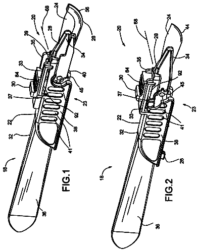
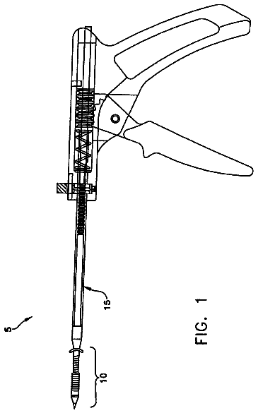
Sheet 1 of 10 from the published document. All sheets in the USPTO PDF
An improved optical fiber comprising a waveguide with an input for coupling focused laser energy into the waveguide and communicating electromagnetic radiation in a propagation direction to an internally reflective tip of the waveguide, a tissue contacting surface wherein the light path from the reflecting surface to the transmitting surface in substantially homogenous in refractive index and cooled by fluid flow. In minimizing the variations in refractive index within the lateral light path, while providing active cooling directly below the tissue contact surface, the invention prevents internal reflections and beam distortion and greatly improves the efficiency and durability of the laterally directing probe. Free rotation of the tissue contact surface, about the lateral tip, may be provided and tissue vaporization efficiency may be improved by providing a morcellating tool on the tissue contact surface.
Treatment of benign prostatic hyperplasia (BPH) with laser energy has a somewhat checkered past, but in recent years advances in the understanding of laser-tissue interactions and laser design have enabled realization of much of the promise of the initial concept. The longstanding “gold standard” for surgical treatment of benign enlarged prostate glands is a technique called TURP: Transurethral Resection of the Prostate. In TURP procedures, an electric current is passed through working tips of various shapes, heating them to permit tissue to be selectively carved or charred away. While TURP is widely used with good clinical results, significant bleeding is common and the procedure can be time consuming, particularly where the glands are large. Post-operative swelling of tissues remaining post-TURP requires catheterization to permit release of urine and hospital recovery can be protracted
1 of 10 drawing sheets so far from the published document, cropped to the drawing. Every sheet is in the USPTO PDF.
What the patent claimed, word for word. All of it is now free to use.
The present invention relates generally to applications of lasers to endosurgery and specifically to delivery of high energy density or high average power to tissues located about the body lumen, such as the prostate gland about the urethra.
Treatment of benign prostatic hyperplasia (BPH) with laser energy has a somewhat checkered past, but in recent years advances in the understanding of laser-tissue interactions and laser design have enabled realization of much of the promise of the initial concept. The longstanding “gold standard” for surgical treatment of benign enlarged prostate glands is a technique called TURP: Transurethral Resection of the Prostate. In TURP procedures, an electric current is passed through working tips of various shapes, heating them to permit tissue to be selectively carved or charred away. While TURP is widely used with good clinical results, significant bleeding is common and the procedure can be time consuming, particularly where the glands are large. Post-operative swelling of tissues remaining post-TURP requires catheterization to permit release of urine and hospital recovery can be protracted. Post-operative pain is often prolonged and complications such as bladder neck strictures and nerve damage are relatively common, leading to a fairly high incidence of retrograde ejaculation, incontinence and temporary impotence among patients.
The VLAP procedure of the 1990s (Visual Laser Ablation of the Prostate) promised solutions to the problems with TURP, but the dominant laser wavelength (1064 nm) penetrated too deeply into the prostate tissue such that deep coagulation (tissue death but not removal) resulted and non-target tissues were often damaged inadvertently. Fluid uptake in these damaged tissues caused post-VLAP complications and it was difficult for surgeons to judge the actual degree of tissue death that would result beneath the surface treatment.
Contributing to this problem was the broad spectrum of lateral fiber function, with fibers manufactured by as many as two dozen companies. With no established minimum performance standards, most lateral emitting fibers of the period delivered relatively diffuse energy with significant scattered radiation such that tissue effects varied widely from surgery to surgery. The vast majority of urologists who experimented with VLAP in the 1990's, and various modifications thereof, abandoned the method and returned to TURP by about 1996.
More recently, a new technique called PVP (Photo Vaporization of the Prostate) has spearheaded resurgence in applications of lasers to BPH surgery, driven more by patients than by the Urological Specialty, for the reportedly very low incidence of side effects. The technique uses 532 nm of light that is strongly absorbed by hemoglobin such that surface vaporization of tissue is the dominant effect. In addition, the lateral fiber used in the procedure (GreenLight™ U.S. Pat. No. 5,428,699, referenced below as prior art) is more efficient than most that were available in the 1990's, such that high energy density spots are presented to tissue with little damaging scatter. Also contributing to the overall high performance of the system (laser plus fiber combination) is the inherently high energy density of the laser itself, as taught by U.S. Pat. No. 6,554,824 (Davenport, et al.). With an average of 80 W of 523 nm of light provided to the fiber, approximately 70 W is delivered laterally to the target tissue within a small diameter, substantially circular. The output spot energy profile produced is such that substantially all of the illuminated tissue is vaporized.
The PVP procedure is popular with patients and surgeons because it is fast, essentially painless (no prolonged, post-operative tissue sloughing), offers immediate relief (often no catheter is required beyond initial recovery), generally requires no hospital stay and has a very low incidence of complications. Such surgeries should also be popular with private and government insurers in that the overall costs associated with treatment are considerably lower for PVP than for TURP in most cases.
While enlarged prostate glands of typical size (30 grams) may be treated sufficiently within as little as 15 minutes with PVP, larger glands prove problematic. As the surgery proceeds, the output “cap” of the lateral fiber degrades: the surface through which the laser light passes becomes opaque or “frosted”, scattering light. The damage accelerates with continued use and eventually the erosion at the surface extends completely through the cap and surgical irrigation fluid leaks into the cap as it heats and cools with each laser pulse.
Since the redirection of light within lateral fibers such as this is based upon total internal reflection (TIR) due to refractive index differences of the fiber core and the air trapped in the protective cap, influx of aqueous solution at a refractive index more similar to the glass than to air disrupts this condition and the fiber fails by firing axially. Such failures can be catastrophic, with uncontrolled laser emission causing bladder neck damage or bladder or urethral perforation. As a minimum inconvenience, at least two GreenLight™ fibers may be required for large glands resulting in delays and added surgical costs.
A competing technique is also gaining some ground: HoLAP or Holmium Laser Ablation of the Prostate. In theory, the holmium wavelength is even more favorable than KTP (532 nm mentioned above, or frequency doubled Nd:YAG) for controlled tissue effect with stronger absorption resulting in even less underlying coagulation. In addition, procedural problems and cost issues with the PVP can be avoided. The bright green KTP laser emission interferes with vision requiring special endoscope camera filters and orange safety goggles for surgical staff and the frequency doubled, 200 W Nd:YAG laser requires non-standard electrical power (50 A, 208 VAC) and high flow cooling water. The KTP laser is costly (about $80,000) and is currently a “single procedure box”, meaning only BPH surgery is done with the appliance, and GreenLight™ fibers are extremely expensive for a disposable device at US $875 each. In contrast, the holmium laser is competent in treating other urological and non-urological conditions from kidney stones to ruptured spinal discs. It would be desirable to provide high performance lateral fibers at reasonable costs for holmium lasers for providing lower cost laser BPH surgery to a broader population.
In the PVP procedure, considerable effort and expense has been expended in producing lasers (Davenport, et al.) and fiber delivery systems (Pon) that minimize irradiation of tissue with low energy density light in that the lower energy densities may cause deep thermal damage without immediate tissue removal. This is not a new observation. Beginning in 1989, the author of the current art personally has experienced laser injuries from a broad range of laser wavelengths and energy profiles and has long noted that higher energy density injuries heal much more rapidly than low energy density injuries, with negligible collateral tissue damage, regardless of the laser wavelength. Unfortunately, in spite of the high energy density light produced by the KTP laser taught in Davenport, the degradation in performance of the fiber taught in Pon is such that the proportion of low energy density light delivered to the tissue steadily grows as the procedure proceeds.
Holmium energy is strongly absorbed by water and is absorbed to a lesser degree by other tissue components, including blood, so where the PVP laser becomes less effective as surface tissue is ablated and underlying tissue are blanched of blood, the holmium continues to work with high efficiency. Owing to the stronger absorption of the holmium laser energy by tissues, the depth of laser energy penetration for the holmium is even lower than that for KTP, so unwanted deep tissue death has the potential to be reduced even further, if high energy density can be reliably delivered to the target tissues over the course of the procedure.
The barriers to holmium applications in BPH are minor but persistent. Protective cap degradation appears to be more pronounced with the holmium wavelength than with KTP, possibly due to a higher degree of interaction of the light with the cap material (silica), the high pulse energy density and the considerable heat generated by interactions of the laser energy with the aqueous irrigation fluid and tissues. In particular, water attacks the hot silica through hydrothermal erosion. This is complicated (and accelerated) by devitrification of the surface that is catalyzed (at elevated temperatures) by ions commonly found in tissues and irrigation fluids: alkali and alkaline earth metal ions such as sodium (I) and calcium (II).
The photo-thermal and/or photo-acoustic shock waves that are generated by the laser pulses in the glass and in the water are so intense that caps of similar dimensions to those used in the PVP fiber can simply shatter to dust at average powers of 40 W or more. Thicker caps resist this damage but remain susceptible to erosion failures in apparent excess of that seen in PVP. (Much of the erosion problem could be surgical technique related, in both procedures, e.g. some surgeons may hold the holmium fiber in closer proximity to tissue than KTP fibers and some may clean the fiber tip intra-operatively while others may not.)
Further, the energy density profile of holmium lasers contains a broader mix of modes than that produced by the KTP laser used in the PVP procedure. As taught by the author of the present art in U.S. Pat. No. 6,282,349 (Griffin) and other publications, holmium lasers are notorious for thermal lensing problems within the lasing medium, resulting in variable mode output beam profile. This broader distribution presents areas of the beam profile that are not of sufficient energy density to cause tissue vaporization and undesirable coagulation is the result. Reducing the high order modes produced by the laser itself by reducing the heating that the pump energy produced in the laser medium, as taught in Davenport, is not the sole means of minimizing this problem, nor is it the most economical or logical means. The higher order modes may be selectively excluded from coupling to the surgical fiber (mode stripping), or preferably, the higher order modes may be converted to lower order modes within the energy delivery fiber, at the laser-to-fiber coupling or at the fiber output.
The energy density profile at the fiber output surface is not only critical for achieving the desired tissue response, but for prolonging the fiber performance. Energy densities presented at the fiber output that are insufficient for tissue vaporization promote tissue adhesions to the fiber tip. Adhered tissues give rise to conditions that promote acceleration of performance degradation. GreenLight™ fibers modified for use on holmium lasers function very well in comparison to other fiber designs, indicating that the superior efficiency of the fiber output is critical to clinical function.
A less efficient fiber design, the (DuoTome™), is sole holmium fiber that is capable of delivering average holmium power equivalent to the GreenLight used with the KTP laser, but it requires a 100 W input to achieve vaporization rates similar to the GreenLight/PVP procedure. At 100 W input, the DuoTome output spot presents lower energy density to tissue than does the GreenLight due to more scatter and cylindrical lens distortion within the lesser fiber design such that, even given the superior absorption of holmium energy by target tissues, more coagulation results than is clinically desirable or necessary.
Inefficiency breeds excess heat at the fiber tip, which promotes tissue adhesions and fiber damage, so fibers are even more prone to premature failure in holmium BPH treatment than they are in PVP. Surgical progress is not quite as rapid nor are target tissues quite as precisely ablated. Further, energy density in the fiber output spot is critical to successful vaporization without significant concomitant damage to critical, non-target structures near the site of therapy.
The DuoTome™ avoids irradiating non-target tissues (with the scattered laser light in the output) by sheathing the cap in stainless steel. Only a tiny window is presented for the laser energy to escape. As a consequence of trapping the undesirable energy within the steel enclosure the fiber tip gets hot in use and hot steel can also cause unwanted tissue damage and complications. Furthermore, the stainless steel containment sacrifices protective cap thickness in an application where total diameter is limited by the size of the working channel provided within the endoscopic device. The maximum diameter limit for the smallest working channel (7.5 Fr.) in rigid cystoscope/resectoscopes is about 2.45 mm where compatibility with most flexible cystoscopes (presenting working channels as small as 6 Fr.) the maximum diameter for the device is about 1.75 mm and the length of rigid section (typically the cap) should not exceed about 12 mm least the device not easily pass the channel in moderate deflection.
At least two other fiber designs have been tried with the holmium lasers, as taught by Griffin and Brekke, referenced below. Both are high efficiency designs that utilize fiber-to-cap fusion to minimize scatter. Both fail at approximately 40 W through catastrophic disintegration. It is thought that the residual stress concentrations in the fiber-to-cap fusion region likely render the fused fibers more susceptible to the thermal shocks encountered in the surgery than non-fused fibers.
All current art lateral fibers based upon total internal reflection (TIR) at tips—polished at the critical angle as defined by Snell's Law as opposed to external reflector designs such as U.S. Pat. No. 5,242,437 (Everett)—that are designed for surgeries such as prostate resection suffer the opacity at output failure mode, where the glass surface in contact with tissue and/or irrigation fluid and/or bodily fluids degrades through hydrolysis and devitrification. Even minor degradation of the output surface quality causes difficulties in surgery. There is typically a coaxial, visible laser wavelength transmitted within the optical fiber that serves the surgeon in orienting the fiber output properly on target tissues: the “aiming beam”. As the output surface degrades, so the clarity of the aiming beam degrades, making precise orientation more difficult.
Accordingly, fibers are also equipped with accessory “orientation markers” that are typically proximal to the output area, generally opposite the fiber output such that they may be visualized when the fiber is properly positioned, pointing generally at tissue rather than generally at the surgeon or endoscopic equipment. These markers are usually ink printed lines or text on transparent heat shrink tubing that are carefully positioned with respect to the fiber output during assembly. As the fiber continues to degrade with use, it becomes more inefficient such that more laser energy is consumed in heating the device, causing more tissue adhesions, more energy absorption and more glass degradation, i.e. the degradation progress is governed by second order kinetics and accelerates. As the output tip heats to greater and greater temperatures, the thermally labile orientation marker becomes damaged, further reducing the ability of the surgeon to properly orient the fiber output.
Where temperatures rise even further, adhesives used to secure the protective cap to the fiber fail, or the fibers' polymer buffer coatings themselves fail, and the cap may dislodge. A dislodged cap is a catastrophic failure (axial emission) and often requires prolonged expeditions within the urinary tract for the purpose of retrieving the loose cap.
In summary, heating of glass tipped fibers in use results in multiple problems far in excess of increasing the depth of tissue death beyond the therapeutic necessity. Elevated temperatures greatly accelerate catalytic devitrification of the silica dissolution surface, resulting in loss of surgical orientation. Reduced surgical efficacy and precision result in increasing scatter at the fiber output. Adhered tissues absorb scattered laser energy and carbonize. Carbonized tissues absorb even more laser energy and may reach temperatures in excess of 750° C. at the protective cap surface. Larger temperature differentials in the low thermal conductivity silica material may produce stresses sufficient to cause fracture.
It would be desirable to address the failure modes and inefficiencies of side fire fibers to permit use with widely deployed lasers to achieve clinical results similar to, and theoretically superior to those achieved with the GreenLight™/PVP procedure, as measured by clinical outcome and speed of surgery, with better ease of use and lower costs. It would also be desirable to improve the performance of the fiber used in PVP and, for that matter, any surgery that is enabled or facilitated by fibers that emit radiation lateral to the fiber axis.
Rowe (U.S. Pat. No. 5,246,436) discloses a low hydroxyl fused silica fiber optic with a metal coated tip, with an opening designed to leak energy in a generally lateral direction that is housed in a hollow tube possessing an opening corresponding to the location of the radiation leak through which cutting energy is to pass. The tip is irrigated by fluid transmitted by one lumen and aspirated by a second lumen. According to Rowe, because the laser energy is highly absorbed by water (the main component of the cooling fluid and the target tissue), air bubbles formed by the laser pulses at the fiber tip permit irreproducible and uncontrolled energy coupling to delicate target tissues.
The fluid flow is designed to sweep those bubbles away as they are formed, providing improved control of laser to tissue interactions. It is unclear if the laser energy exiting the opening in the hollow tube surrounding the fiber tip, or hot gas bubbles formed by the laser energy that are permitted brief contact with tissue, or both, are intended to do the surgical work. Regardless of the intended mechanism, laser energy and fluid are in direct communication with the target tissue in Rowe.
While not specifically addressed in Rowe, the low [OH] fused silica fiber taught is not terribly transparent to the mid-IR energy produced by the Er:YAG laser of the preferred embodiment such that, for the device to function at all, the entire fiber length can not be more than several centimeters. The lateral tip design disclosed in Rowe is also inefficient as much of the laser energy that does manage to reach the lateral tip will convert to heat in multiple reflections therein. Given the delicate surgery addressed by the Rowe design, these inefficiencies may well be intentional, or at least acceptable, but this does not alter the fact that such a design could not possibly function in major surgery, such as prostate resection by Er:YAG infrared energy as disclosed herein. Further, due to the encapsulated volume within which the Rowe device must function, it requires two fluid conduits with metered inflow and metered aspiration of irrigation fluid to prevent inflation or collapse of the eye in use.
Similarly, U.S. Pat. No. 6,802,838 (Loeb, et al.) discloses a device intended to be inserted “interstitially” into tissue, where net fluid flow to or from the target tissue is known to be problematic in the surgical art. Loeb, et al. discloses a more traditional lateral emitting fiber with a bevel tip that is encapsulated to preserve the refractive index barrier needed to cause the light to be redirected off axis (similar to the present invention and other prior art). Loeb, et al., also discloses the lateral fiber tip disposed within a hollow cylinder through which cooling fluid flows, but as in Rowe, Loeb, et al.'s primary purpose is not to cool the fiber tip, but the tissue. Again, there are at least two independent fluid channels within the hollow cylinder with fluid flow propelled by pressure and/or aspiration and both laser energy and irrigation fluids are in direct communication with the target tissue through the common opening (window). As in Rowe, Loeb et al., further discloses that hot gasses generated by the laser interaction with tissue and irrigation fluids are swept away from the surgical site by the fluid flow, preventing excessive heating and suppressing unwanted, deeper coagulation (or other damage) in non-target tissue. Loeb, et al, does suggest that the fiber output may also be kept clean of contaminating tissue by the flowing fluids, but the inverse is true. By providing access to the optical output and further, by preferentially propelling loose tissues through that access port, the output surface is placed in great jeopardy of contamination by tissues.
Similarly, U.S. Pat. No. 5,496,309 (Saadat, et al.) discloses a unidirectional fluid flow system about a light-redirecting prism in communication with the flat tip of an optical fiber. The prism is required to have substantially higher refractive index than the fluid to support reflection of energy in the lateral direction. The light redirection bevel is not formed on the delivery fiber since the fiber material is not of sufficiently higher refractive index. Again, the fluid flow and laser energy are in communication with the target tissue through a common, open port.
The likely reason for teaching an open port through which both laser energy and fluid flow pass or may pass, in Rowe, Loeb, et al., and Saadat, et al., is the prevailing opinion in the field that mid-infrared energy is so strongly absorbed by water that encapsulating a fluid through which IR energy passes will result in structural failures due to the expansion of rapidly vaporizing water. It is the thesis of this art that this is not the case.
The absorption spectrum of liquid water shown in FIG. 20 clearly shows why Rowe teaches not to contain the water within a limited volume: the absorption coefficient of water at Er:YAG wavelengths is very large, meaning very little laser energy beyond the heat of vaporization is required to convert the water to steam. For Ho:YAG, however, the absorption coefficient is more than two orders of magnitude lower meaning the same volume of water would require more than 100-fold more laser energy to vaporize it. It is true that a fiber delivering holmium laser energy into a large volume of water creates bubbles (this bubble formation and collapse was thought to be the source of “acoustic shockwaves” for breaking-up kidney stones years ago, although the mechanism for holmium laser interaction with renal calculi is not known to be photochemical and thermochemical). Proponents of PVP suggest that this bubble formation robs the laser beam of sufficient energy to vaporize tissues. In surgery with holmium lasers, however, the simple fact of the matter is that large bubbles are not formed when the fiber is in close contact with tissue: the path length of travel through the water is not sufficient to absorb enough energy to change phase or such phase change is so limited that it has minimal impact.
Loeb, et al. and Saadat, et al. miss the opportunity to protect the optical surface of the lateral fibers by exposing them directly to tissue contact, most likely for the mistaken impression that to do otherwise is folly. Simply by capturing the fluid flow within a solid, but transmissive window, failure of the art taught in Loeb, et al. and Saadat, et al. would be greatly forestalled.
The fundamental mechanism of bi-directional fluid flow disclosed by Loeb, et al. and Rowe are similar, and both are designed to address similar surgical issues with respect to excess and uncontrolled heating of tissues by steam bubbles generated by the laser interaction with the irrigation fluid and tissues. Further, the laser and irrigation fluid are in direct communication with the target tissue through a common physical opening in the fluid flow-supporting hollow tube, exposing the critical optical output surface to direct contamination. The art disclosed herein utilizes unidirectional fluid flow to cool the tissue contact surface of the device where only laser energy is in direct communication with the target tissue. Cooling fluids exit the device through a remote port or ports formed expressly for that purpose and unidirectional flow greatly reduces the potential for tissue migration through a port to the optical output surface.
Accordingly, the unique strategy disclosed herein is to decouple functions that need not be coupled, thereby enabling optimization of each design for that function alone: the optical output and tissue contact surfaces are separated as are the laser energy and fluid output ports.
Fresnel reflections in lateral devices are a fundamental source of inefficiency, causing unwanted heating, tissue destruction and tissue adhesion. Where the light energy exits the sidewall of a fiber and enters the wall of the protective cap a portion of the energy reflected at each refractive index boundary. The amount of energy that is reflected is proportional to the difference in the refractive index differences acts the boundary as well as the light-to-surface contact angle. As Fresnel reflections increase in intensity with off-normal contact angles, it is desirable to minimize the angle that the worst case ray within a fiber will impart the cylindrical fiber outer diameter and the cylindrical protective cap inner diameter. More critically, in most prior art there are light-to-surface contact angles that exceed the critical angle as defined by Snell's Law, angles where all of the light will reflect rather than just a portion thereof, similarly to the desired 100% reflection provided by the bevel tips on the fiber termini.
Pon (U.S. Pat. No. 5,428,699) discloses a mechanism for reducing reflections within lateral devices by reducing the incident angles of the bulk of the light exiting the fiber cylindrical wall and entering the protective cap cylindrical wall to below the critical angle for reflection. This reduction may be accomplished as simply as utilizing a thicker than standard glass cladding layer over the fiber core. By providing a larger diameter where the light exits the side of the fiber, worst-case rays exiting near the edges of the core encounter much lower angles (relative to normal to the boundary tangent). Pon also teaches alternatives for further reductions in incident angles such as using square and triangular fiber segments at the bevel tip, to provide flat exit surfaces, and forming flat surfaces upon the thickened fiber cladding opposite the output.
Griffin (U.S. Pat. No. 5,562,657) and Brekke (U.S. Pat. No. 5,537,499) disclose another mechanism for reducing Fresnel reflections by substantially eliminating the large difference in refractive indices traversed by the emitted rays through fusion of the beveled fiber tip to the protective cap; through an intermediate layer of glass and directly, respectively. Griffin teaches a fused sleeve about the fiber at the output end, upon which composite the reflective bevel is polished. A subsequent fusion of the sleeved and beveled tip to the protective cap effectively eliminates all reflections by eliminating the air layer between fiber and cap. Brekke teaches direct fusion of the fiber output to the cap inner wall.
A further prior art design is disclosed in U.S. Pat. No. 4,740,047 (Abe, et al.). The Abe, et al. invention seeks to prevent misdirected laser energy from damaging patient tissue by including specially arranged reflective and anti-reflective coatings on the appropriate surfaces. The reflective and anti-reflective coating layers taught by the Abe, et al. patent, however, can melt or carbonize at high temperatures during use. Where the coating layers become damaged or carbonized, the degradation and failure cascade is initiated.
Abe, et al. further teaches flat surfaces within the device, but these flat surfaces are on the protective cap outer diameter where the cylindrical distortions are of far lesser concern than on the fiber outer diameter and cap inner diameter where the curvature is much higher and the refractive index difference in use (glass to air and air to glass) is much greater than for the outer diameter (glass to aqueous solution). While the flat output of Abe, et al. could be construed as an attempt to reduce light to surface angles and cylindrical distortions, in that it appears on both sides of the cap it was likely intended to permit the anti-reflective and reflective coatings to be applied and function independent of angular considerations.
Further compromises to performance result from the highly curved surfaces through which the laser energy must pass. Beyond causing significant scatter within the devices, these surfaces also act as lenses and distort the output profiles, resulting in a loss of energy density at the therapeutic site. These distortions are reduced by art taught in Pon, Griffin and Brekke, but not in Loeb, et al. or Abe, et al.
Prior art that utilizes fluid flow does so with flow about the outer surface that is last traversed by the laser energy and is in direct communication with tissue. This arrangement is logical in that the fluid flows are intended to protect the tissue rather than the device. Fusion of fiber output surfaces to tissue contacting structures is used in prior art for reducing reflections (also known as scatter due to the complex reflection patterns involved where myriad angles and curvatures are involved) and minimizing output spot distortions, but high residual stresses in the protective cap that can not be annealed (due to the presence of low thermal damage polymers in the structures) make fused fibers highly susceptible to the thermal shocks of high energy surgery, particularly pulsed holmium laser surgery.
Anti-reflective coatings are taught these purposes as well, but to limited effect in actual practice. Reduced contact angles, as taught by Pon, have the greatest practical effect in improving fiber performance, when carried out on the surface(s) where contact angles are highest, i.e., the fiber outer diameter and the protective cap inner diameter.
The present invention provides design improvements to minimize reflections within the lateral output structure of surgical fibers, forestalling the onset of protective cap degradation and tissue adhesion, while improving the beam quality (energy density) of the lateral output profile for prolonged high performance lifetime in surgery and enabling precise direction of the vaporization of unwanted tissues without deep tissue coagulation and minimizing damage to important structures near the site of the therapy.
This is achieved in one embodiment by providing two separate protective caps that separate the functions that are in conflict in prior art: one (inner) cap for containing the gas (or vacuum) at the polished bevel surface that maintains the necessary refractive index difference for TIR, and a second cap to absorb the abuse of direct tissue contact. The inner cap may be just thick enough to protect the bevel tip, or thick enough to permit formation of a flat output surface without penetrating the fiber core. Complete cap penetration in erosion will not result in axial transmission as in all prior art in that the inner cap remains to protect the TIR condition, i.e., the new art provides double containment for added safety.
Additional protection may be provided by irrigation flow between the primary (gas containment) cap and the tissue contact cap. The fluid flow between the caps (intra-cap flow) serves well to cool the tissue contact cap from within and mainly serves to reduce the reflections and cylindrical distortions in the inner cap to outer cap transition by more closely matching the refractive index of glass. Flow is not intermittently disrupted due to intimate tissue contact and bubble formation as it is in the prior art, and flow is provided for the purpose of preventing unnecessary heating in the device, as well as some cooling, as opposed to cooling the tissue. It is the thesis of this technology that device efficiency is of paramount import: to reduce, forestall onset or eliminate degradation improves surgical precision and speed, obviating the need of tissue-cooling. Further, flow does not exit the device through a port provided for laser emission, as in prior art, since this does not accomplish the goal of keeping the primary emission window clean while keeping the working surface cool.
A more interesting approach than intra-cap flow cooling, alone, is one that uses a laminar flow (or flows) within a compound outer cap structure for more immediate cooling of the device at the tissue contact point, from within the working cap itself. This concept is really an extension of intra-cap flow cooling, but is a special case where the two cap surfaces are not provided by separate structures, but are afforded within a single, monolithic cap that provides a lumen of some type within to conduct coolant.
The monolithic cap with integral laminar flow channels functions because, though made through laminating layers of silica material, with thermal fusion, the cap is a separate component that may be annealed to remove the stresses of fusion fabrication. It is no more susceptible to thermal shock damage than a standard cap of prior art, dry, and with sufficient fluidic flow within the regions that are subjected to rapid thermal cycling by the interaction of the laser pulses with tissues, thermal stresses are greatly reduced over prior art.
The concept can be extended ad infinitum by utilizing a laminar flow cap with intra-cap flow as described previously, and by adding more layers of laminar flow. At some point, however, one reduces the structural integrity of the device as more and more volume that was previously silica is replaced with fluid flow channels, but even multiple thin wall silica layers, separated by fluid flow channels, may be of interest in some applications. As one silica barrier fails, flow is more concentrated in the damaged area by leakage through the new port formed by failure. Thin-wall failures are less likely to distort the laser output than thick-wall failures, in that perforations through thin walls have more similar opening diameters on the outer and inner diameters.
Device output energy densities may be increased by forming curved bevel reflection surfaces (focusing minors), by reducing the maximum light propagation angles within the base fiber and/or at the fiber tip and by providing additional beam shaping lens surfaces at refractive index barriers throughout the design. A reflective metallic marker may be provided for a thermal stable orientation mark directly opposite the fiber output in fixed cap orientation embodiments.
Free rotation of the outer protective cap is provided in one embodiment to increase the surface area of tissue to cap contact throughout the surgery, further forestalling degradation onset, tissue adhesions and fiber failure. A scraping surface is provided on the tissue contact surface of one embodiment for enhanced removal (mechanical) of coagulated tissues, accumulations of which would otherwise reduce subsequent laser energy to tissue interaction.
Fusion of the inner protective cap is possible in this invention because the thin-wall harbors less internal stress than thicker caps required in single cap designs and because the thin-wall inner cap is not in contact with vaporizing tissues, but is shielded by the thicker working cap and insulated by either air (dry design) or fluid flows (wet designs).
“Fluid flow” embodiments of the new art also anticipate the use of non-aqueous fluids that offer greater transparency at the surgical wavelength, better heat transfer and/or more attractive refractive indices than aqueous fluids, e.g., fluorocarbon solvents. Further, materials that are not fluid or that become fluid at elevated temperature are also anticipated, such as amorphous fluoropolymers (e.g., DuPont Teflon AF 1600) and index matching gels.
Among the objects of the present invention are the following:
To provide a new and useful lateral fiber output termination for resistance to hydrothermal damage to energy delivery devices used in surgical procedures involving vaporization for removal of unwanted tissue;
To provide a new and useful lateral fiber output termination for resistance to devitrification damage to energy delivery devices used in surgical procedures involving vaporization for removal of unwanted tissue;
To provide a new and useful lateral fiber output termination for resistance to tissue adhesions in surgical procedures involving vaporization for removal of unwanted tissue;
To provide a new and useful lateral fiber output termination for resistance to thermal shock damage to devices in surgical procedures involving vaporization for removal of unwanted tissue;
To provide a new and useful lateral fiber output termination for resistance to photo-acoustic shockwave damage to devices in surgical procedures involving vaporization for removal of unwanted tissue;
To provide a new and useful lateral fiber output termination for improved energy to target tissue coupling efficiency in surgical procedures involving vaporization for removal of unwanted tissue;
To provide a new and useful lateral fiber output termination for improved energy density at target tissues in surgical procedures involving vaporization for removal of unwanted tissue;
To provide a new and useful lateral fiber output termination for improved aiming laser energy density at target tissue for enhanced orientation in surgical procedures involving vaporization for removal of unwanted tissue;
To provide a new and useful lateral fiber output termination for improved accessory orientation in surgical procedures involving vaporization for removal of unwanted tissue;
To provide a new and useful lateral fiber output termination for reducing collateral tissue damage about or near target tissue in surgical procedures involving vaporization for removal of unwanted tissue;
To provide a new and useful lateral fiber output termination for reduced scatter of therapeutic energy in surgical procedures involving vaporization for removal of unwanted tissue;
To provide a new and useful structure on lateral fiber output terminations for enabling concomitant mechanical removal of coagulated tissues in surgical procedures involving vaporization for removal of unwanted tissue.
The description continues in the full USPTO document.
About 6,111 words. The USPTO PDF has it with every drawing.
Fees are due 3.5, 7.5 and 11.5 years after grant. This patent expired on September 12, 2025, so the fee marked "not paid" was the one that went unpaid.
Lateral laser fiber for high average power and peak pulse energy
Filed Jun 2005 · published Dec 2006Lateral laser fiber for high average power and peak pulse energy
Filed Jun 2005 · granted Mar 2011Lateral Laser Fiber for High Average Power and Peak Pulse Energy
Filed Feb 2011 · published Sep 2011Lateral laser fiber for high average power and peak pulse energy
Filed Feb 2011 · granted Sep 2013LATERAL LASER FIBER FOR HIGH AVERAGE POWER AND PEAK PULSE ENERGY
Filed Sep 2013 · published Mar 2014Lateral laser fiber for high average power and peak pulse energy
Filed Sep 2013 · granted Sep 2017Earlier publications, parents and continuations. None of them can still be enforced, or this patent would not be listed.
Prior art cited by the examiner or applicant. Useful when you check your own idea for novelty.
Everything on this page comes from the documents linked above.