Field of the invention
This invention relates generally to the field of molecular biology and specifically to antibodies to α.sub.vβ.sub.6 integrins.
Background of the invention
Integrins are a superfamily of cell surface receptors that mediate cell-cell and cell-matrix adhesion. These proteins are known to provide anchorage as well as signals for cellular growth, migration and differentiation during development and tissue repair. Integrins have also been implicated in cell dedifferentiation and invasion, notably where cells lose their specialized form and become metastasizing cancer cells.
Integrins are heterodimeric proteins composed of two noncovalently linked subunits, α and β. The binding specificity of integrins is dictated by the combination of some 18 different α chains with some 8 different β chains. The α.sub.vβ.sub.6 integrin can bind to a number of ligands including fibronectin, tenascin, vitronectin, and the recently identified latency associated peptide “LAP,” a 278 amino acid peptide synthesized as part of the precursor TGF-β protein (Munger et al., Cell 96(3):319-328 (1999)). LAP is cleaved from the mature form of TGF-β as an N-terminal peptide during secretion but remains noncovalently associated with TGF-β to maintain its latent state. This complex cannot bind to the TGF-β receptor and hence is not biologically active. The α.sub.vβ.sub.6 integrin can bind directly to an RGD motif contained within LAP, resulting in release of LAP and activation of TGF-β. Since α.sub.vβ.sub.6's binding to LAP may be important in the conversion of TGF-β to its active state, blocking the binding may result in inhibition of α.sub.vβ.sub.6-mediated activation of TGF-β and the associated fibrotic pathology.
Summary of the invention
This invention is based on the discovery and characterization of high affinity antibodies against α.sub.vβ.sub.6, including the identification and analysis of key amino acid residues in the complementary determining regions (CDRs) of such antibodies.
This invention embraces a monoclonal antibody that (a) specifically binds to α.sub.vβ.sub.6; (b) inhibits the binding of α.sub.vβ.sub.6 to its ligand such as LAP, fibronectin, vitronectin, and tenascin with an IC.sub.50 value lower than that of 10D5 (International Patent Application Publication WO 99/07405); (c) blocks activation of TGF-β; (d) contains certain amino acid sequences in the CDRs (e.g., those shown in FIGS. 7A and 7B ) that provide binding specificity to α.sub.vβ.sub.6; (e) specifically binds to the β.sub.6 subunit; and/or (f) recognizes α.sub.vβ.sub.6 in immunostaining procedures, such as immunostaining of paraffin-embedded tissues.
It has been discovered that antibodies that bind to α.sub.vβ.sub.6 can be grouped into biophysically distinct classes and subclasses. One class of antibodies exhibits the ability to block binding of a ligand (e.g., LAP) to α.sub.vβ.sub.6 (blockers). This class of antibodies can be further divided into subclasses of cation-dependent blockers and cation-independent blockers. Some of the cation-dependent blockers contain an arginine-glycine-aspartate (RGD) peptide sequence, whereas the cation-independent blockers do not contain an RGD sequence. Another class of antibodies exhibits the ability to bind to α.sub.vβ.sub.6 and yet does not block binding of α.sub.vβ.sub.6 to a ligand (nonblockers).
Accordingly, in some embodiments of this invention, some antibodies of this invention are divalent cation-dependent for binding to α.sub.vβ.sub.6, while others are divalent cation-independent. Exemplary cations are Ca.sup.2+, Mg.sup.2+ and Mn.sup.2+.
In some embodiments, the antibody comprises the same heavy and light chain polypeptide sequences as an antibody produced by hybridoma 6.1A8, 6.3G9, 6.8G6, 6.2B1, 6.2B10, 6.2A1, 6.2E5, 7.1G10, 7.7G5, or 7.1C5.
In some embodiments, the antibodies comprise a heavy chain whose complementarity determining regions (CDR) 1, 2 and 3 consist essentially (i.e., with the exception of some conservative variations) of the sequences of SEQ ID Nos:1, 4 and 7, respectively, and/or a light chain whose CDRs 1, 2 and 3 consist essentially of the sequences of SEQ ID NOs:10, 13 and 15, respectively.
In some embodiments, the antibodies comprise a heavy chain whose CDRs 1, 2 and 3 consist essentially of the sequences of SEQ ID NOs:3, 5 and 8, respectively, and/or a light chain whose CDRs 1, 2 and 3 consist essentially of the sequences of SEQ ID NOs:11, 14 and 17, respectively.
In some embodiments, the antibodies comprise a heavy chain whose CDRs 1, 2 and 3 consist essentially of the sequences of SEQ ID NOs:3, 6 and 9, respectively, and/or a light chain whose CDRs 1, 2 and 3 consist essentially of the sequences of SEQ ID NOs:12, 14 and 18, respectively.
In some embodiments, the antibodies comprise a heavy chain whose CDRs 1, 2 and 3 consist essentially of the sequences of SEQ ID NOs:2, 46 and 47, respectively, and/or a light chain whose CDRs 1, 2 and 3 consist essentially of the sequences of SEQ ID NOs:48, 13 and 16, respectively.
In some embodiments, the antibodies comprise a heavy chain whose CDRs 1, 2 and 3 consist essentially of the sequences of SEQ ID NOs:49, 51 and 53, respectively, and/or a light chain whose CDRs 1, 2 and 3 consist essentially of the sequences of SEQ ID NOs:55, 57 and 59, respectively.
In some embodiments, the antibodies comprise a heavy chain whose CDRs 1, 2 and 3 consist essentially of the sequences of SEQ ID NOs:50, 52 and 54, respectively, and/or a light chain whose CDRs 1, 2 and 3 consist essentially of the sequences of SEQ ID NOs:56, 58 and 60, respectively.
In some embodiments, the antibodies comprise a heavy chain variable domain sequence of any one of SEQ ID NOs:19-36 and 61-62.
In some embodiments, the antibodies comprise heavy and light chain variable domain sequences of
SEQ ID NOs:19 and 37;
SEQ ID NO:20 or 21, and SEQ ID NO:38;
SEQ ID NOs:22 and 43;
SEQ ID NOs:23 and 44;
SEQ ID NOs:24 and 45;
SEQ ID NO:25 or 26 and SEQ ID NO:42;
SEQ ID NO:27, 28, or 29, and SEQ ID NO:39;
SEQ ID NO:34 or 35, and SEQ ID NO:40;
SEQ ID NOs:36 and 41;
SEQ ID NOs:61 and 63; or
SEQ ID NOs:62 and 64,
respectively.
In some embodiments, the antibodies specifically binds to α.sub.vβ.sub.6 but does not inhibit the binding of α.sub.vβ.sub.6 to latency associated peptide (LAP). At least some of these antibodies are capable of binding to α.sub.vβ.sub.6 in paraffin-embedded tissue sections and therefore can be used for diagnostic purposes. Exemplary antibodies include 6.2A1 and 6.2E5.
This invention also embraces antibodies that bind to the same epitope as any of the above-described antibodies.
This invention also embraces compositions comprising one or more antibodies of this invention, and a pharmaceutically acceptable carrier. In some of these compositions, the antibodies are conjugated to a cytotoxic agent (i.e., an agent that impairs the viability and/or the functions of a cell) such as a toxin or a radionuclide. The antibodies in these compositions can be cation-dependent antibodies. The compositions can be administered to a subject (e.g., a mammal such as a human) having or at risk of having a disease mediated by α.sub.vβ.sub.6, so as to treat (e.g., alleviating, mitigating, reducing, preventing, postponing the onset of) the disease. Examples of such diseases include, but are not limited to: fibrosis (e.g., scleroderma, scarring, liver fibrosis, lung fibrosis, and kidney fibrosis); psoriasis; cancer (e.g., epithelial cancer; oral, skin, cervical, ovarian, pharyngeal, laryngeal, esophageal, lung, breast, kidney, or colorectal cancer); Alport's Syndrome; acute and chronic injuries of the lung, liver, kidney and other internal organs; and sclerosis of the lung, liver, kidney and other internal organs. Risks of having such diseases may result from genetic predisposition; certain lifestyles such as smoking and alcoholism; exposure to environmental pollutants such as asbestos; physiological conditions such as diabetes, hepatitis viral infection (e.g., hepatitis C viral infection), autoimmune diseases; and medical treatments such as radiation therapy.
This invention also embraces methods of detecting α.sub.vβ.sub.6 in a tissue sample from a mammal (e.g., a human), comprising contacting the tissue sample with the antibody of the invention, such as 6.2A1 and 6.2E5.
This invention also embraces cells of hybridomas 6.1A8, 6.2B10, 6.3G9, 6.8G6, 6.2B1, 6.2A1, 6.2E5, 7.1G10, 7.7G5, and 7.1C5; isolated nucleic acids comprising a coding sequence for any one of SEQ ID NOs:19-45 and 61-64; isolated polypeptides comprising an amino acid sequence of any one of SEQ ID NOs:19-45 and 61-64.
An antibody of this invention refers to a full antibody, e.g., an antibody comprising two heavy chains and two light chains, or to an antigen-binding fragment of a full antibody such as a Fab fragment, a Fab′ fragment, a F(ab′).sub.2 fragment or a F(v) fragment. An antibody of this invention can be a murine antibody or a homolog thereof, or a fully human antibody. An antibody of this invention can also be a humanized antibody, a chimeric antibody or a single-chained antibody. An antibody of this invention can be of any isotype and subtype, for example, IgA (e.g., IgA1 and IgA2), IgG (e.g., IgG1, IgG2, IgG3 and IgG4), IgE, IgD, IgM, wherein the light chains of the immunoglobulin may be of type kappa or lambda.
In some embodiments, the antibody of the invention may comprise a mutation (e.g., deletion, substitution or addition) at one or more (e.g., 2, 3, 4, 5, or 6) of certain positions in the heavy chain such that the effector function of the antibody (e.g., the ability of the antibody to bind to a Fc receptor or a complement factor) is altered without affecting the antibody's antigen-binding ability. In other embodiments, the antibody of this invention may contain a mutation at an amino acid residue that is a site for glycosylation such that the glycosylation site is eliminated. Such an antibody may have clinically beneficial, reduced effector functions or other undesired functions while retaining its antigen-binding affinity. Mutation of a glycosylation site can also be beneficial for process development (e.g., protein expression and purification). In still other embodiments, the heavy or light chains can contain mutations that increase affinity or potency.
Several of the Fusion #6 and Fusion #7 hybridomas were deposited at the American Type Culture Collection (“ATCC”; P.O. Box 1549, Manassas, Va. 20108, USA) under the Budapest Treaty. Hybridoma clones 6.1A8, 6.2B10, 6.3G9, 6.8G6, and 6.2B1 were deposited on Aug. 16, 2001, and have accession numbers ATCC PTA-3647, -3648, -3649, -3645, and -3646, respectively. Hybridoma clones 6.2A1, 6.2E5, 7.1G10, 7.7G5, and 7.1C5 were deposited on Dec. 5, 2001, and have accession numbers ATCC PTA-3896, -3897, -3898, -3899, and -3900, respectively. See Table 1, infra.
The antibodies of the invention are useful for treating any clinically undesirable condition or disease (as discussed herein) that is mediated by binding of α.sub.vβ.sub.6 to its ligand, such as LAP and fibronectin. These antibodies can be more potent, via higher affinity or avidity, and cation dependency or independency of binding to ligand, than previously known α.sub.vβ.sub.6 antibodies.
In addition to therapeutic applications of the antibodies of the invention, especially the blockers, the nonblocker class of antibodies can be used for diagnostic purposes, such as in antigen capture assays, enzyme-linked immunosorbent assays (ELISAs), immunohistochemistry, and the like.
Other features and advantages of the invention will be apparent from the following detailed description, drawings, and claims.
Brief description of the drawings
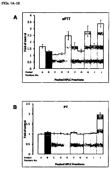
FIGS. 1A and 1B are bar graphs showing the results of a cell capture assay that determined the ability of various anti-α.sub.vβ.sub.6 monoclonal antibodies (“mAb”) to bind β.sub.6-transfected FDC-P1 cells (untransfected cells as control).
FIG. 2A is a graph showing the results of ELISA assays that determined the ability of various purified anti-α.sub.vβ.sub.6 “Fusion 6” monoclonal antibodies to bind soluble recombinant human α.sub.vβ.sub.6 (“hsα.sub.vβ.sub.6”). These antibodies were generated by immunizing β.sub.6−/− mice with soluble human truncated α.sub.vβ.sub.6. The numbers in the legend indicate the clone numbers. For the corresponding clone names, see Table 2.
FIG. 2B is a graph showing the results of ELISA assays that determined the ability of various purified anti-α.sub.vβ.sub.6 “Fusion 7” monoclonal antibodies to bind soluble recombinant hsα.sub.vβ.sub.6. These antibodies were generated by immunizing β.sub.6−/− mice with β.sub.6-transfected NIH 3T3 cells (Fusion #7).
FIGS. 3A-F are graphs showing the differential cation dependence of the binding of various anti-α.sub.vβ.sub.6 monoclonal antibodies to hsα.sub.vβ.sub.6.
FIGS. 4A and 4B are graphs showing that Fusion #6 and Fusion #7 monoclonal antibodies, respectively, inhibit the binding of biotin-hsα.sub.vβ.sub.6 to LAP.
FIGS. 5A-E are graphs showing that exemplary monoclonal antibodies of the invention inhibit the binding of β6-transfected FDC-P1 cells to LAP. FIGS. 5A and 5B display the results from Fusion #6 antibodies. FIGS. 5C-E display the results from Fusion #7 antibodies.
FIGS. 6A and 6B are graphs showing that Fusion #6 and Fusion #7 antibodies, respectively, inhibit the α.sub.vβ.sub.6-mediated activation of TGF-β, using a PAI-1 luciferase reporter gene assay to monitor TGF-β activation.
FIG. 7A depicts the amino acid sequences of the variable domains of the heavy chains of α.sub.vβ.sub.6 monoclonal antibodies 6.1A8, 6.8G6 (subclones A and B), 7.7G5, 6.2B1, 6.3G9, 6.2B10 (subclones A and B), 6.2G2, 6.2A1, 6.4B4 (subclones A, B and C), 7.10H2, 7.9H5, 7.4A3 (subclones A and B), 7.1C5 (subclones A and B) and 7.1G10. Antibodies 6.1A8, 6.8G6 and 7.7G5 are cation-dependent in binding to α.sub.vβ.sub.6, while antibodies 6.2B1, 6.2A1, 6.3G9, 6.2B10, 6.4B4, 7.1C5 and 7.1G10 are cation-independent (infra). The numbers in parentheses denote amino acid residue positions. The CDRs are in the large boxes, while the small boxes containing italicized amino acids represent polymorphism in different clones of a particular antibody.
FIG. 7B depicts the amino acid sequences of the variable domains of the light chains of α.sub.vβ.sub.6 monoclonal antibodies 6.1A8, 6.8G6, 6.4B4, 6.2A1, 7.1C5, 7.1G10, 6.2B10, 7.7G5, 6.2B1 and 6.3G9.
FIG. 8 is a scatter plot showing the expression of α.sub.vβ.sub.6 in human breast cancer and human squamous carcinoma tissue sections. Normal human tissues show only negligible expression levels of α.sub.vβ.sub.6.
FIGS. 9A and 9B are quadratic curve graphs depicting the solution binding affinities of two anti-α.sub.vβ.sub.6 antibodies, 6.8G6 and 6.3G9, respectively, for soluble α.sub.vβ.sub.6.
FIGS. 10A and 10B are bar graphs demonstrating the ability of purified monoclonal antibodies to compete with biotinylated 6.3G9 and biotinylated 6.8G6, respectively, for binding to α.sub.vβ.sub.6.
FIG. 11 is a bar graph showing percent smooth actin staining in kidneys from UUO animals treated with anti-α.sub.vβ.sub.6 mAb treatment.
FIG. 12 shows α.sub.vβ.sub.6 expression on tumor cell lines by FACS analysis (right side of figure) and inhibition of tumor cell lines binding to the LAP ligand by mAbs 6.3G9 and 6.4B4 (left side of figure).
FIG. 13 is a bar graph demonstrating inhibition of three tumor cell lines binding to the LAP ligand by anti-α.sub.vβ.sub.6 mAbs 6.3G9, 6.8G6 and 6.4B4. The mAb binding was compared to total binding without the addition of test mAbs (TB) and nonspecific binding to BSA control alone (NSB).
FIGS. 14A and 14B are graphs showing the effects of anti-α.sub.vβ.sub.6 mAb 6.3G9 and 6.4B4, respectively, over a 33 day study period on tumors arising from subcutaneously implanted Detroit 562 cells.
FIGS. 15A-C are graphs showing the effects of anti-α.sub.vβ.sub.6 mAb on bleomycin-induced lung fibrosis. (A) Antibody treatment using 6.3G9 mAb was started on day 0 at the time of bleomycin administration and was monitored over a 30 day period; (B) Antibody treatment using 6.3G9 mAb was started 15 days after bleomycin treatment and was monitored over a 30 day period; (C) Antibody treatment using 6.3G9, 6.8G6 and 6.4B4 mAbs was started 15 days after bleomycin treatment and was monitored over an extended 60 day period. In both FIGS. 15A and 15B , the bar graphs on the left represent μg hydroxyproline/lung while the bar graphs on right show percent increase in hydroxyproline above saline treated mice (no bleomycin). In FIG. 15C , the graph shows hydroxyproline content per lung.
Detailed description of the invention
This invention features classes and subclasses of antibodies that are specific for the integrin α.sub.vβ.sub.6. At least one class of the antibodies (blockers) are capable of blocking the binding of α.sub.vβ.sub.6 to LAP or preventing the activation of TGF-β.
The following describes the various methods of making the antibodies of this invention. Methods that are known in the art but not specifically described herein are also within the scope of this invention. For instance, antibodies of this invention can also be identified using phage-displayed antibody libraries, such as those described in Smith, Science 228:1315-7 (1985); U.S. Pat. Nos. 5,565,332, 5,733,743, 6,291,650, and 6,303,313. Additional antibodies of this invention can be made by coupling the heavy chains identified herein with a noncognate light chain, e.g., a light chain identified by phage display technology.
Non-Human Hybridoma Antibodies
The monoclonal antibodies of this invention can be generated by well known hybridoma technology. To do so, β.sub.6−/− animals (e.g., mice, rats or rabbits) are immunized with purified or crude α.sub.vβ.sub.6 preparations, cells transfected with cDNA constructs encoding α.sub.v, β.sub.6 or both antigens, cells that constitutively express α.sub.vβ.sub.6, and the like. The antigen can be delivered as purified protein, protein expressed on cells, protein fragment or peptide thereof, or as naked DNA or viral vectors encoding the protein, protein fragment, or peptide. Sera of the immunized animals are then tested for the presence of anti-α.sub.vβ.sub.6 antibodies. B cells are isolated from animals that test positive, and hybridomas are made with these B cells.
Antibodies secreted by the hybridomas are screened for their ability to bind specifically to α.sub.vβ.sub.6 (e.g., binding to β.sub.6-transfected cells and not to untransfected parent cells) and for any other desired features, e.g., having the desired CDR consensus sequences, inhibiting (or not inhibiting in the case of nonblockers) the binding between LAP and α.sub.vβ.sub.6 with an IC.sub.50 value lower than that of known anti-α.sub.vβ.sub.6 antibody 10D5, or inhibiting TGF-β activation.
Hybridoma cells that test positive in the screening assays are cultured in a nutrient medium under conditions that allow the cells to secrete the monoclonal antibodies into the culture medium. The conditioned hybridoma culture supernatant is then collected and antibodies contained in the supernatant are purified. Alternatively, the desired antibody may be produced by injecting the hybridoma cells into the peritoneal cavity of an unimmunized animal (e.g., a mouse). The hybridoma cells proliferate in the peritoneal cavity, secreting the antibody which accumulates as ascites fluid. The antibody may then be harvested by withdrawing the ascites fluid from the peritoneal cavity with a syringe.
The monoclonal antibodies can also be generated by isolating the antibody-coding cDNAs from the desired hybridomas, transfecting mammalian host cells (e.g., CHO or NSO cells) with the cDNAs, culturing the transfected host cells, and recovering the antibody from the culture medium.
Chimeric Antibodies
The monoclonal antibodies of this invention can also be generated by engineering a cognate hybridoma (e.g., murine, rat or rabbit) antibody. For instance, a cognate antibody can be altered by recombinant DNA technology such that part or all of the hinge and/or constant regions of the heavy and/or light chains are replaced with the corresponding components of an antibody from another species (e.g., human). Generally, the variable domains of the engineered antibody remain identical or substantially so to the variable domains of the cognate antibody. Such an engineered antibody is called a chimeric antibody and is less antigenic than the cognate antibody when administered to an individual of the species from which the hinge and/or constant region is derived (e.g., a human). Methods of making chimeric antibodies are well known in the art.
The chimeric antibodies embraced in this invention may contain a heavy chain variable domain having a sequence identical (or substantially so) to any one of SEQ ID NOs:19-36 and/or a light chain variable domain having a sequence identical (or substantially so) to any one of SEQ ID NOs:37-45.
Preferred human constant regions include those derived from IgG1 and IgG4.
Fully Human Antibodies
The monoclonal antibodies of this invention also include fully human antibodies. They may be prepared using in vitro-primed human splenocytes, as described by Boerner et al., J. Immunol. 147:86-95 (1991), or using phage-displayed antibody libraries, as described in, e.g., U.S. Pat. No. 6,300,064.
Some other methods for producing fully human antibodies involve the use of non-human animals that have inactivated endogenous Ig loci and are transgenic for un-rearranged human antibody heavy chain and light chain genes. Such transgenic animals can be immunized with α.sub.vβ.sub.6 and hybridomas are then made from B cells derived therefrom. These methods are described in, e.g., the various GenPharm/Medarex (Palo Alto, Calif.) publications/patents concerning transgenic mice containing human Ig miniloci (e.g., Lonberg U.S. Pat. No. 5,789,650); the various Abgenix (Fremont, Calif.) publications/patents with respect to XENOMICE (e.g., Kucherlapati U.S. Pat. Nos. 6,075,181, 6,150,584 and 6,162,963; Green et al., Nature Genetics 7:13-21 (1994); and Mendez et al., 15(2):146-56 (1997)); and the various Kirin (Japan) publications/patents concerning “transomic” mice (e.g., EP 843 961, and Tomizuka et al., Nature Genetics 16:133-1443 (1997)).
Humanized Antibodies
The monoclonal antibodies of this invention also include humanized versions of cognate anti-α.sub.vβ.sub.6 antibodies derived from other species. A humanized antibody is an antibody produced by recombinant DNA technology, in which some or all of the amino acids of a human immunoglobulin light or heavy chain that are not required for antigen binding (e.g., the constant regions and the framework regions of the variable domains) are used to substitute for the corresponding amino acids from the light or heavy chain of the cognate, nonhuman antibody. By way of example, a humanized version of a murine antibody to a given antigen has on both of its heavy and light chains
constant regions of a human antibody;
framework regions from the variable domains of a human antibody; and
CDRs from the murine antibody. When necessary, one or more residues in the human framework regions can be changed to residues at the corresponding positions in the murine antibody so as to preserve the binding affinity of the humanized antibody to the antigen. This change is sometimes called “back mutation.” Humanized antibodies generally are less likely to elicit an immune response in humans as compared to chimeric human antibodies because the former contain considerably fewer non-human components.
The methods for making humanized antibodies are described in, e.g., Winter E P 239 400; Jones et al., Nature 321:522-525 (1986); Riechmann et al., Nature 332:323-327 (1988); Verhoeyen et al., Science 239: 1534-1536 (1988); Queen et al., Proc. Nat. Acad. Sci. USA 86:10029 (1989); U.S. Pat. No. 6,180,370; and Orlandi et al., Proc. Natl. Acad. Sci. USA 86:3833 (1989). Generally, the transplantation of murine (or other non-human) CDRs onto a human antibody is achieved as follows. The cDNAs encoding heavy and light chain variable domains are isolated from a hybridoma. The DNA sequences of the variable domains, including the CDRs, are determined by sequencing. The DNAs encoding the CDRs are transferred to the corresponding regions of a human antibody heavy or light chain variable domain coding sequence by site directed mutagenesis. Then human constant region gene segments of a desired isotype (e.g., γ1 for CH and k for CL) are added. The humanized heavy and light chain genes are co-expressed in mammalian host cells (e.g., CHO or NSO cells) to produce soluble humanized antibody. To facilitate large scale production of antibodies, it is often desirable to produce such humanized antibodies in bioreactors containing the antibody-expressing cells, or to produce transgenic mammals (e.g., goats, cows, or sheep) that express the antibody in milk (see, e.g., U.S. Pat. No. 5,827,690).
At times, direct transfer of CDRs to a human framework leads to a loss of antigen-binding affinity of the resultant antibody. This is because in some cognate antibodies, certain amino acids within the framework regions interact with the CDRs and thus influence the overall antigen binding affinity of the antibody. In such cases, it would be critical to introduce “back mutations” (supra) in the framework regions of the acceptor antibody in order to retain the antigen-binding activity of the cognate antibody.
The general approach of making back mutations is known in the art. For instance, Queen et al. (supra), Co et al., Proc. Nat. Acad. Sci. USA 88:2869-2873 (1991), and WO 90/07861 (Protein Design Labs Inc.) describe an approach that involves two key steps. First, the human V framework regions are chosen by computer analysis for optimal protein sequence homology to the V region framework of the cognate murine antibody. Then, the tertiary structure of the murine V region is modeled by computer in order to visualize framework amino acid residues that are likely to interact with the murine CDRs, and these murine amino acid residues are then superimposed on the homologous human framework.
Under this two-step approach, there are several criteria for designing humanized antibodies. The first criterion is to use as the human acceptor the framework from a particular human immunoglobulin that is usually homologous to the non-human donor immunoglobulin, or to use a consensus framework from many human antibodies. The second criterion is to use the donor amino acid rather than the acceptor if the human acceptor residue is unusual and the donor residue is typical for human sequences at a specific residue of the framework. The third criterion is to use the donor framework amino acid residue rather than the acceptor at positions immediately adjacent to the CDRs.
One may also use a different approach as described in, e.g., Tempest, Biotechnology 9: 266-271 (1991). Under this approach, the V region frameworks derived from NEWM and REI heavy and light chains, respectively, are used for CDR-grafting without radical introduction of mouse residues. An advantage of using this approach is that the three-dimensional structures of NEWM and REI variable regions are known from X-ray crystallography and thus specific interactions between CDRs and V region framework residues can be readily modeled.
Other Moieties
The monoclonal antibodies of this invention may further comprise other moieties to effect the desired functions. For instance, the antibodies may include a toxin moiety (e.g., tetanus toxoid or ricin) or a radionuclide (e.g., .sup.111In or .sup.90Y) for killing of cells targeted by the antibodies (see, e.g., U.S. Pat. No. 6,307,026) The antibodies may comprise a moiety (e.g., biotin, fluorescent moieties, radioactive moieties, histidine tag or other peptide tags) for easy isolation or detection. The antibodies may also comprise a moiety that can prolong their serum half life, for example, a polyethylene glycol (PEG) moiety.
Diseased Conditions and Animal Models
The antibodies of the invention are useful in the treatment, including prevention, of α.sub.vβ.sub.6-mediated diseases. For example, these antibodies can be used to treat fibrosis (e.g., lung fibrosis, acute lung injury, kidney fibrosis, liver fibrosis, Alport's Syndrome, and scleroderma) by blocking the activation of TGF-β or blocking the binding of α.sub.vβ.sub.6 to any other ligands, such as fibronectin, vitronectin, and tenascin. The novelty of this approach includes:
it blocks the activation of TGF-β rather than the binding of TGF-β to its receptor,
it can inhibit TGF-β locally (i.e., at sites of α.sub.vβ.sub.6 upregulation) rather than systemically, and
it inhibits binding of α.sub.vβ.sub.6 to a ligand. Other than fibrotic diseases or conditions, the antibodies of the invention are useful in treating cancer or cancer metastasis (including tumor growth and invasion), particularly epithelial cancers. A subset of epithelial cancers is squamous cell carcinoma, e.g., head and neck, oral, breast, lung, prostate, cervical, pharyngeal, colon, pancreatic and ovarian cancers. Our studies using the new α.sub.vβ.sub.6 monoclonal antibodies demonstrated that α.sub.vβ.sub.6 is highly expressed in many epithelial cancers, especially on the leading edge of the tumors. The new antibodies can also be used to any other diseases mediated by α.sub.vβ.sub.6, including psoriasis.
The treatments of this invention are effective on both human and animal subjects afflicted with these conditions. Animal subjects to which the invention is applicable extend to both domestic animals and livestock, raised either as pets or for commercial purposes. Examples are dogs, cats, cattle, horses, sheep, hogs and goats.
The efficacy of the antibodies of the invention can be tested in various animal models. Mouse models for lung fibrosis include bleomycin-(Pittet et al., J. Clin. Invest. 107(12):1537-1544 (2001); and Munger et al., supra) and irradiation-inducible lung fibrosis (Franko et al., Rad. Res. 140:347-355 (1994)). In bleomycin-treated mice, the expression of α.sub.vβ.sub.6 increases in the epithelial alveolar cells of the lungs. But β.sub.6 knockout mice are protected from bleomycin-induced injury and fibrosis.
Mouse models for kidney fibrosis include COL4A3−/− mice (see, e.g., Cosgrove et al., Amer. J. Path. 157:1649-1659 (2000), mice with adriamycin-induced injury (Wang et al., Kidney International 58: 1797-1804 (2000); Deman et al., Nephrol Dial Transplant 16: 147-150 (2001)), db/db mice (Ziyadeh et al., PNAS USA 97:8015-8020 (2000)), and mice with unilateral ureteral obstruction (Fogo et al., Lab Investigation 81: 189A (2001); and Fogo et al., Journal of the American Society of Nephrology 12:819A (2001)). In all of these models, the mice develop kidney injury and fibrosis that can progress to renal failure. α.sub.vβ.sub.6 is upregulated in the epithelial lining of the ascending and descending tubules of the kidneys of the COL4A3−/− mice, adriamycin-treated mice, and mice that undergo unilateral ureteral obstruction. It is likely that α.sub.vβ.sub.6 expression also increases in a variety of kidney injury models.
Anti-α.sub.vβ.sub.6 monoclonal antibodies can also be tested for their ability to inhibit tumor growth, progression, and metastasis in such animal models as the standard in vivo tumor growth and metastasis models. See, e.g., Rockwell et al., J. Natl. Cancer Inst. 49:735 (1972); Guy et al., Mol. Cell Biol. 12:954 (1992); Wyckoff et al., Cancer Res. 60:2504 (2000); and Oft et al., Curr. Biol. 8:1243 (1998). Important α.sub.vβ.sub.6 ligands in cancer may include TGF-β, which is involved in metastasis (for review see Akhurst et al., Trends in Cell Biology 11:S44-S51 (2001)), fibronectin and vitronectin.
The efficacy of the treatments of this invention may be measured by a number of available diagnostic tools, including physical examination, blood tests, proteinuria measurements, creatinine levels and creatinine clearance, pulmonary function tests, plasma blood urea nitrogen (BUN) levels, observation and scoring of scarring or fibrotic lesions, deposition of extracellular matrix such as collagen, smooth muscle actin and fibronectin, kidney function tests, ultrasound, magnetic resonance imaging (MRI), and CT scan.
Pharmaceutical Compositions
The pharmaceutical compositions of this invention comprise one or more antibodies of the present invention, or pharmaceutically acceptable derivatives thereof, optionally with any pharmaceutically acceptable carrier. The term “carrier” as used herein includes known acceptable adjuvants and vehicles.
According to this invention, the pharmaceutical compositions may be in the form of a sterile injectable preparation, for example a sterile injectable aqueous or oleaginous suspension. This suspension may be formulated according to techniques known in the art using suitable dispersing, wetting, and suspending agents.
The pharmaceutical compositions of this invention may be given orally, topically, intravenously, subcutaneously, intraperitoneally, intramuscularly, intramedullarily, intra-articularly, intra-synovially, intrasternally, intrathecally, intrahepatically, or intracranially as desired, or just locally at sites of inflammation or tumor growth. The pharmaceutical compositions of this invention may also be administered by inhalation through the use of, e.g., a nebulizer, a dry powder inhaler or a metered dose inhaler.
The dosage and dose rate of the antibodies of this invention effective to produce the desired effects will depend on a variety of factors, such as the nature of the disease to be treated, the size of the subject, the goal of the treatment, the specific pharmaceutical composition used, and the judgment of the treating physician. Dosage levels of between about 0.001 and about 100 mg/kg body weight per day, for example between about 0.1 and about 50 mg/kg body weight per day, of the active ingredient compound are useful. For instance, an antibody of the invention will be administered at a dose ranging between about 0.01 mg/kg body weight/day and about 20 mg/kg body weight/day, e.g., ranging between about 0.1 mg/kg body weight/day and about 10 mg/kg body weight/day, and at intervals of every one to fourteen days. In another embodiment, the antibody is administered at a dose of about 0.3 to 1 mg/kg body weight when administered intraperitoneally. In yet another embodiment, the antibody is administered at a dose of about 5 to 12.5 mg/kg body weight when administered intravenously. In one embodiment, an antibody composition is administered in an amount effective to provide a plasma level of antibody of at least 1 mg/ml.
Diagnostic Methods
The antibodies of this invention can be used to diagnose diseased conditions associated with altered α.sub.vβ.sub.6 expression levels. A tissue sample from a subject, such as a tissue biopsy, body fluid sample or lavage (e.g., alveolar lavage), can be tested in an antigen capture assay, ELISA, immunohistochemistry assay, and the like using the antibodies. A tissue sample from a normal individual is used as control.
Practice of the present invention will employ, unless indicated otherwise, conventional techniques of cell biology, cell culture, molecular biology, microbiology, recombinant DNA, protein chemistry, and immunology, which are within the skill of the art. Such techniques are described in the literature. See, for example, Molecular Cloning: A Laboratory Manual, 2nd edition (Sambrook et al., Eds.), 1989; Oligonucleotide Synthesis , (M. J. Gait, Ed.), 1984; U.S. Pat. No. 4,683,195 to Mullis et al.; Nucleic Acid Hybridization , (B. D. Hames and S. J. Higgins), 1984; Transcription and Translation , (B. D. Hames and S. J. Higgins), 1984; Culture of Animal Cells (R. I. Freshney, Ed.), 1987; Immobilized Cells and Enzymes , IRL Press, 1986; A Practical Guide to Molecular Cloning (B. Perbal), 1984 ; Methods in Enzymology , Volumes 154 and 155 (Wu et al., Eds.), Academic Press, New York; Gene Transfer Vectors for Mammalian Cells (J. H. Miller and M. P. Calos, Eds.), 1987; Immunochemical Methods in Cell and Molecular Biology (Mayer and Walker, Eds.), 1987; Handbook of Experiment Immunology , Volumes I-IV (D. M. Weir and C. C. Blackwell, Eds.), 1986; Manipulating the Mouse Embryo, 1986.
Unless otherwise defined, all technical and scientific terms used herein have the same meaning as commonly understood by one of ordinary skill in the art to which this invention pertains. Exemplary methods and materials are described below, although methods and materials similar or equivalent to those described herein can also be used in the practice of the present invention. All publications and other references mentioned herein are incorporated by reference in their entirety. In case of conflict, the present specification, including definitions, will control. The materials, methods, and examples are illustrative only and not intended to be limiting. Throughout this specification, the word “comprise,” or variations such as “comprises” or “comprising” will be understood to imply the inclusion of a stated integer or group of integers but not the exclusion of any other integer or group of integers.
Examples
The following examples are meant to illustrate the methods and materials of the present invention. Suitable modifications and adaptations of the described conditions and parameters normally encountered in the antibody art that are obvious to those skilled in the art are within the spirit and scope of the present invention.
In the following examples, the β.sub.6−/− mice were generated as described in Huang et al., J. Cell Biol. 133:921 (1996). Recombinant human LAP was purchased from R & D Systems (Minneapolis, Minn.). Antibody 10D5 was purchased from Chemicon (Temecula, Calif.). The L230 hybridoma was purchased from ATCC and the secreted antibody was purified from the supernatant of saturated cultures by affinity chromatography on immobilized protein A. Isotyping of antibodies was carried out using the ISOSTRIP kit (Roche Diagnostics) according to the manufacturer's instructions. The β.sub.6-transfected SW480 cell line was prepared as described in Weinacker et al., J. Biol. Chem. 269:6940-6948 (1994). Example 1: Generation of β6-Transfected Stable Cell Lines
β.sub.6-transfected NIH 3T3 and FDC-P1 cells were generated by electroporating parent cell lines with a DNA construct containing full length murine β.sub.6 cDNA and a neomycin selectable marker. Stably transfected cells were selected by passaging cells in culture medium containing G418 for 14 days followed by fluorescent activated cell sorting (FACS) to isolate cells expressing the highest level of surface β.sub.6. Transfected FDC-P1 cells were cultured in DMEM supplemented with 4 mM L-glutamine adjusted to contain 1.5 g/L sodium bicarbonate, 4.5 g/l glucose, and 1.0 mM sodium pyruvate, 10% FBS, 2.5% mouse IL-3 culture supplement, and 1.5 mg/ml active G418. Transfected NIH 3T3 cells were cultured in DMEM supplemented with 10% FBS, 2 mM L-glutamine, penicillin/streptomycin, and 1 mg/ml active G418. Example 2: Purification of Human Soluble αvβ6
The α.sub.vβ.sub.6 protein was purified essentially as described in Weinacker, supra. A CHO cell line expressing hsα.sub.vβ.sub.6 was cultured, and the resultant supernatant collected by centrifugation. The integrin was purified by affinity chromatography using anti-α.sub.v antibody L230. Purified L230 was cross-linked to CNBr-activated Sepharose 4B (Sigma) at a ratio of 4.8 mg antibody/ml resin. The α.sub.vβ.sub.6 supernatant was loaded at 0.5 mg antibody/ml resin onto the L230 affinity column, and the column was washed with 10 column volumes of each of
50 mM Tris-Cl, pH 7.5, 1 M NaCl, 1 mM MgCl.sub.2;
50 mM Tris-Cl, pH 7.5, 50 mM NaCl, 1 mM MgCl.sub.2; and
The description continues in the full USPTO document.
 Lapsed, fee not paid28 drawings

