 Lapsed, fee not paid2 drawings
Medical Devices · US 8,771,301 B2
Ultrasonic handpiece
A handpiece having a single set of piezoelectric elements polarized to produce longitudinal motion when excited at the relevant resonant frequency.
US 8,771,310 B2 · Assignee: Adenopaint, LLC · Inventors: Forman; Mervyn B. et al.
Sheet 1 of 22 from the published document. All sheets in the USPTO PDF
The present invention relates to compositions and methods for improving outcomes in vascular interventional procedures. In particular, the present invention relates to compositions and methods for improving outcomes in vascular interventional procedures using an anti-no-reflow guide wire that attenuates the "no-reflow" phenomenon that is associated with negative outcomes.
Atherosclerotic vascular disease remains a major cause of morbidity and mortality in spite of numerous advances in pharmacological modalities to modify associated risk factors. Atherosclerotic involvement of the myocardium, brain and kidneys is responsible for the majority of adverse affects of the disorder. Coronary artery disease remains the leading cause of death in the Westernized world. Approximately 1.5 million Americans per year suffer a myocardial infarction with an annual death toll of 400,000 (see e.g., Thom, T., et al., Circulation 113: 85 (2006)). Cerebrovascular disease is the third leading cause of death with stenosis of the internal carotid artery accounting for 20% of strokes and transient ischemic attacks (TIAs) (see e.g., Heart Disease and Stroke Statistics--2006 Update Dallas, Tx; AHA, (2006); Roffie, M., and Yadav, J. S., Circulation 114: el (2006)). Renal artery sten
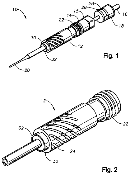
1 of 22 drawing sheets so far from the published document, cropped to the drawing. Every sheet is in the USPTO PDF.
What the patent claimed, word for word. All of it is now free to use.
The present invention relates to compositions and methods for improving outcomes in vascular interventional procedures. In particular, the present invention relates to compositions and methods for improving outcomes in vascular interventional procedures using a guide wire (for example an anti-no-reflow guide wire) that attenuates the "no-reflow" phenomenon that is associated with negative outcomes.
Atherosclerotic vascular disease remains a major cause of morbidity and mortality in spite of numerous advances in pharmacological modalities to modify associated risk factors. Atherosclerotic involvement of the myocardium, brain and kidneys is responsible for the majority of adverse affects of the disorder. Coronary artery disease remains the leading cause of death in the Westernized world. Approximately 1.5 million Americans per year suffer a myocardial infarction with an annual death toll of 400,000 (see e.g., Thom, T., et al., Circulation 113: 85 (2006)). Cerebrovascular disease is the third leading cause of death with stenosis of the internal carotid artery accounting for 20% of strokes and transient ischemic attacks (TIAs) (see e.g., Heart Disease and Stroke Statistics--2006 Update Dallas, Tx; AHA, (2006); Roffie, M., and Yadav, J. S., Circulation 114: el (2006)). Renal artery stenosis due to atherosclerosis is relatively common (6-8%) in patients >65 years of age and often results in progressive renal failure, worsening hypertension and precipitation of congestive heart failure and unstable angina (see e.g., Hansen, K. J., et al., J. Vasc. Surg. 36: 443 (2002); Pasternak, R. C., et al., Circulation 109: 2605 (2004)).
The rapid evolution of percutaneous balloon angioplasty (PTA) followed by the development of stent technology has resulted in increased utilization of vascular interventional procedures in the treatment of atherosclerotic-induced stenosis of the major blood vessels supplying the myocardium, brain, kidneys and peripheral vessels. Coronary artery stenting has surpassed coronary artery bypass surgery (CABG) as a treatment of choice to revascularize patients with a single or double vessel coronary artery disease with more than 700,000 procedures performed in the United States each year. Furthermore, stenting procedures are frequently performed in CABG patients who have degenerative and stenotic saphenous vein grafts (SVG) secondary to atherosclerosis.
In reference to the heart, patients with atherosclerosis may present with stable angina, acute coronary syndromes (ACS), namely unstable angina and non-ST segment elevation myocardial infarction (NSTEMI), or acute ST segment elevation infarction (STEMI). These syndromes occur in the setting of an unstable ulcerative plaque associated with a variable thrombus burden. These conditions can be successfully treated in the majority of patients with stent implantation resulting in restoration of normal vessel caliber. However, the introduction of a guide wire, balloon, stent or embolic protection device (EPD) may compromise tissue blood flow through macro-embolization of thrombotic and atherosclerotic debris into the distal vessel (see e.g., Topol, E. J., and Yadav, J. S., Circulation 101: 570 (2000)). This has been shown to occur in 14% of STEMI patients treated with percutaneous coronary intervention (PCI) and is associated with larger infarct size, worse left ventricular function and higher mortality (see e.g., Henriques, J. P., et al., Eur. Heart J. 23: 1112 (2002)). Embolization with atheromatous debris is even more frequent during PCI of SVG's (.about.80%) and results in increased mortality (see e.g., Trono, R., et al., Cleve. Clin. J. Med. 56:581 (1989)).
Vascular interventional procedures performed in the presence of an occlusive thrombus may also compromise tissue blood flow either through micro-embolization or via microvascular injury induced by the deterious effects of reperfusion (see e.g., Constantini, C. O., et al., J. Am. Coll. Cardiol. 44: 305 (2004); Kenner, M. D., et al., J. Am. Coll. Cardiol. 76: 861 (1995); van't H of A. W., et al., Circulation 97: 2302 (1998); Forman, M. B., et al., Cardiovascular Drug Reviews 24: 116 (2006)). The common occurrence of micro-embolization after interventional procedures has recently been appreciated by the frequent retrieval of atheroembolic material from EPDs. These particles consist of cellular and non-cellular elements with wide variations in size and volume (see e.g., Akbar O., et al., Am. Heart J. 152: 207 (2000)). Although the pathogenesis of reperfusion injury is complex and multi-factorial, the introduction of activated neutrophils and platelets associated with release of various vasoconstrictors produced from circulating cells and dysfunctional endothelial cells may contribute to a progressive decrease in flow during the peri-reperfusion period; defined as the "no-reflow" phenomenon (see e.g., Forman, M. B. et. al, Cardiovascular Drug Reviews 24: 116 (2006)). Impaired tissue perfusion occurs in 29-44% of reperfused patients, the highest incidence with occlusion of the left anterior descending coronary artery (50-80%), and correlates with infarct size, ventricular function and early and late mortality (see e.g., Bax, M., et al., J. Am. Coll. Cardiol. 43: 534 (2004); Ito, H., et al., Circulation 93: 1993 (1996); Deluca, G., et al., Circulation 109: 958 (2004); Wu, K. C., et al., Circulation 97: 765 (1998)). Furthermore, numerous studies have demonstrated that abnormal tissue perfusion, as assessed by myocardial blush grade (MBG), is an independent, multivariate predictor of both early and late mortality in STEMI undergoing thrombolysis or PCI. Abnormal MBG (0-1) results in a 7-fold increase in mortality which is maintained for up to 2 years (see e.g., Gibson, C. M. and Schomig, A., Circulation 109: 3096 (2004); Forman, M. B., and Jackson, E. K., Clin. Cardiol. 30: 583 (2007)). Abnormal tissue perfusion following PCI in patients with NSTEMI is also associated with higher risk of myocardial necrosis and death at 6 months and 1 year (see e.g., Wong, G. C., et al., Circulation 106: 202 (2002)). Therefore, the development of devices or pharmacologic strategies that preserve tissue perfusion by attenuating the no-reflow phenomenon has important clinical implications.
PCI results in myocardial cell necrosis in 22-44% of patients after an otherwise uncomplicated procedure irrespective if the procedure is elective for stable angina or emergent for ACS (see e.g., Ali, O. A., et al., Am. Heart J. 152: 207 (2006); Johansen, O., et al., Eur. Heart J. 19: 112 (1998)). Elevations of cardiac enzymes (for example creatine kinase-myocardial band (CK-MB) and Troponin I) significantly increase the risk of early and late mortality and myocardial infarction (see e.g., Cavallini, C., et al., Eur. Heart J. 26:1494 (2005); Antman, E. M., et al., N. Engl. J. Med. 335: 1342 (1996)). Furthermore increasing levels of cardiac enzymes are associated with parallel increases in mortality (see e.g., Antman, E. M., et al., N. Engl. J. Med. 335: 1342 (1996); Brener, S. J., et al., Eur. Heart J. 23: 869 (2002)). MRI studies have confirmed that mild increases in CK-MB after PCI are due to microinfarction secondary to embolic microvascular obstruction (see e.g., Ricciardi. M. J., et al., Circulation 103:2780 (2001)). PCI with stent deployment amplifies myonecrosis probably secondary to increased vascular trauma and release of vasoconstrictor mediators such as serotonin (see e.g., Lesoco, D., et al., Am J. Cardiol. 84:1317 (1999)). While preloading with a potent thienopyridine platelet inhibitor (600 mg of clopidogrel) reduced cell necrosis in a large study, 14% of patients continued to manifest evidence of micro-embolization (see e.g., Patti, G., et al., Circulation 111: 2099 (2005)). Therefore the common occurrence of embolization during routine interventional procedures provides further impetus to develop improved modalities to reduce this important complication.
In an attempt to reduce the incidence of macro- and micro-embolization after PCI, numerous pharmacological therapies and embolic and thrombectomy devices have been utilized in patients with STEMI. A potent glycoprotein IIa/IIIb inhibitor failed to reduce infarct size and improve tissue perfusion after PTA or primary stenting in STEMI patients (see e.g., Antoniucci, D., et al, J. Am. Coll. Cardiol. 42: 1879 (2003); Constantini, C. O., et al., J. Am. Coll. Cardiol. 44: 30 (2004)). In the EMERALD trial, the GuardWire distal balloon occlusive device did not improve tissue perfusion or decrease infarct size in 501 patients with STEMI undergoing PCI within 6 hours of symptoms (see e.g., Stone, G. W., et al., JAMA 293: 1063 (2005)). This occurred in spite of use of anti-platelet agents and removal of embolic debris in the majority of patients. The FilterWire system, which consists of a guide wire that incorporates a non-occluding polyurethane porous membrane filter in the shape of a wind sock, has been evaluated in two studies. In one study (hereinafter referred to as the "PROMISE study") no difference in infarct size or tissue perfusion assessed with flow velocity was observed in the FilterWire group in 200 patients with NSTEMI and STEMI (see e.g Gick M., et al., Circulation 112:1462 (2005)). Similar findings were observed in a second study (hereinafter referred to as the "DEDICATION study") where 676 patients with STEMI underwent PCI with and without distal protection. The filter wire system failed to improve ST segment resolution, regional ventricular function or major adverse cardiac events at 30 days (see Kelbaek, et al., J. Am. Coll. Cardiol. 51: 899 (2008).
A number of thrombectomy devices have also been utilized as an adjunct to PCI in STEMI. These include the X-Sizer system (consists of wire system with a helical shaped cutter and aspiration catheter), several aspiration devices (Diver, Export, Rescue catheter) and the angiojet rheolytic thrombectomy system in which high-velocity saline is directed back into the catheter. Eight trials have been conducted with the majority being small (see e.g., Brodie, B. R., J. Invasive Cardiol. 18: 24C (2006); Baim, D. S., J. Invasive Cardiol. 18: 28C (2006)). The two largest trials did not show improved tissue perfusion and one showed larger infarct size compared to control (see e.g., Kaltoft, A., et al., Circulation 114: 40 (2006); Ali, A., et al., J. Am. Coll. Cardiol. 48: 244 (2006)). These findings demonstrate that the current protective and embolic devices are not optimal in achieving complete thrombectomy or preventing distal embolization. Furthermore, they support the concept that other mechanisms, such as humoral factors and cytotoxic compounds, may also be playing an important role in microvascular damage after STEMI.
Prior to the introduction of PCI and stents, CABG was the most frequently utilized procedure to relieve myocardial ischemia in patients with coronary artery disease. However, 50-60% of SVG's develop severe significant atherosclerosis within ten years of surgery requiring catheter based intervention (see e.g., Bourasa, M. G., et al., Circulation 72: V71 (1985); Lau, G. T., et al., Semin. Vasc. Med. 4: 153 (2004)). The soft and friable nature of the lipid rich plaque in SVGs contributes to the frequent embolization of atherothrombolic material after PCI (see e.g. Popma, J. J., Cathet. Cardiovasc. Intervent. 57: 125 (2002); Webb, J., et al., J. Am. Coll. Cardiol. 34: 461 (1999); Safian, R. D., Prog. Cardiovasc. Dis. 44; 437 (2002)). Platelet clumping with subsequent activation and release of the potent vasoconstrictor serotonin may also reduce flow in the distal vascular bed. PCI of degenerated SVG's is associated with significant (.about.20%) risk of major adverse clinical events (MACE), predominantly myocardial infarction and no-reflow (see e.g. Piana, R. N., et al., Circulation 89: 2514 (1994); de Feyter, P. J., et al., J. Am. Coll. Cardiol. 21: 1539 (1993)). A 3-fold elevation of CK-MB is associated with a 14% 30 day mortality compared with less than 1% without isoenzyme elevation (see e.g. Hong, et al., Circulation 100: 2400 (1999); Lefkovitz, J., et al., Circulation 92: 734 (1995)). Multi-variable predictors of MACE include the extent of graft disease (graft length disease and plaque volume), number of stents inserted and age of the patient (see e.g., Stone, G. W., et al., Circulation 108: 548 (2003)). The high peri-procedural complication rate has necessitated the use of adjunctive therapies to reduce the high adverse event rates. Administration of the IIb/IIIa platelet inhibitor (abciximab) and the thrombectomy catheter (X-sizer) failed to reduce MACE at thirty days (see e.g. Ellis, S. G., et al., J. Am. Coll. Cardiol. 32: 1619 (1998)). In contrast, the GuardWire, FilterWire and a proximal embolic protection system (Proxis) produced significant and equivalent reductions in peri-procedure complications at 30 days (see e.g., Stone, G. W., et al., Circulation 108: 548 (2003); Baim, D. S. et al., Circulation 105: 1285 (2003); Mauri, L., et al., J. Am. Coll. Cardiol. 50: 1442 (2007)). However, peri-procedural complications still occurred in .about.10% of patients with these devices emphasizing the need for complimentary devices and/or new pharmacologic interventions.
PCI (PTA, stents) may also compromise blood flow in the subacute and chronic phase following an uneventful procedure. Abnormal vasomotor responses are invariably present after PTA and life threatening vasospasm has been observed at variable times after stent implantation (see e.g., Fischell, T. A., et al., J. Clin. Invest. 86:575 (1990); Brott, B. C., et al., J. Invasive Cardiol. 18:584 (2006)). Drug eluting stents (DESs) are currently the most frequently deployed stent in the USA and consist of a metal stent, polymer and impregnated drug; either an antineoplastic agent, paclitaxel (Taxus) or antiproliferative agents such as sirolimus (Cypher) and zotarolimus (Endeavor). DESs are associated with a small but potentially lethal increase in sub-acute and chronic thrombosis when compared with bare metal stents (BMSs) (see e.g., Camenzind, E., et al., Circulation 115:1440 (2007)). DESs activate pro-coagulant factors and may diminish the ability to develop collateral vessels with stent thrombosis (see e.g., Salloum, J. et al., J. Intervent. Cardiol. 17:575 (2005); Meier, P., et al., J. Am. Coll. Cardiol. 40:21 (2007)). A recent study demonstrates long term adverse effects of DES on endothelial cell function. Vasodilatory responses to acetylcholine were significantly impaired in segments distal to both paclitaxel and sirolimus stents when compared to BMS or a reference non stented vessel 6 months after implantation (see e.g., Kim, J. W., et al., J. Am. Coll. Cardiol. Intv. 1:65 2008). The hypothesis that chronic endothelial dysfunction may result in recurrent ischemia and late stent thrombosis is supported by two studies. In the BASKET study utilizing Taxus stents, cardiac death and infarction at 6 to 18 months was approximately four times greater in the DES arm compared with BMSs (see e.g., Pfasterer, M., et al., J. Am. Coll. Cardiol. 48:2584 (2006)). Recently, a post hoc analysis of the RRISC Trial revealed a significant increase in mortality with Cypher stents implanted in SVGs after a median follow-up of 32 months (see e.g., Vermeersch, P. et al., J. Am. Coll. Cardiol. 50:261 (2007)). Pathological studies with DESs have invariably shown incomplete endothelialization on strut surfaces extending beyond 40 months after implantation with extensive fibrin deposition (see e.g., Joyner, M., et al., J. Am. Coll. Cardiol. 48:193 (2006)). Chronic inflammatory cells (lymphocytes, macrophages and eosinophils) in the intima and media are also present in late stent thrombosis. Release of numerous vasoconstrictors and platelet aggregatory substances by these cells may also contribute to late stent thrombosis. The histological changes observed have been attributed to either direct toxicity and/or delayed hypersensitivity reaction to the drug or polymer, or excessive barotraumas (see e.g., Togni, M., et al., J. Invasive Cardiol. 18:593 (2006)). Local delivery of high concentrations of the physiological nucleotide adenosine that rapidly accelerates endothelial healing, prevents thrombus formation, reduces inflammatory cell infiltration and promotes new vessel formation would have important clinical implications.
While newer DESs are currently undergoing safety and efficacy trials, it appears likely that they will be associated with comparable side effects to the two currently approved stents due to their similar structure and mode of action. In Europe, BMSs are being increasingly utilized in large vessels (>3 mm) and in non-diabetic patients. While late stent thrombosis is rare, restenosis remains a significant problem with BMSs with an incidence of 17 to 25% in non-diabetics and 23 to 33% in diabetics with vessels of 3 mm. Since adenosine is a potent inhibitor of vascular smooth muscle proliferation and extracellular matrix production, it may prove useful in preventing restenosis following BMS implantation.
Carotid revascularization utilizing surgically performed carotid endarterectomy (CEA) has been shown to reduce stroke rate compared with medical therapy in patients with significant (greater than 50%) atherosclerotic narrowing of the carotid bifurcation and internal carotid artery (see e.g. Halliday, A., et al., Lancet 363: 1491 (2004); North American Symptomatic Trial Collaborators, N. Engl. J. Med. 325: 445 (1991)). The rapid development of catheter based technology has resulted in the evaluation of carotid artery stenting (CAS) as an alternative therapy to CEA (see e.g., Roubin, G. S., et al., Circulation 113: 2021 (2006)). CAS has now been approved by the Center for Medicare and Medicaid for patients who are at high risk for CEA (see e.g., Yadav, J. S., J. Am. Coll. Cardiol. 47: 2397 (2006)). Peri-procedural neurological and cardiovascular events remain the main complication of both procedures. Clinically silent micro-embolization occurred in 92% of patients undergoing CEA utilizing transcranial Doppler studies (see e.g., Grant, M., et al., Br. J. Surg. 8: 1435 (1994)). Similarly, 29% of patients manifested silent embolic events after CAS utilizing MRI (see e.g., Jaeger, H., Am. J. Neuroradiol. 23: 200 (2002)). The introduction of EPDs, which are now considered the standard of care, have reduced (by 50%), but not eliminated, embolization of atheromatous debris into the cerebral circulation after stenting (see e.g., Wholey, M. H., and Al-Mubarek, N., Catheter Cardiovasc. Intervent. 60: 259 (2003)). CAS with EPD in high risk patients, in whom a non-surgical approach is preferred due to lower morbidity and mortality, results in approximately 6-12% incidence of major adverse cardiac and cerebral vascular events (see e.g., Safian, R. D., et al., J. Am. Coll. Cardiol. 47: 2384 (2006); Yadav, J. S., et al., N. Eng. J. Med. 351: 1493 (2004)). Low risk patients undergoing CAS with EPD manifest an event rate of 2-3% (see e.g., Zahn, R., et al., Eur. Heart J. 25: 1550 (2004); Kastrur, A., et al., Stroke 34: 813 (2003)). Complications remain high in the elderly (>80 years) with approximately 17.1% incidence of death or stroke at 30 days (see e.g., Hobson, R. W., et al., J. Vasc. Surg. 40: 1106 (2004)).
The reasons why EPDs are not fully protective are multifactorial. Atheromatous material may be dislodged from the aortic arch or common carotid artery during manipulation of the guide catheter and wire prior to the insertion of the EPD. The EPDs are bulky and may induce further embolization during deployment in calcified and tortuous vessels. While EPD devices are universally beneficially, the duration of the deployment significantly increase the risks of complications. For example, deployment of the Filter protection device greater than 20 minutes has been shown to double the risk of death and stroke compared with deployment times of less than 20 minutes (see e.g., Yadav, J. S., Circulation 47: 2397 (2006)). The device may also produce vascular damage (endothelial dysfunction and dissection) and result in incomplete capture or retrieval of debris. Finally, humoral factors released during deployment of the EPD and stent may result in vasospasm or hyperperfusion syndrome, the latter being responsible for 1.3% of intracranial hemorrhage in high risk patients (see e.g., Abou-Chebl, et al., J. Am. Coll. Cardiol. 43: 1596 (2004)).
Renal artery stenosis is a progressive disease associated with high morbidity and mortality and therefore mandates the use of aggressive treatment to improve prognosis (see e.g., Hansen, K. J., et al., J. Vasc. Surg. 36: 443 (2002); Pasternak, K. J. et al., Circulation 109: 2605 (2004)). Renal artery stenting (RAS) has emerged as the treatment of choice due to its excellent success rate and good long term patency (see e.g., Isles, C. G, et al., Q. J. M. 92: 159 (1999)). A major concern is the 20-30% deterioration of renal function after RAS, the highest incidence in patients with underlying renal dysfunction and in those undergoing stent placement compared to PTA (see e.g., Dorros, G., et al., Am. J. Cardiol. 75: 1051 (1995); Leertouwer, T. C., et al., Radiology 216: 7885 (2000); Guerrero, B., et al., Am. J. Cardiol. 90: 63H (2002)). While the etiology of renal dysfunction after RAS is multifactorial, athero-embolism plays an important role over the 3-8 weeks after the procedure (see e.g., Scolari, F., et al., Am. J. Kidney Dis. 36: 1089 (2000)). Most renal lesions involve extensive atheromatous disease of the aorta which amplifies the chance of plaque detachment during the interventional procedure through cholesterol crystal embolization. The high occurrence of embolization has recently been confirmed with EPD where atheromatous debris is captured in greater than 80% of cases (see e.g., Henry, M., et al., J. Endovasc. Ther. 8: 227 (2001)). Atheroembolism has also been shown to adversely affect survival. Since EPDs have only been used in a few small non-randomized series, their long-term effects on renal function and mortality are unknown (see e.g., Henry, et al., Catheter Cardiovasc. Interv. 60: 299 (2003); Hayspiel, K. D., et al., J. Vasc. Interv. Radiol. 16: 125 (2005)). Numerous limitations are present in deployment of EPDs in renal vessels compared with coronary and carotid vessels. Deployment may be difficult due to the sharp angulation of the renal artery from the aorta and its early bifurcation. Furthermore, incomplete capture of embolic debris is more likely in the renal vasculature due to the high incidence of cholesterol crystal embolizations which due to their small size are not captured by EPDs and by the frequent occurrence of branching of the renal vessels.
Vascular occlusive disease of the femoropopliteal system is a frequent cause of claudication and critical limb ischemia in patients with peripheral arterial disease. Percutaneous interventional procedures are frequently utilized in patients with peripheral vascular disease and are complicated by significant peripheral emboli in up to 5% of cases which may lead to serious complications such as amputation or emergency bypass surgery (see e.g., Lin, P. H., et al., J. Surg. Res. 103: 153 (2002); Uher, P., et al., J. Endovasc. Ther. 9: 67 (2002)). Embolization occurs more frequently in high risk patients (up to 37%) such as after thrombolytic therapy or with mechanical thrombectomy (see e.g., Rickard, M. J., et al., Cardiovasc. Surg. 5: 634 (1997)). Macroscopic debris was retrieved in all cases in a small series undergoing an interventional procedure for femoral occlusion (see e.g., Siablis, D., et al., Eur. J. Radiol. 55: 243 (2005)). This has resulted in the use of EPDs in a few high risk patients undergoing interventions (see e.g., Wholey, M. H., et al., Catheter Cardiovasc. Inter. 64:227 (2005)). The current role of EPDs in peripheral vascular disease remains to be determined. The limitations of these devices are likely to be comparable to interventions in other vascular beds. Additional disadvantages include loss of lesion location and potentiation of thrombus formation with occlusive balloon devices, incomplete sealing and excessive movement with subsequent vasospasm with filter devices and technical inability to place the device due to the small size of the femoropopliteal system (see e.g., Wholey, M., et al., Endovascular Today, June: 67 (2007)). These limitations support the need for further technical advances in this field.
Adenosine is an endogenous nucleoside that functions as a local hormone and is found in numerous tissues and organs throughout the body. Adenosine, through activation of four well characterized receptors (A.sub.1, A.sub.2A, A.sub.2B and A.sub.3), ameliorates many of the adverse processes activated during vascular interventional procedures and thereby exerts multiple protective effects. The protective effects of adenosine include: (a) preservation of microcirculatory flow by reversing the affects of numerous potent vasoconstrictors present in the atherosclerotic ischemic vessel through adenosine's powerful vasodilatory properties; (b) inhibition by adenosine of vascular thrombosis and embolization via adenosine's anti-platelet effects and its ability to restore the profibrinolytic activity of endothelial cells; (c) reduction by adenosine of the cytotoxic effects of free radicals and activated neutrophils; (d) restoration by adenosine of cellular calcium homoestasis; (e) promotion by adenosine of vessel repair (vasculogenesis) and acceleration of the development of new blood vessels (angiogenesis); (f) preservation of vascular patency of interventional site (PTC and/or stent) by limiting intimal hyperplasia via inhibition of vascular smooth muscle cell proliferation and extracellular matrix production (see e.g., Forman, M. B., et al., Cardiovasc Res. 27: 9 (1993); Forman, M. B., et al Cardiovasc. Drug Reviews 24: 116 (2006)). Thus adenosine would be expected to attenuate the no-reflow phenomenon via multiple mechanisms with reversal of vasoconstriction and anti-platelet activity being paramount. The latter is supported by the experimental observation that adenosine functions as an antithrombotic in the ischemic myocardium. Following low flow ischemia, endogenous adenosine inhibits the formation of thromboemboli formed by platelets and platelet-neutrophil aggregates via inhibition of P-selectin receptors on these cells (see e.g., Minamino, T., et al., J. Clin. Invest., 101: 1643 (1998)).
Two small studies have evaluated the effect of intracoronary adenosine on myocardial cell necrosis following non-urgent PCI in stable and unstable angina. Both an intracoronary infusion or bolus administered via the guide catheter prior to the procedure significantly attenuated the rise in creatine kinase-myocardial band (CK-MB) and Troponin 124 hours after PCI (see e.g., Lee. C-H., et al., Eur. Heart J. 28:19 (2007); Desmet, W. J., et al., Heart 88: 293 (2002)). The extremely short half plasma life (.about.1-2 secs) of adenosine coupled with dilutional effects of ostial administration, likely diminished its vascular protective effects when the PCI was performed. Medicating the distal vascular bed before and throughout the procedure with concentrated amounts of the drug would optimize its vascular and cardioprotective effects.
Large doses of intravenous adenosine have been shown to have cardioprotective affects with reperfusion therapies in STEMI (see e.g., Maffey, K. W., et al., J. Am. Coll. Cardiol. 34: 1711 (1999); Ross, A. M., et al., J. Am. Coll. Cardiol. 45: 1775 (2005); Kloner, R. A., et al., Eur. Heart J. 27: 2400 (2006)). However, due to the rapid clearance of the drug, large doses are required to obtain an adequate blood level at the target organ, and results in significant side effects. Therefore, there is a need in the art to provide methods and compositions using adenosine-based technology for attenuating the no-reflow phenomenon and reducing or preventing vascular and organ damage during vascular interventions on various organ systems.
Disclosed herein are guide wires that attenuate the no-reflow phenomenon by releasing one or more medications into the downstream microvascular bed during a vascular interventional procedure. The medications that can be released from the guide wire include, but are not limited to, adenosine, adenosine analogues, an agonist of one or more of the adenosine receptors, a substance that increases endogenous levels of adenosine by inhibiting adenosine metabolism, a substance that increases endogenous levels of adenosine by inhibiting adenosine transport or any combination of the above.
Also disclosed are other devices that can be impregnated or coated with the above listed medications to improve vascular outcomes. For example, disclosed are catheters, balloons, stents (bare metal or drug-eluting), stent grafts, vascular grafts and patches and intraluminal paving systems that can be impregnated or coated with the above listed medications to improve vascular outcomes.
Also disclosed are various vascular interventional procedures in which the disclosed guide wires can be used. These include, but are not limited to, a vascular interventional procedure that involves the native coronary arteries, involves a saphenous vein bypass graft, involves arteries supplying blood to the brain, involves arteries supplying blood to the kidneys, involves arteries supplying blood to the limbs, involves percutaneous balloon angioplasty, involves stent implantation (both bare metal and drug-eluting stents), involves laser angioplasty devices, involves atherectomy devices, involves rotoblader devices, involves a balloon occlusion system, involves a clot protection system, involves a balloon-drug delivery system or involves a clot removing device.
Also disclosed are guide wires that attenuate the no-reflow phenomenon by releasing one or more medications into the downstream microvascular bed during a vascular interventional procedure wherein the guide wire is coated with a medication-releasing preparation in which the medication(s) is (are) incorporated. The medications can be released from any of the disclosed devices when the guide wire or medication(s) comes into contact with body fluids. Alternatively, the medication(s) can be released thorough other physical means.
Also disclosed are guide wires that attenuate the no-reflow phenomenon by releasing one or more medications into the downstream microvascular bed during a vascular interventional procedure, wherein the guide wire is coated with a polymer to which the medication is covalently linked and released when in contact with body fluids.
Also disclosed are guide wires that attenuate the no-reflow phenomenon by releasing adenosine (or adenosine analogues) into the downstream microvascular bed during a vascular interventional procedure, wherein the guide wire is coated with a polymer of adenosine (or adenosine analogues), lysine methyl ester and glycerol of the general chemical structure shown in FIG. 6.
Also disclosed are guide wires that attenuate the no-reflow phenomenon by releasing adenosine (or adenosine analogues) into the downstream microvascular bed during a vascular interventional procedure, wherein the guide wire is coated with a polymer of adenosine (or adenosine analogues), lysine methyl ester and cysteine ethyl ester of the general chemical structure shown in FIG. 16.
Also disclosed are guide wires that attenuate the no-reflow phenomenon by releasing adenosine (or adenosine analogues) into the downstream microvascular bed during a vascular interventional procedure, wherein the guide wire is coated with a monomer containing two or more molecules of adenosine (or adenosine analogues), for example the monomers shown in FIGS. 4, 11 and 21.
Also disclosed are guide wires that attenuate the no-reflow phenomenon by releasing adenosine (or adenosine analogues) into the downstream microvascular bed during a vascular interventional procedure, wherein the guide wire is coated with a monomer containing two or more molecules of adenosine (or adenosine analogues) or with a polymer containing adenosine (or adenosine analogues) and in which additional non-covalently linked ("free") adenosine molecules or molecules of adenosine analogues are incorporated.
Also disclosed are guide wires that attenuate the no-reflow phenomenon by releasing adenosine (or adenosine analogues) into the downstream microvascular bed during a vascular interventional procedure, wherein the guide wire is coated with any combination of any of the following: 1) monomers containing two or more molecules of adenosine (or adenosine analogues); 2) polymers containing adenosine (or adenosine analogues); 3) non-covalently linked ("free") adenosine molecules or free molecules of adenosine analogues; 4) medications either free or in monomers or polymers that limit adenosine metabolism or uptake.
Also disclosed is a polymer that releases adenosine (or adenosine analogues) upon contact with body fluids. For example, disclosed is a polymer of adenosine (adenosine analogues), lysine methyl ester and glycerol that releases adenosine (or adenosine analogues) upon contact with body fluids in which the chemical structure can be that shown in FIG. 6.
Also disclosed is a polymer of adenosine (or adenosine analogues), lysine methyl ester and cysteine ethyl ester that releases adenosine (or adenosine analogues) upon contact with body fluids in which the chemical structure is shown in FIG. 16.
Also disclosed are monomers of adenosine (or adenosine analogues) and lysine methyl ester that releases adenosine (or adenosine analogues) upon contact with body fluids in which the chemical structures are shown in FIGS. 4 and 21
Also disclosed is a monomer of adenosine (or adenosine analogues) and lysine methyl ester that releases adenosine (or adenosine analogues) upon contact with body fluids in which the chemical structure is shown in FIG. 11.
FIG. 1 shows the synthesis of LDI-Glycerol Monomer. Shown in FIG. 1 is nucleophilic oxygen in all three hydroxy moieties of glycerol attacking the electrophilic carbons in one or the other isocyanate groups of LDI to form LDI-glycerol. Three moles of LDI per 1 mole of glycerol ensures monomer of glycerol substituted at all three hydroxy groups. Linkage with LDI can be in one of two orientations, but unimportant with regard to activity. The reaction was monitored by FT-IR.
FIG. 2 shows the general structure of LDI-Glycerol Monomer.
FIG. 3 shows the structure of a specific example of a LDI-Glycerol Monomer.
FIG. 4 shows the synthesis of LDI-Adenosine Monomer. Nitrogen in primary amine is much stronger nucleophil than oxygen in hydroxy so linkage is with amino group of adenosine. Two moles of adenosine per 1 mole of LDI ensures linkage of LDI with two molecules of adenosine. The reaction was monitored by FT-IR.
FIG. 5 shows the polymerization between LDI-Adenosine Monomer and LDI-Glycerol Monomer to form a polymer of adenosine, lysine methyl ester and glycerol.
FIG. 6 shows the general structure of the polymer of adenosine, lysine methyl ester and glycerol. The R.sub.2 at each indicated site can be different or same. When the R.sub.2 at each indicated site is the same, the result can be intramolecular or intermolecular crosslinking resulting in polymer of adenosine, lysine methyl ester, and glycerol. Additionally, adenosine analogues can be substituted for adenosine. Furthermore, the R group in the general structure of R.sub.2 can be in either orientation. The ester linkages to R.sub.2 in the general structure of the polymer can also be at any of the three ester linkage sites in R.sub.2.
FIG. 7 shows the structure of a specific example of a polymer of adenosine, lysine methyl ester and glycerol.
FIG. 8 shows a line graph illustrating the time-related release of adenosine from a wire coated with a polymer of adenosine, lysine methyl ester and glycerol which has adenosine molecules covalently linked to the polymer. A recirculating system pumped phosphate-buffered saline (physiological pH and temperature) from a reservoir, through a tubing and back to the reservoir. The wire coated with the adenosine-containing polymer was inserted into the inline tubing, and samples of phosphate-buffered saline were taken periodically from the reservoir and analyzed for adenosine concentration by liquid chromatography-mass spectrometry. Data were analyzed by analysis of variance followed by a Fisher's Least Significant Difference test.
FIG. 9 shows a line graph illustrating the effects of an adenosine-releasing wire (a wire coated with a polymer that has adenosine molecules covalently linked to the polymer in the form of an adenosine, lysine methyl ester and glycerol polymer) or a control wire (a wire coated with a monomer not containing adenosine, i.e., the LDI-Glycerol Monomer) on changes in mesenteric blood flow (MBF) with respect to time. Rats were anesthetized, and the distal aorta was ligated as were both renal arteries. A transit-time flow probe was placed around the superior mesenteric artery and connected to a transit-time flow meter. A 30-gauge needle was placed in the superior mesenteric artery and both methoxamine (3 .mu.g/minute) and angiotensin II (3 ng/minute) were infused into the superior mesenteric artery to cause intense vasoconstriction (increased vascular tone) to mimic the no-reflow phenomenon. The wires were inserted into the aorta just past the ligation and at the level of the superior mesenteric artery. Data were analyzed by analysis of variance followed by a Fisher's Least Significant Difference test.
FIG. 10 shows the synthesis of Adenosine-LDI Monomer.
FIG. 11 shows synthesis of Adenosine-LDI-Cysteine Monomer from Adenosine, Adenosine-LDI Monomer and cysteine.
FIG. 12 shows FT-IR spectrum of Adenosine-LDI Monomer.
FIG. 13 shows FT-IR spectrum of Adenosine-LDI-Cysteine Monomer.
FIG. 14 shows electro-polymerization method of coating wire with polymer of adenosine, lysine methyl ester and cysteine ethyl ester.
FIG. 15 shows synthesis of polymer of adenosine, lysine methyl ester and cysteine ethyl ester from Adenosine-LDI-Cysteine Monomer.
FIG. 16 shows general structure of the polymer of adenosine, lysine methyl ester and cysteine ethyl ester.
FIG. 17 shows effect of electrical current on deposition of polymer of adenosine, lysine methyl ester and cysteine ethyl ester on wire surface.
FIG. 18 shows effect of time on deposition of polymer of adenosine, lysine methyl ester and cysteine ethyl ester on wire surface.
FIG. 19 shows morphology of coating of polymer of adenosine, lysine methyl ester and cysteine ethyl ester on copper wire surface. The surface of the wire was coated by electro-polymerization process using 1 mA for 2 min. Panels A and B show that a smooth film was coated on the wire surface. Panel C shows surface using a scanning electron micrograph of the same sample (magnification=500.times.).
FIG. 20 shows morphology of coating of polymer of adenosine, lysine methyl ester and cysteine ethyl ester on cardiac guide wire surface. The surface of guide wire was coated by electro-polymerization process using 1 mA for 2 min. Image A showed a smooth film was coated on the guide wire surface. Some nodules were observed by scanning electron micrograph of the same sample at higher magnification (B and C).
FIG. 21 shows the synthesis of the Pentameric Adenosine-LDI Monomer.
The description continues in the full USPTO document.
About 5,994 words. The USPTO PDF has it with every drawing.
Fees are due 3.5, 7.5 and 11.5 years after grant. This patent expired on July 8, 2026, so the fee marked "not paid" was the one that went unpaid.
ANTI-NO-REFLOW GUIDE WIRE FOR VASCULAR INTERVENTIONAL PROCEDURES
Filed May 2008 · published Feb 2010Anti-no-reflow guide wire for vascular interventional procedures
Filed May 2008 · granted Jul 2014Earlier publications, parents and continuations. None of them can still be enforced, or this patent would not be listed.
Prior art cited by the examiner or applicant. Useful when you check your own idea for novelty.
Everything on this page comes from the documents linked above.


