Background
1. Field of the invention
The present invention relates to medical devices and methods. More particularly, the present invention relates to electrosurgical instruments and methods for sealing and transecting tissue.
2. Description of the related art
In various open, endoscopic, and/or laparoscopic surgeries, for example, it may be necessary to coagulate, seal, and/or fuse tissue. One means of sealing tissue relies upon the application of electrical energy to tissue captured within an end effector of a surgical instrument in order to cause thermal effects within the tissue. Various mono-polar and bi-polar radio frequency (Rf) surgical instruments and surgical techniques have been developed for such purposes. In general, the delivery of Rf energy to the captured tissue elevates the temperature of the tissue and, as a result, the energy can at least partially denature proteins within the tissue. Such proteins, such as collagen, for example, may be denatured into a proteinaceous amalgam that intermixes and fuses, or "welds", together as the proteins renature. As the treated region heals over time, this biological "weld" may be reabsorbed by the body's wound healing process.
In certain arrangements of a bi-polar radiofrequency (Rf) surgical instrument, the surgical instrument can comprise opposing first and second jaws, wherein the face of each jaw can comprise an electrode. In use, the tissue can be captured between the jaw faces such that electrical current can flow between the electrodes in the opposing jaws and through the tissue positioned therebetween. Such instruments may have to seal or "weld" many types of tissues, such as anatomic structures having walls with irregular or thick fibrous content, bundles of disparate anatomic structures, substantially thick anatomic structures, and/or tissues with thick fascia layers such as large diameter blood vessels, for example. With particular regard to sealing large diameter blood vessels, for example, such applications may require a high strength tissue weld immediately post-treatment.
The foregoing discussion is intended only to illustrate various aspects of the related art in the field of the invention at the time, and should not be taken as a disavowal of claim scope.
Summary
In at least one form, a surgical instrument can comprise a handle comprising a trigger, and, in addition, a shaft extending from the handle, wherein the shaft comprises a firing member operably coupled with the trigger, wherein the firing member is sequentially movable between an initial position, a first deployed position, and a second deployed position, and wherein an actuation of the trigger is configured to impart a firing motion to the firing member and move the firing member between the initial position and the first deployed position and between the first deployed position and the second deployed position. The surgical instrument can further comprise a first conductor, a second conductor, and a return conductor, wherein the return conductor is electrically insulated from the first conductor and the second conductor, and wherein the second conductor is electrically insulated from the first conductor. The surgical instrument can further comprise a cutting member operably coupled with the firing member, and, in addition, an end effector comprising a proximal end operably engaged with the shaft, a distal end, and a first jaw comprising a first electrode comprised of a positive temperature coefficient material, wherein the first conductor is electrically coupled with the first electrode. The end effector can further comprise a second electrode comprised of a positive temperature coefficient material, wherein the first electrode is positioned proximally with respect to the second electrode, and wherein the second conductor is electrically coupled with the second electrode, and, in addition, a second jaw, wherein one of the first jaw and the second jaw is movable relative to the other of the first jaw and the second jaw, wherein the second jaw further comprises a return electrode, and wherein the return conductor is electrically coupled to the return electrode. The surgical instrument can further comprise a controller configured to, one, electrically couple the first conductor with a power source when the firing member is moved into the first deployed position, and, two, electrically couple the second conductor with a power source when the firing member is moved into the second deployed position.
In at least one form, a surgical instrument can comprise a handle comprising a trigger, and, in addition, a shaft extending from the handle, wherein the shaft comprises a firing member operably coupled with the trigger, wherein the firing member is sequentially movable between an initial position, a first deployed position, and a second deployed position, and wherein at least one actuation of the trigger is configured to impart a firing motion to the firing member and move the firing member between the initial position and the first deployed position and between the first deployed position and the second deployed position. The surgical instrument can further comprise a first conductor, a second conductor, wherein the second conductor is electrically insulated from the first conductor, and a cutting member operably coupled with the firing member. The surgical instrument can further comprise an end effector, comprising a proximal end operably engaged with the shaft, a distal end, and a first jaw comprising a first electrode comprised of a positive temperature coefficient material, wherein the first conductor is electrically coupled with the first electrode, and, in addition, a second electrode comprised of a positive temperature coefficient material, wherein the first electrode is positioned proximally with respect to the second electrode, and wherein the second conductor is electrically coupled with the second electrode. The surgical instrument can further comprise a second jaw, wherein one of the first jaw and the second jaw is movable relative to the other of the first jaw and the second jaw, at least one sensor configured to detect when the firing member is in the first deployed position and the second deployed position, and, in addition, a microprocessor configured to, one, electrically couple the first conductor with a power source when the firing member is moved into the first deployed position, and, two, electrically couple the second conductor with a power source when the firing member is moved into the second deployed position.
In at least one form, a surgical instrument can comprise a handle comprising a trigger sequentially movable between an unactuated position, a first actuated position, and a second actuated position, and in addition, a shaft extending from the handle, wherein the shaft comprises a firing member operably coupled with the trigger, wherein the firing member is sequentially movable between an initial position, a first deployed position, and a second deployed position, and wherein at least one actuation of the trigger is configured to impart a firing motion to the firing member and move the firing member between the initial position and the first deployed position and between the first deployed position and the second deployed position. The surgical instrument further comprises a first conductor, a second conductor, wherein the second conductor is electrically insulated from the first conductor, and a cutting member operably coupled with the firing member. The surgical instrument further comprises an end effector comprising a proximal end operably engaged with the shaft, a distal end, and a first jaw, comprising a first electrode, wherein the first conductor is electrically coupled with the first electrode, and, in addition, a second electrode, wherein the first electrode is positioned proximally with respect to the second electrode, and wherein the second conductor is electrically coupled with the second electrode. The surgical instrument further comprises a second jaw, wherein one of the first jaw and the second jaw is movable relative to the other of the first jaw and the second jaw, detection means for detecting the position of one of the trigger and the firing member, and operating means for electrically coupling the first conductor with a power source when one of the trigger is moved into the first actuated position and the firing member is moved into the first deployed position, and for electrically coupling the second conductor with a power source when one of the trigger is moved into the second actuated position and the firing member is moved into the second deployed position.
The foregoing discussion should not be taken as a disavowal of claim scope.
Brief description of the drawings
Various features of the embodiments described herein are set forth with particularity in the appended claims. The various embodiments, however, both as to organization and methods of operation, together with advantages thereof, may be understood in accordance with the following description taken in conjunction with the accompanying drawings as follows.
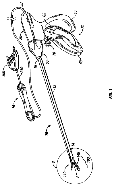
FIG. 1 is a perspective view of an electrosurgical instrument.
FIG. 2 is a side view of a handle of the surgical instrument of FIG. 1 with a half of a handle body removed to illustrate some of the components therein.
FIG. 3 is a perspective view of an electrosurgical instrument.
FIG. 4A illustrates an end effector of an electrosurgical instrument in an open configuration.
FIG. 4B illustrates the end effector of FIG. 4A in a closed configuration.
FIG. 4C is a sectional view of a translatable member shaped like an I-beam which is configured to close the end effector of the surgical instrument of FIG. 3.
FIG. 5 is a perspective view of an end effector of an electrosurgical instrument illustrated with components removed.
FIG. 6 is a top view of the end effector of FIG. 5 illustrated with components removed.
FIG. 7 is a schematic of the end effector of FIG. 5.
FIG. 8 is a partial cross-sectional view of the end effector of FIG. 5.
FIG. 9 is a diagram illustrating a temperature-resistance curve of a positive temperature coefficient material.
Corresponding reference characters indicate corresponding parts throughout the several views. The exemplifications set out herein illustrate various embodiments of the invention, in one form, and such exemplifications are not to be construed as limiting the scope of the invention in any manner.
Detailed description
Various embodiments are directed to apparatuses, systems, and methods for the treatment of tissue. Numerous specific details are set forth to provide a thorough understanding of the overall structure, function, manufacture, and use of the embodiments as described in the specification and illustrated in the accompanying drawings. It will be understood by those skilled in the art, however, that the embodiments may be practiced without such specific details. In other instances, well-known operations, components, and elements have not been described in detail so as not to obscure the embodiments described in the specification. Those of ordinary skill in the art will understand that the embodiments described and illustrated herein are non-limiting examples, and thus it can be appreciated that the specific structural and functional details disclosed herein may be representative and do not necessarily limit the scope of the embodiments, the scope of which is defined solely by the appended claims.
Reference throughout the specification to "various embodiments," "some embodiments," "one embodiment," or "an embodiment", or the like, means that a particular feature, structure, or characteristic described in connection with the embodiment is included in at least one embodiment. Thus, appearances of the phrases "in various embodiments," "in some embodiments," "in one embodiment," or "in an embodiment", or the like, in places throughout the specification are not necessarily all referring to the same embodiment. Furthermore, the particular features, structures, or characteristics may be combined in any suitable manner in one or more embodiments. Thus, the particular features, structures, or characteristics illustrated or described in connection with one embodiment may be combined, in whole or in part, with the features structures, or characteristics of one or more other embodiments without limitation.
It will be appreciated that the terms "proximal" and "distal" may be used throughout the specification with reference to a clinician manipulating one end of an instrument used to treat a patient. The term "proximal" refers to the portion of the instrument closest to the clinician and the term "distal" refers to the portion located furthest from the clinician. It will be further appreciated that for conciseness and clarity, spatial terms such as "vertical," "horizontal," "up," and "down" may be used herein with respect to the illustrated embodiments. However, surgical instruments may be used in many orientations and positions, and these terms are not intended to be limiting and absolute.
The entire disclosures of the following non-provisional United States patents are hereby incorporated by reference herein:
U.S. Pat. No. 7,381,209, entitled ELECTROSURGICAL INSTRUMENT;
U.S. Pat. No. 7,354,440, entitled ELECTROSURGICAL INSTRUMENT AND METHOD OF USE;
U.S. Pat. No. 7,311,709, entitled ELECTROSURGICAL INSTRUMENT AND METHOD OF USE;
U.S. Pat. No. 7,309,849, entitled POLYMER COMPOSITIONS EXHIBITING A PTC PROPERTY AND METHODS OF FABRICATION;
U.S. Pat. No. 7,220,951, entitled SURGICAL SEALING SURFACES AND METHODS OF USE;
U.S. Pat. No. 7,189,233, entitled ELECTROSURGICAL INSTRUMENT;
U.S. Pat. No. 7,186,253, entitled ELECTROSURGICAL JAW STRUCTURE FOR CONTROLLED ENERGY DELIVERY;
U.S. Pat. No. 7,169,146, entitled ELECTROSURGICAL PROBE AND METHOD OF USE;
U.S. Pat. No. 7,125,409, entitled ELECTROSURGICAL WORKING END FOR CONTROLLED ENERGY DELIVERY; and
U.S. Pat. No. 7,112,201, entitled ELECTROSURGICAL INSTRUMENT AND METHOD OF USE.
The entire disclosures of the following co-owned non-provisional United States patent applications filed on even date herewith are hereby incorporated by reference herein:
U.S. patent application Ser. No. 12/797,866, entitled HEAT MANAGEMENT CONFIGURATIONS FOR CONTROLLING HEAT DISSIPATION FROM ELECTROSURGICAL INSTRUMENTS;
U.S. patent application Ser. No. 12/797,861, entitled COOLING CONFIGURATIONS FOR ELECTROSURGICAL INSTRUMENTS; and
U.S. patent application Ser. No. 12/797,853, entitled ELECTROSURGICAL INSTRUMENT EMPLOYING A THERMAL MANAGEMENT SYSTEM.
Various embodiments of systems and methods of the invention relate to creating thermal "welds" or "fusion" within native tissue volumes. The alternative terms of tissue "welding" and tissue "fusion" may be used interchangeably herein to describe thermal treatments of a targeted tissue volume that result in a substantially uniform fused-together tissue mass, for example, in welding blood vessels that exhibit substantial burst strength immediately post-treatment. The strength of such welds is particularly useful for (i) permanently sealing blood vessels in vessel transection procedures; (ii) welding organ margins in resection procedures; (iii) welding other anatomic ducts wherein permanent closure is required; and also (iv) for performing vessel anastomosis, vessel closure or other procedures that join together anatomic structures or portions thereof. The welding or fusion of tissue as disclosed herein is to be distinguished from "coagulation", "hemostasis" and other similar descriptive terms that generally relate to the collapse and occlusion of blood flow within small blood vessels or vascularized tissue. For example, any surface application of thermal energy can cause coagulation or hemostasis--but does not fall into the category of "welding" as the term is used herein. Such surface coagulation does not create a weld that provides any substantial strength in the treated tissue.
At the molecular level, the phenomena of truly "welding" tissue as disclosed herein may not be fully understood. However, the authors have identified the parameters at which tissue welding can be accomplished. An effective "weld" as disclosed herein results from the thermally-induced denaturation of collagen and other protein molecules in a targeted tissue volume to create a transient liquid or gel-like proteinaceous amalgam. A selected energy density is provided in the targeted tissue to cause hydrothermal breakdown of intra- and intermolecular hydrogen crosslinks in collagen and other proteins. The denatured amalgam is maintained at a selected level of hydration---without desiccation--for a selected time interval which can be very brief. The targeted tissue volume is maintained under a selected very high level of mechanical compression to insure that the unwound strands of the denatured proteins are in close proximity to allow their intertwining and entanglement. Upon thermal relaxation, the intermixed amalgam results in protein entanglement as re-crosslinking or renaturation occurs to thereby cause a uniform fused-together mass.
A surgical instrument can be configured to supply energy, such as electrical energy, ultrasonic energy, and/or heat energy, to the tissue of a patient. For example, various embodiments disclosed herein provide electrosurgical jaw structures adapted for transecting captured tissue between the jaws and for contemporaneously welding the captured tissue margins with controlled application of RF energy. In more detail, in various embodiments, referring now to FIG. 1, an electrosurgical instrument 100 is shown. Surgical or electrosurgical instrument 100 can comprise a proximal handle 105, a distal working end or end effector 110 and an introducer or elongate shaft 108 disposed in-between. End effector 110 may comprise a set of openable-closeable jaws with straight or curved jaws--an upper first jaw 120A and a lower second jaw 120B. First jaw 120A and second jaw 120B may each comprise an elongate slot or channel 242 (see FIG. 4A), therein disposed along their respective middle portions along axis 125, for example. First jaw 120A and second jaw 120B may be coupled to an electrical source or RF source 145 and a controller 150 through electrical leads in cable 152. Controller 150 may be used to activate electrical source 145. In various embodiments, the electrical source 145 may comprise an RF source, an ultrasonic source, a direct current source, and/or any other suitable type of electrical energy source, for example.
Moving now to FIG. 2, a side view of the handle 105 is shown with half of a first handle body 106A (see FIG. 1) removed to illustrate some of the components within second handle body 106B. Handle 105 may comprise a lever arm 128 which may be pulled along a path 129. Lever arm 128 may be coupled to a movable cutting member disposed within elongate shaft 108 by a shuttle 146 operably engaged to an extension 127 of lever arm 128. The shuttle 146 may further be connected to a biasing device, such as spring 141, for example, which may also be connected to the second handle body 106B, in order to bias the shuttle 146 and thus the cutting member in a proximal direction, thereby urging the jaws 120A and 120B to an open position as seen in FIG. 1. Also, referring to FIGS. 1 and 2, a locking member 131 (see FIG. 2) may be moved by a locking switch 130 (see FIG. 1) between a locked position, where the shuttle 146 is substantially prevented from moving distally as illustrated, and an unlocked position, where the shuttle 146 may be allowed to freely move in the distal direction, toward the elongate shaft 108. The handle 105 can be any type of pistol-grip or other type of handle known in the art that is configured to carry actuator levers, triggers and/or sliders for actuating the first jaw 120A and second jaw 120B. Elongate shaft 108 may have a cylindrical or rectangular cross-section and can comprise a thin-wall tubular sleeve that extends from handle 105. Elongate shaft 108 may include a bore extending therethrough for carrying actuator mechanisms configured to actuate the jaws and/or for carrying electrical leads for delivery of electrical energy to electrosurgical components of end effector 110.
End effector 110 may be adapted for capturing, welding and transecting tissue. First jaw 120A and second jaw 120B may close to thereby capture or engage tissue therebetween. First jaw 120A and second jaw 120B may also apply compression to the tissue. Elongate shaft 108, along with first jaw 120A and second jaw 120B, can be rotated a full 360.degree. degrees, as shown by arrow 117, relative to handle 105 through one or more rotary contacts, for example. First jaw 120A and second jaw 120B can remain openable and/or closeable while rotated. Referring now to FIG. 1, end effector 110 may be coupled to electrical source 145 and controller 150. Controller 150 can regulate the electrical energy delivered by electrical source 145 which in turn delivers electrosurgical energy to electrodes within the jaws 120A, 120B. The energy delivery may be initiated by an activation button 124 operably engaged with lever arm 128 and in electrically communication with controller 150 via cable 152. As mentioned above, the electrosurgical energy delivered by electrical source 145 may comprise radiofrequency (RF) energy. As described in greater detail below, the electrodes of the jaw members may carry variable resistive positive temperature coefficient (PTC) bodies that are coupled to electrical source 145 and controller 150. Additional details regarding electrosurgical end effectors, jaw closing mechanisms, and electrosurgical energy-delivery surfaces are described in the following U.S. patents and published patent applications, all of which are incorporated herein in their entirety by reference and made a part of this specification: U.S. Pat. Nos. 7,381,209; 7,311,709; 7,220,951; 7,189,233; 7,186,253; 7,125,409; 7,112,201; 7,087,054; 7,083,619; 7,070,597; 7,041,102; 7,011,657; 6,929,644; 6,926,716; 6,913,579; 6,905,497; 6,802,843; 6,770,072; 6,656,177; 6,533,784; and 6,500,176; and U.S. Pat. App. Pub. Nos. 2010/0036370 and 2009/0076506.
FIG. 3 illustrates an electrosurgical instrument 200 comprising a handle end 205, a shaft, or introducer, 206, and an end effector, or working end, 210. Shaft 206 can comprise any suitable cross-section, such as a cylindrical and/or rectangular cross-section, for example, and can comprise a tubular sleeve that extends from handle 205. End effector 210 can extend from shaft 206 and may be adapted for welding and transecting tissue. In various embodiments, end effector 210 can comprise an openable and closeable jaw assembly which can, in various embodiments, comprise straight, curved, and/or any other suitably configured jaws. In various embodiments, the end effector 210 can comprise a first jaw 222a and a second jaw 222b, wherein at least one of the jaws 222a and 222b can move relative to the other. In at least one embodiment, the first jaw 222a can be pivoted about an axis relative to the second jaw 222b in order close onto, capture, and/or engage tissue positioned between the jaws and apply a compression force or pressure thereto. In various embodiments, the handle 205 can comprise a lever arm 228 adapted to actuate a translatable member 240 (FIG. 4A). More particularly, referring to FIGS. 4A and 4B, the lever arm 228 can be actuated in order to move member 240 distally toward the distal end 211 of end effector 210 and, when member 240 is advanced distally, member 240 can contact first jaw 222a and move it downwardly toward second jaw 222b, as illustrated in FIG. 4B. In at least one embodiment, the translatable member 240 can comprise a proximal rack portion and the lever arm 228 can comprise a plurality of gear teeth which can be configured to drive the proximal rack portion of translatable member 240 distally. In certain embodiments, rotation of the lever arm 228 in the opposite direction can drive the translatable member 240 proximally.
As described above, the translatable member 240 can be configured to contact first jaw 222a and pivot jaw 222a toward second jaw 222b. In various embodiments, referring now to FIGS. 4A-4C, the distal end of reciprocating member 240 can comprise a flanged "I"-beam configured to slide within a channel 242 in the jaws 222a and 222b. Referring primarily to FIG. 4C, the I-beam portion of member 240 can comprise an upper flange 250A, a lower flange 250B, and a center, or intermediate, portion 251 connecting the flanges 250A and 250B. In at least one embodiment, the flanges 250A and 250B and the center portion 251 can define "c"-shaped channels on the opposite sides of member 240. In any event, in various embodiments, the flanges 250a and 250b can define inner cam surfaces 252a and 252b, respectively, for slidably engaging outward-facing surfaces 262A and 262B of jaws 222a and 222b, respectively. More particularly, the inner cam surface 252A can comprise a suitable profile configured to slidably engage the outer surface 262A of first jaw 222a and, similarly, the inner cam surface 252B can comprise a suitable profile configured to slidably engage the outer surface 262B of second jaw 222b such that, as translatable member 240 is advanced distally, the cam surfaces 252A and 252B can co-operate to cam first jaw member 222a toward second jaw member 222b and configure the end effector 240 in a closed configuration. As seen in FIG. 4B, jaws 222a and 222b can define a gap, or dimension, D between the first and second electrodes 265A and 265B of jaws 222a and 222b, respectively, when they are positioned in a closed configuration. In various embodiments, dimension D can equal a distance between approximately 0.0005'' to approximately 0.005'', for example, and, in at least one embodiment, between approximately 0.001'' and approximately 0.002'', for example.
As discussed above, the translatable member 240 can be at least partially advanced in order to move the first jaw 222a toward the second jaw 222b. Thereafter, the movable member 240 can be advanced further distally in order to transect the tissue positioned between the first jaw 222a and the second jaw 222b. In certain embodiments, the distal, or leading, end of the I-beam portion of 240 can comprise a sharp, or knife, edge which can be configured to incise the tissue. Before, during, and/or after the member 240 is advanced through the tissue, electrical current can be supplied to the electrodes in the first and second jaw members in order to weld the tissue as described in greater detail further below. In various circumstances, the operation of the trigger 228 can advance the knife edge of the cutting member 240 to the very distal end of slot or channel 242. After the cutting member 240 has been sufficiently advanced, the trigger 228 can be released and moved into its original, or unactuated, position in order to retract the cutting member 240 and allow first jaw 222a to move into is open position again. In at least one such embodiment, the surgical instrument can comprise a jaw spring configured to bias the first jaw 222a into its open position and, in addition, a trigger spring configured to bias the trigger 288 into its unactuated position.
In various embodiments, further to the above, the surgical instrument can comprise a first conductor, such as an insulated wire, for example, which can be operably coupled with the first electrode 265A in first jaw member 222a and, in addition, a second conductor, such as an insulated wire, for example, which can be operably coupled with the second electrode 265B in second jaw member 222b. In at least one embodiment, referring again to FIGS. 3 and 4A, the first and second conductors can extend through shaft 206 between an electrical connector in handle 205 and the electrodes 265A and 265B in the end effector 210. In use, the first and second conductors can be operably coupled to electrical source 245 and controller 250 by electrical leads in cable 252 in order for the electrodes 265A and 265B to function as paired bi-polar electrodes with a positive polarity (+) and a negative polarity (-). More particularly, in at least one embodiment, one of the first and second electrodes 265A and 265B can be operably coupled with a positive (+) voltage terminal of electrical source 245 and the other of the first and second electrodes 265A and 265B can be electrically coupled with the negative voltage (-) terminal of electrical source 245. Owing to the opposite polarities of electrodes 265A and 265B, current can flow through the tissue positioned between the electrodes 265A and 265B and heat the tissue to a desired temperature. In certain embodiments, the cutting member 240 can act as an electrode when it is electrically coupled to a positive terminal or negative terminal of the source 245, and/or any suitable ground.
As discussed above, a surgical instrument can comprise an end effector including first and second jaws each having one or more electrodes positioned therein. As also discussed above, one or more of such electrodes can be electrically coupled to a power source and polarized such that current can flow between the electrodes. In various embodiments, referring now to FIG. 5, a surgical instrument can comprise an end effector, such as end effector 310, for example, comprising one or more movable jaws. In at least one embodiment, end effector 310 can comprise a first jaw 322a and a second jaw 322b wherein the first jaw 322a is movable relative to the second jaw 322b, although other embodiments are envisioned where the second jaw 322b is movable relative to the first jaw 322a, for example. In various embodiments, referring now to FIG. 8, the first jaw 322a can comprise a first frame 323a and a plurality of electrodes, such as electrodes 364a-364e and 365a-365e, for example, mounted thereto. More particularly, in at least one embodiment, the frame 323a can comprise a plurality of pockets 366 wherein each of the pockets 366 can be configured to receive one of electrodes 364a-364e and 365a-365e therein. In various embodiments, referring again to FIGS. 5 and 6, each of the electrodes 364a-364e and 365a-365e can comprise a mounting portion 367 positioned within a pocket 366 and, in addition, an extended portion 368 extending from the mounting portion 367. In order to electrically insulate the electrodes 364a-364e and the electrodes 365a-365e from one another, the frame 323a can be comprised of an electrically non-conductive or an at least substantially non-conductive insulative material. In addition, the electrodes 364a-364e and 365a-365e can be sufficiently spaced apart from one another such that they are not in contact with one another and/or such that current does not arc between the electrodes 364a-364e and 365a-365e during use.
In various embodiments, referring now to FIGS. 5 and 8, the second jaw 322b can comprise a second frame 323b and at least one electrode, such as return electrode 363, for example, mounted thereto. In at least one embodiment, the second jaw 322b can comprise a first electrode 363 positioned opposite the electrodes 364a-364e and a second electrode 363 positioned opposite the electrodes 365a-365e. In certain embodiments, the frame 323b can be comprised of an electrically non-conductive, or at least substantially non-conductive, insulative material while, in other embodiments, the second frame 323b can be comprised of an electrically conductive material. In use, as described in greater detail below, current can flow between the electrodes 364a-364e, 365a-365e and the return electrodes 363 when they are electrically coupled with a power source. In various embodiments, further to the above, each of the electrodes 364a-364e, 365a-365e can comprise a tissue-contacting surface 369 which can be positioned against tissue positioned intermediate the first and second jaws 322a, 322b. Similarly, the electrodes 363 can each comprise a tissue-contacting surface 362 which can also be positioned against the tissue. In various embodiments, the tissue-contacting surfaces 362 and 369 can be flat, or at least substantially flat, for example, while, in at least some embodiments, the tissue-contacting surfaces 362 and 369 can comprise one or more arcuate surfaces, for example. In certain embodiments, each of the tissue-contacting surfaces 369 can be co-planar, or at least substantially co-planar, with one another, for example, while, in at least some embodiments, the tissue-contacting surfaces 369 may be parallel, or at least substantially parallel, with one another, for example. In various embodiments, each of the tissue-contacting surfaces 362 can be co-planar, or at least substantially co-planar, with one another, for example, while, in at least some embodiments, the tissue-contacting surfaces 362 may be parallel, or at least substantially parallel, with one another, for example.
In various embodiments, further to the above, the surgical instrument can comprise a firing member, such as cutting member 340, for example, which can be moved relative to the electrodes 363, 364a-364e, and 365a-365e by a trigger, such as trigger 228, for example, in order to, one, compress the tissue-contacting surfaces of such electrodes against the tissue and, two, transect the tissue positioned intermediate the first and second jaws 322a, 322b. Similar to the above, referring now to FIG. 5, the cutting member 340 can comprise a first flange 350a which can be configured to engage the first jaw 322a and, in addition, a second flange 350b which can be configured to engage the second jaw 322b. In certain embodiments, the flanges 350a, 350b of cutting member 340 can be configured to engage the jaws 322a, 322b, respectively, and move the jaws 322a, 322b toward one another as the cutting member 340 is advanced from a proximal position to a distal position within the end effector 310. In certain embodiments, referring again to FIG. 5, the electrodes 364a-364e and the electrodes 365a-365e can define a compression channel, or slot, 359 which can be configured to receive the first flange 350a of cutting member 340 and, in addition, a cutting slot 358 defined between the electrodes 364a-364e and 365a-365e configured to receive a cutting edge 351 extending between the flanges 350a, 350b. In various embodiments, similar to the above, the second jaw 322b can comprise a compression channel, or slot, 357 configured to receive the second flange 350b and, in addition, a cutting slot 356 defined between the electrodes 363 configured to receive the cutting edge 351. In use, in at least one embodiment, the flanges 350a, 350b of cutting member 340 can directly engage the electrodes 364a-364e, 365a-365e and 363 and move the respective tissue-contacting surfaces 362 and 369 toward each other. In certain embodiments, the first flange 350a can comprise a first leading, or compression, portion 355a and the second flange 350b can comprise a second leading, or compression, portion 355b which can be positioned distally with respect to the cutting edge 351.
In various embodiments, the electrodes 364a-364e and 365a-365e can be polarized simultaneously such that current can flow from the electrodes 364a-364e and 365a-365e in the first jaw 322a to the electrodes 363 in the second jaw 322b at the same time. In at least one such embodiment, each of the electrodes 364a-364e and 365a-365e can be polarized with the same, or at least substantially the same, voltage potential while, in other embodiments, the electrodes 364a-364e and 365a-365e can be polarized with different voltage potentials. In various circumstances, the electrodes 364a-364e and 365a-365e can be polarized before the cutting member 340 is advanced relative to the electrodes. In at least some such circumstances, the current flowing between the electrodes can weld the tissue positioned between the jaws 322a, 322b before the tissue is incised. In certain circumstances, the cutting member 340 can be advanced at the same time that current is flowing between the electrodes 364a-364e, 365a-365e and electrodes 363, for example. In various embodiments, the electrodes 364a-364e and 365a-365e can be polarized sequentially. In at least one such embodiment, a first pair of electrodes comprising first electrodes 364a and 365a can be polarized by an RF source, such as RF source 145, for example, such that current can flow between the first electrodes 364a, 365a and the return electrode 363. In certain embodiments, a power source, such as a battery, for example, can be positioned within the handle of the surgical instrument, for example. When the first electrodes 364a and 365a are polarized by the RF source, in various embodiments, the electrodes 364b-364e and 365b-365e may not be polarized by the RF source. In at least one such embodiment, as a result, current may not flow between the electrodes 364b-364e, 365b-365e and electrodes 363 eventhough current may be flowing between the first electrodes 364a, 365a and the electrodes 363. In various embodiments, as described in greater detail below, an electrosurgical instrument can comprise a controller, computer, and/or any suitable microprocessor, such as controller 380 (FIG. 7), for example, which can be configured to selectively electrically couple one or more of the electrodes of end effector 310 with the RF source.
The description continues in the full USPTO document.
 Lapsed, fee not paid10 drawings