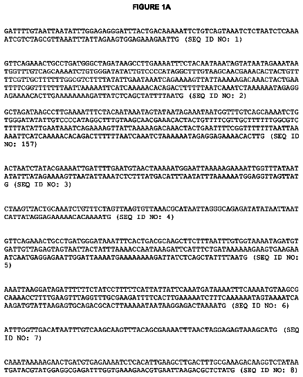
Sequence listing
A paper copy of the sequence listing and a computer readable form of the same sequence listing are appended below and herein incorporated by reference. The information recorded in computer readable form is identical to the written sequence listing, according to 37 C.F.R. 1.821 (f).
Background
1. Field of the invention
The invention is directed generally to genetic engineering methods and systems, and specifically to efficient methods and systems of gene targeting and integration of genes into a genome.
2. Description of the related art
Homologous recombination (HR) is at the heart of gene-targeting technologies. Technologies that increase the frequency of homologous recombination increase gene-targeting efficiency [1]. Applications of gene targeting technologies include gene knockouts and gene replacements in animals, cell model systems and gene therapies involving modification(s) of defective genes. Targeted homologous recombination (gene targeting) is widely used in murine embryonic stem (ES) cells as a method to ablate or introduce mutations into endogenous genes [2]. The subsequent transmission of the targeted alleles in ES cells into the mouse germ line provides a powerful method for studying gene function and has resulted in the creation of several critical mouse models for a variety of diseases [3, 4].
Development of efficient gene targeting methods for ES and somatic cells will greatly expand the use of gene replacement as a tool for researchers in their studies. Replacement of gene segment(s) known to cause disease in humans or gene ablation in human ES cells or adult somatic cells will pave the way to understanding biological processes and help in identifying causes and cures of diseases. Gene targeting in human ES and somatic cells has important applications in areas where rodent models do not adequately recapitulate human biology or disease progression. However, those applications require targeting frequencies much above the 0.1% levels achievable with today's technology. Currently, gene therapy mostly uses viral mediated approaches, which although successful, also can lead to serious complications [5]. While viral vectors provide efficient gene delivery, their main limitations are in areas of safety, in part because random integration of the vector may cause inactivation or activation of endogenous genes leading to potentially serious side effects [5, 6]. For example, in a retroviral mediated gene therapy clinical trial of ten children with X-linked SCID (IL-2 receptor .gamma. chain defect), three of the children developed acute lymphoblastic leukemia due to insertion of the vector close to the LMO2 proto-oncogene [7, 8]. Therefore, the best biological approach for gene therapy is HR.
When DNA is introduced into mammalian cells by transfection, the cell machinery integrates transfected DNA into the genome by one of two routes:
a HR pathway in which the introduced DNA replaces the endogenous genomic sequences; and
a non-homologous pathway leading to random integration. The RAD51 gene of eukaryotes is a homologue of the E. coli recA gene and plays a crucial role in HR [12, 13]. The HR pathway entails pairing of homologous DNAs, strand exchange between them, and resolution of one or more Holliday junctions. A network of interacting proteins catalyzes each step [14]. RAD51 provides the enzymatic functions for recognition of homology and DNA strand exchange in HR. RAD51 binds and polymerizes onto the introduced DNA in a step called presynapsis. In the next step, the RAD51 nucleoprotein filament searches for homologous regions in chromosomal DNA, catalyzes pairing between introduced and endogenous DNA, and promotes strand exchange [13, 15].
The amino acids at the N-terminal region of RAD51 are highly conserved among species and form a domain involved in the oligomerization of RAD51 for nucleoprotein filament formation [16]. Besides RAD51, RAD52 and Replication Protein A (RPA), together known as "the catalytic triad" of proteins, are also involved in filament formation [17]. HR is mediated by double strand breaks in the recombining DNAs and RPA binds and protects the single strand ends until they are coated with RAD51. RAD51 then forms a helical nucleoprotein filament on the single strand DNA--a process facilitated by RAD52 displacement of RPA. Genetic and biochemical studies indicate that RAD51 interacts with both RAD52 and RPA [14, 18]. Another protein involved in RAD51 function is BRCA2. The conserved BRC domains within BRCA2 bind RAD51 and it is thought that this interaction recruits RAD51 to sites of DNA damage, thereby promoting repair and/or recombination [19]. Cells transiently over-expressing either recA or RAD51 (2-fold over-expression) show some 20-fold elevation in the frequencies of HR, suggesting that efficient nucleoprotein filament formation is a rate limiting step in HR [1, 20]. The novel approach taken in this proposal to increase the frequency of HR involves covalently attaching a RAD51 binding peptide to the targeting vector so as to promote nucleation and polymerization of RAD51 onto the transfected DNA. By analogy to the function of BRCA2, the recruitment of RAD51 to the targeting DNA is expected to increase nucleoprotein filament formation and thereby HR (FIG. 1).
Three proteins, RAD52, RPA and BRCA2 have been shown to interact physically with RAD51 [19, 21, 22]. Most of the studies regarding RAD52 function come from studies done in yeast, where both genetic and molecular studies clearly demonstrate its binding to the N-terminal region of RAD51 and its role in HR and repair of double strand DNA breaks [23]. Interaction between the human RAD52 and RAD51 proteins has been demonstrated in the yeast two hybrid system, by their co-immunoprecipitation when expressed in either HeLa or insect cells, and by affinity column chromatography [21, 24, 25]. By deletion analysis, the domain within RAD52 that binds RAD51 has been mapped to amino acid residues 291-330 of the human RAD52 [21].
RPA is a heterotrimer consisting of 70, 32 and 14 kDa subunits and it is the 70 kDa subunit which binds to RAD51 [17, 22]. At sites of DNA damage, RPA binds to single strand DNA and protects the exposed DNA ends until they can be coated by RAD51. RPA also removes secondary structures that prevent extension of nucleoprotein filament formation by RAD51. NMR chemical shift mapping indicates that residues 1-93 at the N terminus of RAD51 interact with the DNA binding region of the 70 kDa subunit of RPA in a domain defined by residues 181-326 [26].
Human BRCA2 is a very large protein (3418 amino acids) but its RAD51 binding domains appear to reside within a set of eight conserved repeats (termed BRC1-8) each about 70 amino acids in length [27, 28]. Mutations within this eight-repeat region are associated with a predisposition to cancer and reduced DNA repair [29, 30]. Recent reports indicate that although the repeats are homologous, they appear to bind RAD51 at different regions of the protein. BRC3 (residues 1415-1483) binds to the N-terminal region of RAD51, whereas BRC4 (residues 1511-1579) binds to the nucleotide binding core of RAD51 located in the middle of that protein (residue 127-135:GEFRTGKT [SEQ ID NO:9] and 228-232:LLIVD [SEQ ID NO:10]) [27, 28]. That finding is important for this proposal because it suggests that a targeting vector containing both the BRC3 and BRC4 domains may recruit RAD51 in vivo much more efficiently due to cooperative binding. Furthermore, BRC5 (residues 1618-1670) does not appear to interact with RAD51, despite its homology to the other BRC repeats. The interaction of BRC repeats with RAD51 was shown using peptides (.about.69 amino acids in length) corresponding in sequence to a given BRC repeat. These studies also demonstrated that shorter peptides (.about.30 amino acids in length) corresponding to the central conserved motif of the BRC repeats were not effective for RAD51 binding [27]. This observation forms the rationale for coupling the full-length RAD51 binding domain to the targeting DNA as "bait" for the recruitment of RAD51.
Blocking DNA ends so as to prevent exonucleolytic degradation has been shown to reduce unwanted random integration, and in Dictyostelium discoideum, to increase site-specific targeting events [31]. According to the present invention, the ends of the targeting DNA are blocked by ligation of a hairpin oligonucleotide so as to eliminate free 3' or 5' ends. Another avenue for increased gene targeting is based on more effective gene delivery to the nucleus where HR takes place. Several attempts to improve the entry of plasmid DNA into the nucleus have been reported including "piggyback" techniques like electrostatic binding of DNA to cationic proteins containing a nuclear localization signal (NLS) [32, 33], NLS-containing peptides [34], lipids [35] and karyophilic proteins [36, 37]. A major drawback of this "piggyback" nuclear transport is that it relies upon the unpredictable stability of the complex in the cytoplasm [35]. In our case, we will covalently couple an NLS-containing peptide to one end of the targeting DNA. Interestingly, DNAs tagged with a single NLS-peptide show enhanced delivery to the nucleus, but the presence of more than one NLS-peptide tag on a DNA molecule prevents gene delivery. This observation suggests that a DNA molecule with two NLS tags threads through two adjacent nuclear pores in a manner that leads to entrapment of the DNA at the nuclear membrane and consequent decreased gene delivery into the nucleus [35].
Targeting of a gene to the HPRT locus is desirable for gene therapy not only because it avoids the random insertional mutagenesis associated with viral mediated gene delivery, but also because extensive experience exists relevant to targeting genes to that locus. The HPRT gene is expressed in all cells and during all stages of development and is a locus that constitutively remains in an open chromatin configuration. Transgenic mice expressing human angiotensinogen from a gene targeted to the HPRT locus showed normal tissue expression and functionality at physiological levels [38].
The protein encoded by the HPRT gene (nine exons spread over a 33 Kb region of the X chromosome) is involved in the salvage pathway of nucleotide metabolism [9]. Cells with functional HPRT incorporate the nucleotide analogue 6-thioguanine (6-TG) into DNA, which leads to cell death. In the absence of HPRT, there is no incorporation of 6-TG into the nucleotide pool, thus, HPRT disrupted cells survive in the presence of 6-TG [9]. The HPRT locus has been used as a target for the study of various aspects of HR. In one study, the influence of homology length in the targeting vector and its targeting efficiency was compared using the HPRT locus. Deng and Capecchi [39] demonstrated similar targeting frequencies in vectors of different homology lengths. Zhang et al [40] evaluated HR frequencies as a function of the endogenous size of the deletion region that occurs upon insertion of targeting vectors. Hatada et al used targeting to the HPRT locus to show that HR frequencies are similar in ES cells versus hematopoietic progenitor cells [11].
Since the HPRT targeting vector contains a neomycin resistance marker, both random and targeted integration events will confer resistance to the antibiotic G418. However, when cells are grown in the presence of G418 plus 6-TG, only cells with a targeted disruption of the HPRT locus will survive. Therefore, there are two ways recombination frequencies can be expressed:
targeted cells (those that are G418 and 6-TG resistant) divided by the total number of cells subjected to selection provides the overall recombination frequency; and
targeted cells (those that are G418 and 6-TG resistant) divided by the number of G418 resistant cells (i.e., both random and targeted recombination events) provides the targeted recombination frequency. Both methods of expression of recombination frequency appear in the literature. For example, a study using murine ES-D3 cells for HPRT gene targeting had overall recombination frequencies of 0.4.times.10.sup.-6 and 1.6.times.10.sup.-6 in two different experiments with targeted recombination frequencies of 1.3.times.10.sup.-5 and 5.3.times.10.sup.-5 respectively [41]. Whereas the targeted recombination frequency reports on the ratio of site-specific to random integration events, the overall recombination frequency reports on the total number of targeted integration events in the cell population. The invention uses both measures to evaluate the effects of the proposed modification to the targeting vector on HR. So, for example, we expect that attaching an NLS-signal peptide to the targeting vector will increase the overall recombination frequency but not necessarily site-specific targeting. On the other hand, vectors with an attached peptide bait that binds RAD51 should increase both site-specific targeting--i.e., increase the targeted recombination frequency--as well as the overall recombination frequency.
Current gene therapy using viruses has advantages, such as efficient gene delivery, but their main limitations are in the areas of safety and random integration events causing inactivation or activation of endogenous genes [6, 8, 42, 43]. The best biological approach for gene therapy therefore is HR where one either replaces the defective region of a gene with its normal counterpart or expresses a normal gene at a known locus thereby avoiding random insertional mutagenesis. Development of a method for efficient gene targeting will allow for rapid advancements in the creation of cells or cell lines containing modified genes for the purpose of studying the biological function of specific genes. Drawbacks to the use of HR in mammalian cells for gene targeting purposes are its inherent inefficiency and relatively low frequency of targeted integration.
Cited references
The follow references are cited throughout this disclosure. They are incorporated herein in their entirety by reference. They are used for illustration purposes. Applicant reserves the right to challenge the veracity of any statement made therein.
[1] S. Vispe, C. Cazaux, C. Lesca and M. Defais, Overexpression of Rad51 protein stimulates homologous recombination and increases resistance of mammalian cells to ionizing radiation. Nucleic Acids Res 26, 2859-2864 (1998).
[2] M. R. Capecchi, Altering the genome by homologous recombination. Science 244, 1288-1292 (1989).
[3] T. E. Willnow and J. Herz, Homologous recombination for gene replacement in mouse cell lines. Methods Cell Biol 43 Pt A, 305-334 (1994).
[4] S. Thompson, A. R. Clarke, A. M. Pow, M. L. Hooper and D. W. Melton, Germ line transmission and expression of a corrected HPRT gene produced by gene targeting in embryonic stem cells. Cell 56, 313321 (1989).
[5] Y. Yi, S. H. Hahm and K. H. Lee, Retroviral gene therapy: safety issues and possible solutions. Curr Gene Ther 5, 25-35 (2005).
[6] S. Laufs, B. Gentner, K. Z. Nagy, A. Jauch, A. Benner, S. Naundorf, K. Kuehlcke, B. Schiedlmeier, A. D. Ho, W. J. Zeller and S. Fruehauf, Retroviral vector integration occurs in preferred genomic targets of human bone marrow-repopulating cells. Blood 101, 2191-2198 (2003).
[7] D. B. Kohn, M. Sadelain and J. C. Glorioso, Occurrence of leukaemia following gene therapy of X-linked SCID. Nat Rev Cancer 3, 477-488 (2003).
[8] H. B. Gaspar and A. J. Thrasher, Gene therapy for severe combined immunodeficiencies. Expert Opin Biol Ther 5, 1175-1182 (2005).
[9] D. W. Melton, D. S. Konecki, J. Brennand and C. T. Caskey, Structure, expression, and mutation of the hypoxanthine phosphoribosyltransferase gene. Proc Natl Acad Sci USA 81, 2147-2151 (1984).
[10] S. K. Bronson, E. G. Plaehn, K. D. Kluckman, J. R. Hagaman, N. Maeda and O. Smithies, Single-copy transgenic mice with chosen-site integration. Proc Natl Acad Sci USA 93, 9067-9072 (1996).
[11] S. Hatada, K. Nikkuni, S. A. Bentley, S. Kirby and O, Smithies, Gene correction in hematopoietic progenitor cells by homologous recombination. Proc Natl Acad Sci USA 97, 13807-13811 (2000).
[12] T. Ogawa, A. Shinohara, A. Nabetani, T. Ikeya, X. Yu, E. H. Egelman and H. Ogawa, RecA-like recombination proteins in eukaryotes: functions and structures of RAD51 genes. Cold Spring Harb Symp Quant Biol 58, 567-576 (1993).
[13] P. Baumann and S.C. West, Role of the human RAD51 protein in homologous recombination and double-stranded-break repair. Trends Biochem Sci 23, 247-251 (1998).
[14] D. Jackson, K. Dhar, J. K. Wahl, M. S. Wold and G. E. Borgstahl, Analysis of the human replication protein A:Rad52 complex: evidence for crosstalk between RPA32, RPA70, Rad52 and DNA. J Mol Biol 321, 133-148 (2002).
[15] J. M. Stark, P. Hu, A. J. Pierce, M. E. Moynahan, N. Ellis and M. Jasin, ATP hydrolysis by mammalian RAD51 has a key role during homology-directed DNA repair. J Biol Chem 277, 20185-20194 (2002).
[16] K. A. Miller, D. Sawicka, D. Barsky and J. S. Albala, Domain mapping of the Rad51 paralog protein complexes. Nucleic Acids Res 32, 169-178 (2004).
[17] M. S. Park, D. L. Ludwig, E. Stigger and S. H. Lee, Physical interaction between human RAD52 and RPA is required for homologous recombination in mammalian cells. J Biol Chem 271, 18996-19000 (1996).
[18] T. Sugiyama and S. C. Kowalczykowski, Rad52 protein associates with replication protein A (RPA)single-stranded DNA to accelerate Rad51-mediated displacement of RPA and presynaptic complex formation. J Biol Chem 277, 31663-31672 (2002).
[19] B. J. Orelli and D. K. Bishop, BRCA2 and homologous recombination. Breast Cancer Res 3, 294-298 (2001).
[20] O. G. Shcherbakova, V. A. Lanzov, H. Ogawa and M. V. Filatov, Overexpression of bacterial RecA protein stimulates homologous recombination in somatic mammalian cells. Mutat Res 459, 65-71 (2000).
[21] Z. Shen, K. G. Cloud, D. J. Chen and M. S. Park, Specific interactions between the human RAD51 and RAD52 proteins. J Biol Chem 271, 148-152 (1996).
[22] E. I. Golub, R. C. Gupta, T. Haaf, M. S. Wold and C. M. Radding, Interaction of human rad51 recombination protein with single-stranded DNA binding protein, RPA. Nucleic Acids Res 26, 5388-5393 (1998).
[23] Y. Aylon and M. Kupiec, New insights into the mechanism of homologous recombination in yeast. Mutat Res 566, 231-248 (2004).
[24] J. H. New, T. Sugiyama, E. Zaitseva and S. C. Kowalczykowski, Rad52 protein stimulates DNA strand exchange by Rad51 and replication protein A. Nature 391, 407-410 (1998).
[25] T. Ogawa, A. Shinohara and T. Ikeya, A species-specific interaction of rad51 and rad52 proteins in eukaryotes. Adv Biophys 31, 93-100 (1995).
[26] M. E. Stauffer and W. J. Chazin, Physical interaction between replication protein A and Rad51 promotes exchange on single-stranded DNA. J Biol Chem 279, 25638-25645 (2004).
[27] V. E. Galkin, F. Esashi, X. Yu, S. Yang, S. C. West and E. H. Egelman, BRCA2 BRC motifs bind RAD51DNA filaments. Proc Natl Acad Sci USA 102, 8537-8542 (2005).
[28] M. K. Shivji, O. R. Davies, J. M. Savill, D. L. Bates, L. Pellegrini and A. R. Venkitaraman, A region of human BRCA2 containing multiple BRC repeats promotes RAD51-mediated strand exchange. Nucleic Acids Res 34, 4000-4011 (2006).
[29] A. Tutt and A. Ashworth, The relationship between the roles of BRCA genes in DNA repair and cancer predisposition. Trends Mol Med 8, 571-576 (2002).
[30] C. F. Chen, P. L. Chen, Q. Zhong, Z. D. Sharp and W. H. Lee, Expression of BRC repeats in breast cancer cells disrupts the BRCA2-Rad51 complex and leads to radiation hypersensitivity and loss of G(2)/M checkpoint control. J Biol Chem 274, 32931-32935 (1999).
[31] N. Shah-Mahoney, T. Hampton, R. Vidaver and D. Ratner, Blocking the ends of transforming DNA enhances gene targeting in Dictyostelium. Gene 203, 33-41 (1997).
[32] Y. Kaneda, K. Iwai and T. Uchida, Increased expression of DNA cointroduced with nuclear protein in adult rat liver. Science 243, 375-378 (1989).
[33] J. D. Fritz, H. Herweijer, G. Zhang and J. A. Wolff, Gene transfer into mammalian cells using histone condensed plasmid DNA. Hum Gene Ther 7, 1395-1404 (1996).
[34] P. Collas, H. Husebye and P. Alestrom, The nuclear localization sequence of the SV40 T antigen promotes transgene uptake and expression in zebrafish embryo nuclei. Transgenic Res 5, 451-458 (1996).
[35] M. A. Zanta, P. Belguise-Valladier and J. P. Behr, Gene delivery: a single nuclear localization signal peptide is sufficient to carry DNA to the cell nucleus. Proc Natl Acad Sci USA 96, 91-96 (1999).
[36] J. Fominaya and W. Wels, Target cell-specific DNA transfer mediated by a chimeric multidomain protein. Novel non-viral gene delivery system. J Biol Chem 271, 10560-10568 (1996).
[37] D. A. Dean, Peptide nucleic acids: versatile tools for gene therapy strategies. Adv Drug Deliv Rev 44, 81 95 (2000).
[38] B. Cvetkovic, B. Yang, R. A. Williamson and C. D. Sigmund, Appropriate tissue- and cell-specific expression of a single copy human angiotensinogen transgene specifically targeted upstream of the HPRT locus by homologous recombination. J Biol Chem 275, 1073-1078 (2000).
[39] C. Deng and M. R. Capecchi, Reexamination of gene targeting frequency as a function of the extent of homology between the targeting vector and the target locus. Mol Cell Biol 12, 3365-3371 (1992).
[40] H. Zhang, P. Hasty and A. Bradley, Targeting frequency for deletion vectors in embryonic stem cells. Mol Cell Biol 14, 2404-2410 (1994).
[41] T. Doetschman, N. Maeda and O, Smithies, Targeted mutation of the Hprt gene in mouse embryonic stem cells. Proc Natl Acad Sci USA 85, 8583-8587 (1988).
[42] E. Check, Cancer fears cast doubts on future of gene therapy. Nature 421, 678 (2003).
[43] C. Baum, J. Dullmann, Z. Li, B. Fehse, J. Meyer, D. A. Williams and K. C. von, Side effects of retroviral gene transfer into hematopoietic stem cells. Blood 101, 2099-2114 (2003).
[44] D. R. Greaves, P. Fraser, M. A. Vidal, M. J. Hedges, D. Ropers, L. Luzzatto and F. Grosveld, A transgenic mouse model of sickle cell disorder. Nature 343, 183-185 (1990).
[45] A. Mortellaro, R. J. Hernandez, M. M. Guerrini, F. Carlucci, A. Tabucchi, M. Ponzoni, F. Sanvito, C. Doglioni, S. C. Di, L. Biasco, A. Follenzi, L. Naldini, C. Bordignon, M. G. Roncarolo and A. Aiuti, Ex vivo gene therapy with lentiviral vectors rescues adenosine deaminase (ADA)-deficient mice and corrects their immune and metabolic defects. Blood 108, 2979-2988 (2006).
[46] S. Sekhsaria, J. I. Gallin, G. F. Linton, R. M. Mallory, R. C. Mulligan and H. L. Malech, Peripheral blood progenitors as a target for genetic correction of p47phox-deficient chronic granulomatous disease. Proc Natl Acad Sci USA 90, 7446-7450 (1993).
[47] S. I. Grewal and A. J. Klar, A recombinationally repressed region between mat2 and mat3 loci shares homology to centromeric repeats and regulates directionality of mating-type switching in fission yeast. Genetics 146, 1221-1238 (1997).
[48] X. Liu and D. F. Barker, Evidence for effective suppression of recombination in the chromosome 17q21 segment spanning RNU2-BRCA1. Am J Hum Genet 64, 1427-1439 (1999).
[49] A. K. Wong, R. Pero, P. A. Ormonde, S. V. Tavtigian and P. L. Bartel, RAD51 interacts with the evolutionarily conserved BRC motifs in the human breast cancer susceptibility gene brca2. J Biol Chem 272, 31941-31944 (1997).
[50] T. A. Tatusova and T. L. Madden, BLAST 2 Sequences, a new tool for comparing protein and nucleotide sequences. FEMS Microbiol Lett 174, 247-250 (1999).
[51] C. Gauthier-Rouviere, M. Vandromme, N. Lautredou, Q. Q. Cai, F. Girard, A. Fernandez and N. Lamb, The serum response factor nuclear localization signal: general implications for cyclic AMP-dependent protein kinase activity in control of nuclear translocation. Mol Cell Biol 15, 433-444 (1995).
Summary of the invention
The inventor has succeeded in developing methods and systems for high homologous recombination ("HR") targeting efficiency with relative technical ease of use. The methods and systems comprise the use of and inclusion of (respectively) targeting vectors that comprises (a) an insertion polynucleotide sequence encoding a gene-product-of-interest (e.g., an enzyme for gene therapy; a selectable marker such as neomycin resistance [Neo.sup.r]; gene-product-of-interest is not limiting in its scope beyond genes, thus it can be any polynucleotide, polypeptide, regulatory sequence, intron, exon, RNAi locus, et cetera), (b) a proximal target polynuceotide, which has homology to a proximal portion of a target polynucleotide sequence, (c) and a distal target polynucleotide, which has homology to the distal portion of the target polynucleotide sequence, (d) a nuclear localization signal ("NLS") sequence, which allows the entire targeting vector to enter the nucleus of a transfected cell. In a preferred embodiment, the ends of the targeting vector are blocked to prevent exonucleolysis. Preferred blocking groups are hair pin loops of nucleic acid, but other means may be used in the practice of the invention. In a more preferred embodiment, the targeting vector comprises a bait peptide attached to the end of the targeting vector that is distal to the end to which the NLS is attached. The bait peptide is an amino acid sequence that enables the targeting vector to bind to the homology recognition machinery (which includes the catalytic triad [supra]) of the cell. Preferred bait peptides recognize RAD51 (recA). More preferred bait peptides comprise or consist of, or consist essentially of peptides having a sequence that is at least 70% identical to SEQ ID NOs. 1-3. In yet another aspect, the targeting vector may comprise additional bait polypeptides, to enhance cooperativity and even greater efficiency of homologous recombination. Bait peptides can be a "universal" bait that works in a variety of systems, or species-specific bait polypeptides.
Thus the invention is directed to a targeting vector for efficient homologous recombination ("HR"). In another embodiment, the invention is directed to a kit, which comprises an hp-peptide ready for ligation to targeting vectors for the purpose of efficient targeting. The kit further comprises instructions, buffers and enzymes required for the construction of a custom end-user targeting vector. In yet another embodiment, the invention is directed to an efficient non-viral targeting method to create vectors and cells for gene therapy. The method is also applicable to engineer "humanized" mice, that is, mice in which a human gene has been "knocked in" to replace the corresponding mouse gene; to target the animal genome with known modifications is of interest to individuals in the cattle and dairy as well as other animal-related industries; and other applications for directed gene insertion.
The invention herein discloses improved HR by increasing HR frequency at the HPRT locus so that the HPRT locus may be used as a universal docking site for gene therapy. However, the invention shall not be limited to dropping in genes at the HPRT site. The HPRT site is useful as an example and as a handy "universal" target site. For example, in hematopoietic monogenic diseases such as sickle cell anemia [44], SCID (due to deaminase deficiency) [45] and chronic granulomatous disease (due to defective NADP oxidase) [46], according to this invention, expression of the respective normal gene at the HPRT locus is reasonably expected to significantly improve disease symptoms. Bone marrow or hematopoietic cells are isolated from a patient and used in vitro for targeting of the normal functioning gene to the HPRT docking site. Targeted cells are then selected by growth in 6-TG, and after expansion, transferred back into the patient. Mouse models for single gene diseases (such as SCID and NADP oxidase deficiency) can be used to test the feasibility of autologous gene targeting at the HPRT locus.
Another non-limiting application for the present invention is in targeting genes to genetic loci that are refractory to HR. If the bait present on the targeting vector shows an increase in HR frequency at the HPRT locus, then that bait may also be useful in gene targeting to loci that are usually refractory for HR. For example, genes located in repeat scattered regions or close to centromeric regions do not respond well to gene targeting strategies since HR in repeat regions is suppressed [47, 48]. This HR suppression may be the reason that certain mouse models are difficult to make by currently available gene targeting methodologies. An embodiment of the instant invention is a commercial targeting kit that contains two hairpin oligonucleotides, one with the NLS-peptide and the other with the bait peptide. All that will remain for the consumer in the practice of the invention is the ligation of those peptide-containing oligonucleotides to a linear targeting vector followed by transfection.
Brief description of the drawings
FIG. 1. Diagram for the enhancement of homologous recombination at the HPRT locus in ES cells by attachment of a bait peptide to the targeting vector. The bait peptide binds RAD51 thereby enhancing nucleation and polymerization of RAD51 onto the targeting DNA. A peptide containing a nuclear localization signal (NLSp) enhances trafficking of the vector into the nucleus. The homologous recombinant is resistant to cell death by 6-thioguanine because it lacks HPRT enzymatic activity and is resistant to G418 because of the chromosomal integration of neomycin resistant gene (Neo) carried in the targeting vector.
FIG. 2. Structure of the HPRT targeting vector. The 5' and 3' genomic regions that flank the HPRT promoter-exon 1 region are represented in the vector. The neomycin resistance gene (NEO) contained within the targeting vector is driven by the PGK1 promoter. To the ends of the targeting vector are ligated hairpin oligonucleotides via sticky ends created by FseI (left) and NotI (right) digestion of the targeting DNA. Hairpin oligonucleotides contain an alkylamino modified T residue for coupling to the various peptides discussed in the text. Peptides are ligated to hairpin oligonucleotides before their ligation to the targeting vector.
FIG. 3. Gene Targeting at the HPRT locus. Homologous recombination mediated at the HPRT genomic locus results in the deletion of the .about.4 kbp-promoter exon 1 region (green box) with concomitant replacement by the .about.2 kbp neomycin cassette (NEO). The crosses (x) between the targeting vector and the genomic locus depict homologous recombination. The expected DNA fragment sizes after HindIII (H) digestion of wild type versus recombinant DNA are shown (10.2 kbp and 8.2 kbp, respectively). The position of a 5' probe to detect wild-type versus recombinant HindIII digestion products by Southern analysis is depicted by short box in bold.
FIG. 4. Screening strategy for homologous recombination at the HPRT locus. HindIII digestion of wild-type (wt) genomic DNA produces a 10.2 kbp DNA fragment (lane 1), whereas HindIII digestion of recombinant 10.2 Kbp (Rec) DNA resulting from gene targeting at the HPRT locus produces an 8.2 Kbp 8.2 kb DNA fragment (lane 2). Arrows point to bands of 10.2 kbp and 8.2 kbp sizes. F9 embryonic carcinoma cells used in this experiment were of normal karyotype with respect to XY chromosomes. Since the HPRT gene is located in the X chromosome, only an 8.2 kbp band is seen in the Southern blot.
FIG. 5. Structure of control and bait vectors. The hairpin oligonucleotide in red is ligated to the NotI end of the vectors, and the hairpin oligonucleotide in green is ligated to the FseI end of the vectors. All vectors have a nuclear localization signal peptide (NLSp-11 amino acids in length) ligated to the NotI end. All bait vectors have bait peptides ligated to the FseI end. BRC3, BRC4, and RAD52 indicate peptides derived from BRCA2 and RAD52 that bind RAD51. BRC5 indicates a peptide derived from BRCA2 which does not bind RAD51. All bait peptides are between 52-69 amino acids in length.
Description of a preferred embodiment of the invention
The invention is directed to kits, vectors and methods for high HR targeting efficiency with technical ease of performance. Since several studies have reported successful targeting in mouse ES cells, those cell types are used to exemplify in a non-limiting way, the practice of this invention. HR frequency is generally assessed by targeting to the hypoxanthine phosphoribosyl transferase (HPRT) locus [9]. The HPRT locus offers several advantages as a target docking site for gene therapy applications [10, 11]:
Its open chromatin configuration ensures expression of genes integrated there;
Since HPRT enzymatic activity is dispensable (HPRT knockout mice display normal phenotypes), integration of a targeted gene into that locus is not in and of itself deleterious; and
Powerful selection methods are available to select for and isolate cells having the targeted gene integrated at the HPRT locus--and likewise to select against integration events outside of that locus. HPRT functions in the salvage nucleotide pathway. Cells with a mutated HPRT gene survive when grown in the presence of the nucleotide analogue, 6-thioguanine (6-TG), but cells with a wild-type HPRT do not. Thus, growth in 6-TG selects for cells with HPRT-specific gene integration and against cells with gene integration elsewhere. If the targeted gene cassette also includes the neomycin resistance gene, then growth in G418 (a neomycin analogue) may also be utilized to first select for cells with an integrated target gene before negative selection using 6-TG. Therefore increasing HR frequencies at the HPRT locus can make this locus a universal docking site for chromosomal integration of a gene of interest.
While not wishing to be limited by theory, the invention is based upon the concept that HR frequencies can be greatly increased by using targeting vectors that are modified so as to contain two different peptides that in concert function to promote HR. The first peptide consists of a nuclear localization signal which increases the amount of transfected DNA trafficked into the nucleus. The second peptide acts to bind and recruit onto the targeting DNA RAD51 (the mammalian analogue of the bacterial recA protein), thereby promoting sequence-specific synapsis between the targeting DNA and homologous chromosomal sequences, and in turn HR.
The inventor has established an experimental system to assess frequency of recombination in a linear targeting vector with modified ends, which clearly demonstrated that a targeting vector with one end covalently attached to a nuclear localization peptide (NLSp) has an increased frequency of HR. A reasonable explanation for the increased HR frequency is that the targeting vector with NLSp enters the nucleus more frequently than does the control thereby making more substrate available in the nucleus for use by the HR machinery.
The following examples provided the current best mode and preferred embodiment of the instant invention. They are meant to illustrate the invention and not meant to limit the scope of the invention. The skilled artisan in the practice of this invention will readily recognize that other vectors, constructs, cell lines, and HR sites can be used while remaining within the spirit and scope of the invention, which is set forth in the claims which follow.
Example 1
Targeting Vectors
Modified targeting vectors contain hairpin oligonucleotides on each end with or without attached peptides. Briefly, hairpin oligonucleotides are made with a central T nucleotide containing an alkyamino group to which peptides are attached by a chemical method. The peptide NLSp was made with a C terminal cysteine. The amino group in the hairpin region is linked to the peptide containing a C terminal cysteine by a heterobifunctional cross linker. Before ligation, the hairpin structure of the oligonucleotide presenting a 5' sticky end was obtained by boiling and cooling on ice. The hairpin was ligated to the ends of the vector containing the corresponding sticky ends of the restriction site (FIG. 2). The reason for blocking the linear ends of the vector with hairpin oligonucleotides is to prevent the linear plasmid from forming concatemeric structures which will lead to spurious modification at the targeting locus. Additionally, blocking ends of a targeting vector has been suggested to help in preventing the targeting vector from entering into the nonhomologous recombination pathway [31]
HPRT Targeting Vector Construction
Modified pUC 19 plasmids have been made and used as the backbone for target vector construction. Plasmid pUC19 was digested by AatII and AflII restriction enzymes. This digestion produced two DNA fragments: 1) a 0.875 Kb fragment that contains the polylinker cloning site and lacZ gene of pUC19; and 2) a 1.81 Kb fragment which contains the .beta.-lactamase gene and the origin of replication. To the 1.81 Kb DNA fragment, several restriction sites were added by sequential ligation of oligolinkers thereby creating plasmid pUT1. FIG. 2 shows the HPRT targeting vector. The pUT1 backbone was used to construct the targeting vector. To make the targeting vector, the 5' and 3' homologous regions flanking the 5' region of the promoter (3.5 Kb, red line) and the 3' region of exon 1 (4.8 Kb, blue line) were used as a homologous arm in the targeting vector (FIG. 2). Since transcription of the HPRT gene requires the promoter region, the targeting vector was designed such that HR of this vector at the HPRT locus eliminates the promoter-exon 1 region thereby completely eliminating HPRT gene expression. The 5' XbaI fragment (3.5 Kbp) was cloned into a pUT1 plasmid at the Xba1 site and 3' DNA fragment EcoRV-EcoRI (4.8 Kbp) subjected to a fill-in reaction and cloned at the Ec1136II site. At the EcoRV site of pUT1 containing 5' and 3' arms, a Neo.sup.r selection gene was cloned. The presence of the phosphoglycerate kinase 1 (PGK1) promoter allows for constitutive expression of the Neo.sup.r gene.
HPRT Targeting Vector Modification
The description continues in the full USPTO document.
 Lapsed, fee not paid2 drawings