Field of the invention
The present invention relates to methods, compositions and articles of manufacture for contributing to the treatment of cancers including solid tumors through administration of an endothelin agonist and a chemotherapeutic agent.
Background of the invention
Successful treatment of cancers, including solid tumors, remains an unfulfilled medical goal, despite increased understanding of the molecular biology of tumor cells and the availability of an increased number of potential therapeutic agents. For example, breast cancer incidence has increased substantially in the last 10 years, and is the single leading cause of death for women ages 40-49 years in the United States.
One problem in the treatment of cancers is that an effective dose of a wide variety of potential chemotherapeutic agents is restricted by these agents' non-selective, highly toxic effect on normal tissues. As a result, many patients suffer from the side effects of chemotherapy without reaping the benefits of the treatment. For example, the chemotherapeutic agent paclitaxel inhibits cellular proliferation and induces apoptosis of tumor cells. The clinical utility of paclitaxel has been hampered, however, by its dose limiting toxicities including hypersensitivity, neutropenia and peripheral neuropathy. Thus, there is a necessity to develop more specific and less toxic cancer therapies.
Targeted delivery of chemotherapeutic agents to tumors could have the advantage of enhancing the benefit of chemotherapeutic agents while minimizing their systemic toxic effects. Such targeted delivery could also serve to lower the required dose of chemotherapeutic agents thus potentially reducing the unacceptable adverse effects of these agents. One possible way to achieve targeted delivery of chemotherapeutic agents is to utilize the distinctive features of tumor vasculature.
Tumors greater than a few millimeters in size require a constant nutrient supply, and, therefore, develop their own vascular bed and blood flow. Folkman, Cancer Res, 46:467 (1986). Without constant nourishment from these developing blood vessels, the tumors become hypoxic and subsequently die. Recruitment of new vasculature from preexisting blood vessels is termed "angiogenesis."
During angiogenesis, tumor blood vessels develop substantially differently from normal vasculature, and have different properties. Single layered epithelial cells are the first hastily formed tumor blood vessels. These newly formed tumor blood vessels do not have a smooth muscle layer or innervation. Tumors also incorporate mature blood vessels that possess all their autoregulatory functions. Mattsson et al., Tumor Blood Circulation, CRC Press, Boca Raton, pg. 129 (1979); Reinhold, Tumor Blood Circulation, CRC Press, Boca Raton, pg. 115 (1979); Warren, Tumor Blood Circulation, CRC Press, Boca Raton, pg. 26 (1979).
Vascular tone (the degree to which blood vessels are dilated or constricted) is governed by a host of endogenous factors including H.sup.+, K.sup.+, Ca.sup.2+, pO.sub.2, pCO.sub.2 and nitric oxide (NO), as well as other regulatory substances such as endothelin (ET-1). Secombe et al., Landes, Austin, pg. 40 (1994); Luscher et al., The endothelium: modulator of cardiovascular function, CRC Press, Boca Raton, pg. 61 (1990). ET-1 contributes significantly to regulating vascular tone (Yanagisawa et al., Nature, 332:411 (1988)) and investigators have shown an increase in ET1 and ET.sub.B receptor expression in solid tumors including breast carcinomas. Alanen et al., Histopathology, 36:161 (2000); Nelson et al., Cancer Res, 56:663 (1996); Kar et al., Biochem Biophys Res Commun 216:514 (1995); Pagotto et al., J Clin Invest, 96:2017 (1995); Yamashita et al., Cancer Res, 52:4046 (1992); Yamashita et al., Res Commun Chem Pathol Pharmacol, 74:363 (1991). Further, stimulation of ET.sub.B receptors causes an increase in blood supply to tumors through vasodilation of tumor blood vessels. The present invention takes advantage of this fact by using ET.sub.B receptor agonists to selectively increase blood flow to tumors to enhance the targeted delivery of chemotherapeutic agents.
Summary of the invention
The present invention is directed to the administration of endothelin agonists and a chemotherapeutic agent to contribute to the treatment of cancers including solid tumors. In particular, tumors have distinctive vasculature including an increased number of ET.sub.B receptors which, when bound, cause vasodilation. Because ET.sub.B receptors are vasodilators, an ET.sub.B receptor agonist, in combination with a chemotherapeutic agent, is useful in the treatment of a solid tumor, such as those found in breast cancers. The ET.sub.B receptor agonist can more effectively deliver chemotherapeutic agents to tumors resulting in enhanced treatment.
Specifically, one embodiment according to the present invention includes a method of contributing to the treatment of a cancer comprising administering an ET.sub.B agonist and a chemotherapeutic agent. In various embodiments of the methods according to the present invention, the ET.sub.B agonist and the chemotherapeutic agent can be administered substantially simultaneously or can be administered sequentially (with the chemotherapeutic agent administered prior to the ET.sub.B agonist or the ET.sub.B agonist administered prior to the chemotherapeutic agent). In certain embodiments according to the present invention when the ET.sub.B agonist and the chemotherapeutic agent are administered substantially simultaneously, they can be administered as a single composition.
Another embodiment according to the present invention includes a composition comprising a chemotherapeutic agent, an ET.sub.B agonist, and an optional excipient. Another embodiment according to the present invention includes an article of manufacture comprising a composition comprising an ET.sub.B agonist, and instructional information directing the administration of the composition with a chemotherapeutic agent to treat a solid tumor. Articles of manufacture according to the present invention can further comprise one or more chemotherapeutic agents. When articles of manufacture according to the present invention include one or more chemotherapeutic agents, the ET.sub.B agonist and the chemotherapeutic agent can be part of the same composition, can be provided as separate compositions, or both.
Cancers that are treated with the methods, compositions or articles of manufacture according to the present invention can include solid tumors including, without limitation, ovarian tumors, colon tumors, Kaposi's sarcoma, breast tumors, melanoma, prostate tumors, meningiomas, liver tumors, breast phyllode tumors and combinations thereof.
Endothelin B agonists used in accordance with the methods, compositions or articles of manufacture of the present invention can selectively increase blood supply to solid tumors thus increasing the delivery of chemotherapeutic agents to the solid tumor. Endothelin B agonists that can be used in accordance with the present invention can include, without limitation, one or more of ET-1, ET-2, ET-3, BQ3020, IRL1620 (N-suc-[Glu9, Ala11, 15]ET-1 (8-21)), sarafotoxin 56c, [Ala1, 3, 11, 15]ET-1, and combinations thereof. Chemotherapeutic agents can include, without limitation, one or more of adriamycin, camptothecin, carboplatin, cisplatin, daunorubicin, doxorubicin, alpha interferon, beta interferon, gamma interferon, interleukin 2, irinotecan, docetaxel, paclitaxel, topotecan, 5-fluorouracil, and combinations thereof. Particular methods, compositions or articles of manufacture according to the present invention will include IRL1620 as the ET.sub.B agonist with a chemotherapeutic agent selected from the group consisting of paclitaxel, doxorubicin, 5-fluorouracil, and combinations thereof.
Brief description of the drawings
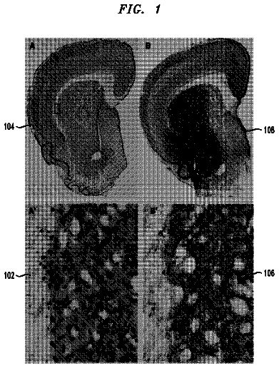
FIG. 1 shows the effect of IRL1620 on paclitaxel-induced changes in tumor perfusion;
FIGS. 2A-2E show the effect of ET-1 on systemic hemodynamics of cancer-free and breast tumor-bearing rats;
FIGS. 3A-3B show the effect of ET-1 on blood flow and regional vascular resistance in the breast tissue of cancer-free and breast tumor-bearing rats;
FIGS. 4A-4C show the effect of ET-1 on perfusion, concentration of moving blood cells (CMBC), and velocity of blood cells in breast tissue of cancer-free and breast tumor-bearing rats;
FIGS. 5A-5C show the effect of BQ788 on ET-1-induced changes in blood perfusion, CMBC, and velocity of blood cells in breast tissue of cancer-free and breast tumor-bearing rats;
FIG. 6 shows the effect of vehicle or IRL1620 on plasma pharmacokinetics of paclitaxel analysis in normal and tumor bearing rats as determined by HPLC;
FIGS. 7 and 8 show the effect of vehicle or IRL1620 on plasma pharmacokinetics of [.sup.3H]-paclitaxel as determined by liquid scintillation counting;
FIGS. 9A and 9B show the effect of IRL1620 on breast tumor perfusion as measured by Laser Doppler Flowmetry;
FIG. 10 shows the time dependent effect of IRL1620 administration on [.sup.3H] paclitaxel concentration in tumor and major organs of breast tumor bearing rats;
FIG. 11 shows the percentage difference in the body weight of breast tumor bearing rats compared to the beginning of treatment;
FIG. 12 shows the effect of IRL1620 administration on the tumor volume of breast tumor bearing rats;
FIG. 13 shows the effect of IRL1620 administration on tumor progression, stasis and regression in breast tumor bearing rats;
FIGS. 14A and 14B show the effects of different doses or IRL1620 on prostate tumor perfusion as measured by Laser Doppler Flowmetry (14A) and the percent change in perfusion of prostate tumor from baseline following administration of IRL1620 (14B);
FIG. 15 shows the effect of IRL1620 on [.sup.14C]-doxorubicin (DOX) concentration in tumor and other major organs of prostate tumor bearing rats;
FIG. 16 shows the body weight (16A); tumor volume (16B); and tumor weight (16C) of prostate tumor bearing rats following administration of IRL1620 and DOX;
FIG. 17 shows the body weight (17A); tumor volume (17B); and tumor weight (17C) of prostate tumor bearing rats following administration of IRL1620 and 5-Fluorouracil (5-FU);
FIGS. 18A and 18B show the effect of IRL1620 on melanoma tumor perfusion as measured by Laser Doppler Flowmetry (18A) and the percent change in perfusion of melanoma tumor from baseline following administration of IRL1620 (18B);
FIG. 19 shows the effect of IRL1620 on [.sup.3H]-paclitaxel concentration in tumor and other major organs of melanoma tumor bearing rats.
Detailed description
I. Definitions
Instructional Information: As used herein, the term "instructional information" shall mean material accompanying a pharmaceutical product that provides a description of how to administer the product, along with the safety and efficacy data required to allow the physician, pharmacist, and patient to make an informed decision regarding use of the product. This instructional information generally is regarded as the "label" for a pharmaceutical product. Instructional information can come in many forms including, without limitation, a paper insert, c.d. rom or directions to a web site containing information relating to the pharmaceutical product.
Prodrug: As used herein, the term "prodrug" shall mean compounds that transform rapidly in vivo to a compound useful in the invention, for example, by hydrolysis. A thorough discussion of prodrugs is provided in Higuchi et al., Prodrugs as Novel Delivery Systems, Vol. 14, of the A.C.S.D. Symposium Series, and in Roche (ed.), Bioreversible Carriers in Drug Design, American Pharmaceutical Association and Pergamon Press, 1987.
Treat, Treatment or Contributing to the Treatment Of: As used herein, the terms "treat", "treatment" and "contributing to the treatment of" shall mean preventing, retarding the progression or growth of, shrinking, or eliminating a cancer including a solid tumor. As such, these terms include both medical therapeutic and/or prophylactic administration, as appropriate.
Substantially Simultaneously: As used herein, the term "substantially simultaneously" shall mean that two pharmaceutical preparations (i.e. an ET.sub.B agonist and a chemotherapeutic agent) are administered at the same time. According to this definition, "same time" should be read to include exactly simultaneously as well as within about ten minutes.
Most chemotherapeutic agents have cytotoxic properties that are targeted to destroy cancer cells, but in the process inflict considerable damage to the body's normal physiological systems. It would be of great advantage, therefore, to selectively deliver chemotherapeutic agents to solid tumors thus helping to avoid these negative effects of cancer treatment.
The angioarchitecture of tumor blood vessels is different from that of normal blood vessels. Carmeliet & Jain, Nature, 407:249 (2000). Therefore, the vascular reactivity of tumors differs from that of normal tissue. For example, the administration of nitric oxide donors, nicotinamide and bradykinin agonists modulate blood flow to tumors. Jordan et al., Int J Radiat Oncol Biol Phys, 48:565 (2000); Fukumura et al., Am J Pathol, 150:713 (1997); Hirst et al., Br J Radiol, 67: 795 (1994).
Endothelin is a vasoactive substance that modulates blood flow and is present in large concentrations in breast carcinoma tissues compared to normal breast tissue (specifically, endothelin can be present in an amount of about 12 pg/mg in breast carcinoma tissues as compared to about 0.12 pg/mg in normal breast tissue). Kojima et al., Surg Oncol, 4(6):309 (1995); Kurbel et al., Med Hypotheses, 52(4):329 (1999); Patel et al., Mol Cell Endocrinol, 126(2):143 (1997); Yamashita et al., Cancer Res, 52(14):4046 (1992); Yamashita et al., Res Commun Chem Pathol Pharmacol, 74(3):363 (1991). Endothelins are a family of cyclic peptides with 21 amino acids, comprising three isoforms in mammals, ET-1, ET-2 and ET-3. Inoue et al., Proc Natl Acad Sci USA 86:2863 (1989); Yanagisawa et al., Nature, 332:411 (1988). Endothelins exert their effects by binding to two distinct cell surface receptors, ET.sub.A and ET.sub.B. The ET.sub.B receptor binds the three peptide isotypes with equal affinity. In contrast, the ET.sub.A receptor binds ET-1 with higher affinity than the other isoforms. Both receptors belong to the G protein-coupled receptor system and mediate biological responses from a variety of stimuli, including growth factors, vasoactive polypeptides, neurotransmitters and hormones. Masaki, J Cardiovasc Pharmacol, 35:S3 (2000); Gulati, Preface. Adv Drug Deliv Rev, 40:129 (2000); Gulati et al., Am J Physiol, 273:H827 (1997); Levin, N Engl J Med, 333:356 (1995). ETB receptors, a focus of the present invention, are present on both endothelial cells (ECs) and vascular smooth muscle cells (VSMCs) and are increased in breast cancer tissue (including in invasive as well as in ductal and lobular breast carcinoma tissue in humans) when compared to normal breast tissue. Wulfing et al., Oncol Rep, 11:791 (2004); Wulfing et al., Clin Cancer Res, 9:4125 (2003); Alanen et al., Histopathology, 36(2):161 (2000). Endothelin acts on ET.sub.B receptors to produce vascular dilation and increase blood flow to breast tumor tissue. ET.sub.B receptors predominating on ECs, produce vasodilatation via the release of factors such as prostacyclin and nitric oxide. de Nucci et al., Proc Natl Acad Sci USA, 85:9797 (1988). Because ET-1 produces an increase in blood flow to tumors by stimulating ET.sub.B receptors, an ET.sub.B receptor agonist can be used to selectively increase blood supply to tumors, thus increasing the targeted delivery and resulting efficacy of chemotherapeutic agents.
ET.sub.B receptors have been shown in, for example and without limitation, ovarian cancers, myofibroblasts, Kaposi's sarcoma tumor and intratumoral vessels, breast cancers and melanomas. Bagnato et al., Am J Pathol, 158:841 (2001); Alanen et al., Histopathology, 36(2):161 (2000); Bagnato et al., Cancer Res, 59:720 (1999); Kikuchi et al., Biochem Biophys Res Comm, 219:734 (1996). Therefore, administration of an ET.sub.B receptor agonist in combination with a chemotherapeutic agent can be used to contribute to the treatment of solid tumors, including, without limitation, ovarian cancer, colon carcinoma, Kapoli's sarcoma, breast cancer, and melanomas.
ET.sub.B agonists useful in accordance with the present invention include, without limitation, ET-1, ET-2, ET-3, BQ3020, IRL1620 (N-suc-[Glu.sup.9, Ala.sup.11,15]ET-1 (8-21)), sarafotoxin 56c, [Ala.sup.1,3,11,15]ET-1, and combinations thereof. [Ala.sup.1,3,11,15]ET-1 is a linear analog of ET-1 in which the disulfide bridges have been removed by substitution of Ala for Cys residues. Saeki et al., Biochem Biophys Res Commun, 179:286 (1991). BQ3020 and IRL1620 are truncated linear synthetic analogs of ET-1 and are the most widely used selective synthetic agonists. IRL1620 is a linear ET-analog whose structure is based on the carboxy terminal end of ET-1 and has 120,000 fold selectivity for the ET.sub.B receptors. Okada & Nishikibe, Cardiovasc Drug Rev, 20:53 (2002); Douglas et al., Br J Pharmacol, 114:1529 (1995). IRL1620 is a highly selective and potent ET.sub.B agonist, with evidence being reported of its selectivity for the ET.sub.B1 receptor subtype in preference over the ET.sub.B2 subtype. Brooks et al., J Cardiovasc Pharmacol, 26 Suppl 3:S322 (1995).
Chemotherapeutic agents useful in accordance with the present invention include, for example and without limitation, alkylating agents, antimetabolites, hormones and antagonists thereof, radioisotopes, antibodies, as well as natural products, and combinations thereof. For example, an ET.sub.B agonist can be administered with antibiotics, such as doxorubicin and other anthracycline analogs, nitrogen mustards, such as, without limitation, cyclophosphamide, pyrimidine analogs such as, without limitation, 5-fluorouracil, cisplatin, hydroxyurea, and its natural and synthetic derivatives, and the like. As another example, in the case of mixed tumors, such as adenocarcinoma of the breast, where the tumors include gonadotropin-dependent and gonadotropin-independent cells, the ET.sub.B agonist can be administered in conjunction with, without limitation, leuprolide or goserelin (synthetic peptide analogs of LH-RH). Additional non-limiting examples of chemotherapeutic agents that can be used with the present invention include adriamycin, camptothecin, carboplatin, cisplatin, daunorubicin, doxorubicin, interferon (alpha, beta, and/or gamma), interleukin 2, irinotecan, docetaxel, paclitaxel, topotecan, and therapeutically effective analogs and derivatives of the same.
In one embodiment of the present invention, an endothelin agonist is used in conjunction with a chemotherapeutic agent to contribute to the treatment of a solid tumor. In this method, the endothelin agonist, notably an ET.sub.B agonist, increases blood flow to the tumor, which is rich in ET.sub.B receptors. The ET.sub.B agonist, therefore, provides a more selective target for the chemotherapeutic agent and improves the chemotherapeutic effect of the agent.
It is theorized, but not relied upon herein, that endothelin agonists stimulate ET.sub.B receptors to dilate tumor blood vessels, thereby increasing blood flow and the resultant delivery of chemotherapeutic agents to the tumor. The increased blood perfusion of tumors caused by endothelin agonists also increases oxygenation of the tissue. Improved oxygenation can enhance the therapeutic action of chemotherapeutic agents. Endothelin also can have mitogenic properties. The mitogenic actions of endothelin can help increase the action of chemotherapeutic agents, when administered together. The mitogenic action of an endothelin agonist can increase the action of chemotherapeutic agents by improving their incorporation into dividing cells, thus increasing their efficacy.
Chemotherapy is frequently indicated as an adjuvant to surgery in the treatment of a cancer. The goal of chemotherapy in the adjuvant setting is to reduce the risk of recurrence and enhance disease-free survival when the primary tumor has been controlled. Chemotherapy is utilized as a treatment adjuvant for a cancer, frequently when the disease is metastatic. An ET.sub.B agonist, therefore, is particularly useful before or following surgery in the treatment of a solid tumor in combination with chemotherapy.
Breast Tumor Model
Example 1
Effect of IRL1620 and Paclitaxel on Breast Tumor Perfusion
The following studies were conducted to examine the systemic hemodynamics and regional circuitry effects of ET-1 in normal and breast tumor-bearing rats.
One extensively studied breast tumor model is the chemically induced rat mammary carcinogenesis model. van Zwieten, The rat as animal model in breast cancer research. Martinus Nijhoff Publishers, Boston, pg. 206 (1984); Dao et al., J Natl Cancer lnst, 71:201 (1983); Russo et al., J Natl Cancer lnst, 61:1439 (1978); Huggins et al., Science, 137 (1962); Huggins et al., Proc Natl Acad Sci USA, 45:1294 (1959). Chemically induced mammary tumorigenesis in rats is the model most closely resembling a human cancer. Russo et al., Lab Invest, 62:244 (1990). In terms of tissue architecture, the mammary gland of a rat is comparable to that of human women. It is formed by an epithelium that covers the ducts and alveoli and a stroma, the connective tissue scaffolding of this organ. These two compartments are in continuous interaction during embryonic development and throughout adulthood. Therefore, this autochthonous experimental model was selected as a model in the presently described studies as it most closely resembles human cancer. Id.
Chemically induced rat mammary carcinogenesis typically is achieved by administration of 7,12-dimethylbenzene(a)anthracene (DMBA) or N-methylnitrosourea (MNU). Rogers et al., Chemically induced mammary gland tumors in rats: modulation by dietary fat. Alan R. Liss, Inc., New York 255 (1996). Tumors induced by DMBA or MNU have different morphological characteristics. In particular, tumors induced by MNU are more localized at the breast and are less likely to metastasize. Macejova et al., Endocr Regul, 35:53 (2001). Therefore, MNU often is chosen as the chemical agent for the specific induction of breast tumors in rats. These breast tumors can be benign with fibroadenomas and papillomas, or they can be malignant. van Zwieten, Martinus Nijhoff Publishers, Boston, pg. 206 (1984). Rats have six pairs of mammary glands, one in the cervical region, two in the thoracic region, one in the abdominal region, and two in the ingual region. Id.; Astwood et al., Am J Anat, 61 (1937). Virgin rats treated with MNU develop more tumors in the thoracic region than the abdominal region. Russo et al., Lab Invest, 57:112 (1987).
Female Sprague Dawley rats (Harlan Co., Madison, Wis.) weighing 180-200 grams (g) were used. All animals were housed, three to a cage, in a temperature controlled room (23.+-.1.degree. C.), humidity (50.+-.10%), and artificial light (0600-1800 hr). The animals were given food and water ad libitum. The experiments were conducted after the animals had been acclimatized to the environment for at least four days.
N-methylnitrosourea (MNU) was purchased from Ash Stevens Inc. (Detroit, Mich.). IRL1620 and Endothelin-1 (ET-1) were obtained from American Peptide Company Inc. (Sunnyvale, Calif.). ET-1 was dissolved in 0.1% albumin.
MNU (50 mg/kg) or saline (1 ml/kg) was administered intraperitoneally (i.p.) to the female Sprague Dawley rats. After tumors reached about 2-4 cm in diameter, blood flow experiments were performed.
During blood flow experiments, rats were anesthetized with urethane (1.5 g/kg, i.p.) (Sigma Chemicals, St. Louis, Mo.), and the left femoral vein was cannulated (PE 50 tubing, Clay Adams, Parsipanny, N.J.) for drug administration.
Animals were divided into the following groups: Group I: Saline+paclitaxel (taxol; 3 mg/kg; 15 minutes after saline administration) in normal rats (N=4); Group II: IRL1620 (3 nmol/kg)+paclitaxel (3 mg/kg; 15 minutes after IRL1620 administration) in normal rats (N=4); Group III: Saline+paclitaxel (3 mg/kg; 15 minutes after saline administration) in tumor bearing rats (N=4); and Group IV: IRL1620 (3 nmol/kg)+paclitaxel (3 mg/kg; 15 minutes after IRL1620 administration) in tumor bearing rats (N=4).
Blood perfusion to the mammary gland of the rats was measured using laser Doppler flowmetry. See Song et al., Int J Radiat Oncol Biol Phys, 18:903 (1990); Song et al., Int J Radiat Oncol Biol Phys, 17:1041 (1989). In this procedure, the animals were shaved around the nipples and the skin surrounding the mammary glands was dissected out. A standard model fiber optic probe was secured to the mammary artery and connected to a Periflux PF2b 4000 Laser Doppler Flowmetry (Perimed K B, Stockholm, Sweden). The time constant was set to 1.5 seconds, and the band width was set to 4 KHz. Data were analyzed using analysis of variance (ANOVA) followed by Duncan's test. A level of p<0.05 was considered significant.
No change in blood flow to the breast tissue of normal rats was observed following the administration of saline or IRL1620 and paclitaxel. Significant differences were observed between the blood flow in tumor tissue after IRL1620 injection (36.3%, p<0.05) and after paclitaxel following IRL1620 administration (51.9%, p<0.0.5) from baseline (see FIG. 1). This study thus demonstrates that IRL1620 can provide an important adjuvant to cancer treatments including the administration of chemotherapeutic agents.
Example 2
Effect Of ET-1 Infusion on Systemic Hemodynamics and Blood Flow to the Mammary Tissue of Normal and Tumor-Bearing Rats
MNU and saline treatments were performed as i.p. injections three months prior to the studies. Rats were palpated regularly starting four weeks after the treatments. Once tumors reached about 4-8 mm in diameter, experiments were initiated.
Rats were anesthetized with urethane (1.5 g/kg, i.p.) (Sigma Chemicals, St. Louis, Mo.). All surgical areas were shaved and cleaned with alcohol swabs. The left femoral vein was cannulated (PE 50 tubing, Clay Adams, Parsipanny, N.J.) for drug administration. The left femoral artery was cannulated (PE 50 tubing) and was used for withdrawal of reference blood sample in microsphere studies using a withdrawal pump (Model 22, Harvard Apparatus, South Natick, Mass.). The right femoral artery was cannulated (PE 50 tubing) and connected to a Gould P23 ID pressure transducer for recording the blood pressure on a Grass P7D polygraph (Grass Instrument Co., Quincy, Mass., USA) through a 7PI preamplifier. The heart rate (HR) was recorded through a 7P4B Grass tachograph (Grass Instrument Co., Quincy, Mass.) triggered from blood pressure signals. The right carotid artery was exposed and a PE 50 tubing was guided through the common carotid artery into the left ventricle. The presence of the cannula in the left ventricle was confirmed by recording the pressure on the Grass polygraph using the Statham P23 DC pressure transducer (Grass Instrument Co., Quincy, Mass.). When the cannula reached the left ventricle; the diastolic pressure dropped to zero. In order to maintain the blood pO.sub.2, pCO.sub.2, and pH constant, and to avoid the effect of respiration on blood pressure and HR, animals were kept on constant rate artificial respiration by inserting an endotracheal cannula connected to a rodent ventilator (Model 683, Harvard Apparatus Inc., South Natick, Mass.).
Rats were initially divided into two groups, each receiving one of the following treatments: Group I: ET-1 (50 ng/kg/min) infusion for 30 minutes in rats treated with saline (normal rats) (N=6); and Group II: ET-1 (50 ng/kg/min) infusion for 30 minutes in treated with MNU (50 mg/kg, i.p.; tumor rats) (N=6).
Systemic hemodynamic and regional circulation parameters were determined at baseline, 30, 60, and 120 minutes after starting ET-1 (50 ng/kg/min) infusion. Because ET-1 infusion was performed for 30 minutes, the 30-minute data shows the effect of ET-1, and the 60- and 120-minute data indicates duration of the ET-1 effect.
Systemic hemodynamics and regional blood circulation were determined using a literature described procedure. See Gulati et al., J Lab Clin Med, 126:559 (1995); Gulati et al., Life Sci., 55:827 (1994); Sharma et al., Artif Cells Blood Substit Immobil Biotechnol, 22:593 (1994). At each measurement, a thoroughly mixed suspension of approximately 100,000 microspheres (15.+-.1 .mu.m diameter) labeled with .sup.46Sc (scandium), .sup.113Sn (tin), .sup.141Ce (cerium), or .sup.95Nb (niobium) (New England Nuclear Corporation, Boston, Mass., USA) in 0.2 ml saline were injected into the left ventricle and flushed with 0.3 ml saline over a 15 second period. In order to calculate blood flow, arterial blood was withdrawn at a rate of 0.5 ml/min through the right femoral artery. Blood was withdrawn for 90 seconds starting about 5-10 seconds before microsphere injection.
Blood perfusion to the mammary gland of the rats was measured using laser Doppler flowmetry. See Song et al., Int J Radiat Oncol Biol Phys, 18:903 (1990); Song et al., Int J Radiat Oncol Biol Phys, 17:1041 (1989). The animals were shaved around the nipples. The skin surrounding the mammary glands was dissected out as a lambeau about 6 cm wide and about 4 cm long. A standard model fiber optic probe was applied to the surface of the lambeau, and secured to the tissue by double stick tape. The lambeau was placed in a metal holder and taped down to prevent movement, then connected to a Periflux PF2b 4000 Laser Doppler Flowmetry (Perimed K B, Stockholm, Sweden). The time constant was set at 1.5 seconds and the bandwidth was set at 4 KHz. Data were analyzed using analysis of variance followed by Duncan's test. A level of p<0.05 was considered significant.
At the end of the experiment, animals were sacrificed with an overdose of pentobarbital sodium. All tissues and organs were dissected out, weighed, and placed in vials. The radioactivity in the standards, the blood samples, and the tissue samples were counted in a Packard Minaxi Auto-Gamma 5000 series gamma counter (Packard Instruments Co., Downers Grove, Ill.) with preset windows discriminating the isotope energies. The following parameters were calculated:
cardiac output (CO) ((radioactivity injected.times.withdrawal rate of arterial blood)/radioactivity in sampled arterial blood),
stroke volume (SV) (CO/HR),
total peripheral resistance (TPR) (mean arterial pressure (MAP)/CO),
regional blood flow ((radioactivity in tissue.times.withdrawal rate of arterial blood)/radioactivity in sampled arterial blood), and
regional vascular resistance (MAP/regional blood flow). The data were calculated using computer programs described in the literature. Saxena et al., Comput Programs Biomed, 12:63 (1980).
The baseline systemic hemodynamic parameters in normal (saline treated) rats were MAP: 111.1.+-.4.8 mmHg; CO:268.6.+-.17.6 ml/min; SV:0.87.+-.0.06 ml; TPR:419.6.+-..24.37 mmHg.min/ml; and HR:312.5.+-.20.2 beats/min. In normal rats, a significant increase in MAP was observed at 30 minutes (14.5%; p<0.05), and a decrease at 120 minutes (17.8%; p<0.05) following ET-1 infusion. TPR increased at 120 minutes (49.2%; p<0.05). CO decreased at 60 and 120 minutes (22.9% and 42.5% respectively; p<0.05) after ET-1 infusion. SV decreased at 60 and 120 minutes (20.9% and 36% respectively; p<0.05). No significant change in HR was observed (FIGS. 2A-2E).
The baseline systemic hemodynamic parameters in tumor-bearing (MNU treated) rats were similar to that in normal rats. A significant increase in MAP was observed at 30 minutes (19.1%; p<0.05) and at 60 minutes (15.3%; p<0.05) following ET-1 infusion in tumor-bearing rats. TPR increased at 30 minutes (73.9%; p<0.05), 60 minutes (39.7%; p<0.05), and 120 minutes (71.4%; p<0.05) following administration of ET-1. CO decreased at 30, 60 and 120 minutes (29.4%, 16.7% and 36.1% respectively; p<0.05). SV decreased significantly at 30, 60 and 120 minutes (31.1%, 17.9% and 32.1% respectively; p<0.05). No change in HR was observed (FIGS. 2A-2E).
No significant change in blood flow to the breast tissue or change in vascular resistance of normal saline-treated rats was observed following the administration of ET-1. Significant differences were observed between the blood-flow and the regional vascular resistance in the breast tissue of tumor-bearing (MNU treated) when compared to normal (saline treated) rats. A significant increase (153%; p<0.05) in blood flow to the breast tissue of tumor-bearing rats as compared to normal rats was observed at 60 minutes following administration of ET-1. The vascular resistance in the tumor-bearing rats was significantly different at baseline (102%; p<0.05) and at 60 minutes (147%; p<0.05) following ET-1 administration compared to normal rats (FIGS. 3A-3B).
FIGS. 4A-4C show the changes in perfusion, concentration of moving blood cells (CMBC), and velocity of red blood cells (RBC) in the breast tissue of tumor-bearing and normal rats. Blood perfusion in the breast tissue of normal rats did not significantly change after ET-1 administration. Perfusion in the breast tissue of tumor-bearing rats at 30 minutes following ET-1 administration increased significantly (176%; p<0.05) compared to normal rats. This increase in perfusion returned to baseline at 60 and 120 minutes following ET-1 administration in tumor-bearing rats.
The CMBC in tumor-bearing rats increased significantly (54%; p<0.05) at 60 minutes post ET-1 administration as compared to normal rats. CMBC returned to baseline at 120 minutes after ET-1 administration. The velocity of RBC increased significantly (252%; p<0.05) at 30 minutes post ET-1 administration compared to normal rats. Two hours (120 minutes) after ET-1 administration, the velocity of RBC in tumor-bearing rats returned to baseline (FIGS. 4A-4C).
Another study evaluated the role of ET.sub.B receptors on the changes induced by ET-1 infusion on the systemic hemodynamics and blood flow to the mammary tissue of normal rats and rats with breast tumors. BQ788 (i.e., N-cis-2,6-dimethylpiperidinocarbonyl-L-gamma-methyll-eucyl-D-1-met- hoxycarbonyltrptophanyl-D-Nle) is a specific ET.sub.B receptor antagonist that inhibits binding to ET.sub.B receptors with an IC.sub.50 value of 1.2 nM. BQ788 was therefore used to determine the role of ET.sub.B receptors in ET-1 induced vasodilation in the breast tumor. This study employed the methods described in the previous study except that animals were divided into the following groups: Group I: BQ788 (American Peptide Company Inc. (Sunnyvale, Calif.) dissolved in saline at 0.5 .mu.mol/kg) infusion for 20 minutes followed by ET-1 (50 ng/kg/min) infusion for 30 minutes in normal saline-treated rats (N=5); and Group II: BQ788 (0.5 pmol/kg) infusion for 20 minutes followed by ET-1 (50 ng/kg/min) infusion for 30 minutes in tumor-bearing MNU-treated rats (50 mg/kg, i.p.) (N=5).
FIGS. 5A-5C show the effect of BQ788 on changes induced by ET-1 in blood perfusion, CMBC, and velocity of RBC in tumor-bearing and normal rats, respectively. Blood perfusion in the breast tissue of normal rats did not change significantly after BQ788 administration or ET-1 infusion. However, perfusion in the breast tumor tissue of tumor-bearing rats decreased significantly at 30 (25.25.+-.5.7%; P<0.05) and 60 minutes (25.17.+-.2.8%; P<0.05) following ET-1 infusion in BQ788 pretreated rats. Pretreatment with BQ788 attenuated the increase in perfusion induced by ET-1 in tumor-bearing rats. No difference between the perfusion in breast tissue of tumor-bearing rats and normal rats was observed following ET-1 administration in BQ788 pretreated rats. This result suggests that ET-1-induced vasodilatory responses are mediated through ET.sub.B receptors.
The baseline CMBC in tumor-bearing rats was significantly higher than the baseline CMBC of breast tissue of normal rats (42.4%; P<0.05). However, after BQ788 infusion, no difference between CMBC of tumor-bearing and normal rats was observed. In addition, no difference in velocity of RBC between the two groups was observed (FIGS. 5A-5C).
The above tests show the effect of ET-1 on systemic hemodynamics and blood flow to the breast tissue of saline-treated and MNU-treated tumor-bearing rats. It is known that ET-1 stimulates angiogenesis by promoting production of VEGF. Studies have shown that ET-1 is increased in many cancer tissues like breast carcinoma (Yamashita et al., Res Commun Chem Pathol Pharmacol, 74:363 (1991)), breast phyllode tumor (Yamashita et al., Cancer Res, 52:4046 (1992)), prostate carcinoma (Nelson et al., Cancer Res, 56:663 (1996)), liver carcinoma (Kar et al., Biochem Biophys Res Commun 216:514 (1995)), and some meningiomas (Pagotto et al., J Clin Invest, 96:2017 (1995)). The above tests demonstrate changes in ET-1-induced vascular responses in the breast tumor. The method used in these tests was a well-established radioactive microsphere technique to study the systemic hemodynamics and regional blood circulation. Gulati et al., Am J Physiol, 273:H827 (1997); Gulati et al., Crit. Care Med, 24:137 (1996); Gulati et al., J Lab Clin Med, 126:559 (1995); Gulati et al., Life Sci, 55:827 (1994).
Example 3
Effect of IRL1620 on Pharmacokinetics of Paclitaxel
Altering blood flow dynamics in the body can significantly affect the pharmacokinetics of a therapeutic moiety, and paclitaxel is known to have complex pharmacokinetic properties. See, for example, Sparreboom et al., Cancer Res 56:2112 (1996a); Gianni et al., J Natl Cancer Inst 87:1169 (1995b); Sonnichsen & Relling, Clin Pharmacokinet 27:256 (1994); Huizing et al., J Clin Oncol 11:2127 (1993); Brown et al., J Clin Oncol 9: 1261 (1991); Longnecker et al., Cancer Treat Rep 71:53 (1987); Wiemik et al., Cancer Res 47:2486 (1987b). It is therefore important to understand the impact of IRL1620 on the plasma pharmacokinetics of paclitaxel. The presently described study was therefore conducted to determine whether IRL1620, a selective ET.sub.B receptor agonist, alters the pharmacokinetics of paclitaxel in breast tumor bearing rats.
Virgin female Sprague Dawley rats (Harlan Co., Madison, Wis.), 48 days old (120-140 g) were used for this study. Upon arrival, all rats were housed three to a cage, in a room with controlled temperature (23.+-.1.degree. C.), humidity (50.+-.10%) and artificial light (0600-1800 hr). The rats were given food and water ad libitum. The experiments were begun only after the rats have been acclimatized to the environment for at least 4 days.
IRL1620 was purchased from Sigma-Aldrich (St. Louis, Mo.). Paclitaxel (6 mg/mL solution) was purchased from Ben Venue Laboratories Inc. (Bedford Ohio). Ketamine and xylazine were purchased from Phoenix Scientific, Inc. (St. Joseph, Mo.). [.sup.3H]-paclitaxel (ImCi, 6.4 Ci/mmol, specific activity) was purchased from Moravek Biochemicals (Moravek Biochemicals, CA). Urethane was purchased from Sigma Aldrich (Sigma Chemicals, St. Louis, Mo.).
N-methyl-n-nitrosourea (MNU) was administered at a dose of 50 mg/kg, i.p. and rats were palpated twice weekly. Once tumors reached about 75-100 mm.sup.3, pharmacokinetic studies were performed.
The description continues in the full USPTO document.
 Lapsed, fee not paid6 drawings