 Lapsed, fee not paid7 drawings
Hardware & Electronics · US 8,718,309 B2
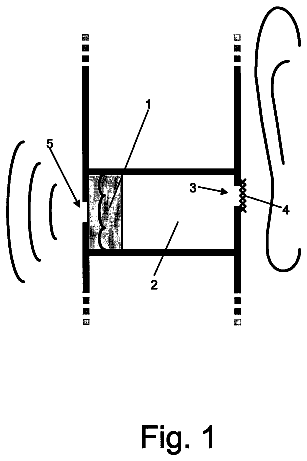
Speaker apparatus combining earpiece and hands free functions
In the invention a speaker apparatus acoustically combines the earpiece and hands free functions.
US 8,718,345 B2 · Assignee: Shengzhen Institutes of Advanced Technology Chinese Academy of Sciences · Inventors: Hu; Qingmao
Sheet 1 of 12 from the published document. All sheets in the USPTO PDF
Method and system for obtaining brain characteristic parameters, thrombolysis decision guideline system and method thereof, provide middle of reference data for early diagnosis and treatment of acute ischemic brain death, and provide effective reference data for thrombolytic therapy. The present invention uses diffusion-weighted imaging, comparing to the clinical programs of diffusion-weighted imaging and perfusion weighted imaging, perfusion weighted imaging can omitted and the degree of functional characterization of perfusion abnormalities is reflected without the perfusion weighted imaging, to provide middle data of brain tissue can be saved and the risk of thrombolytic therapy. The present invention based on ADC low signal constraint and DWI high signal constraint DWI binarization processing to obtain the core area and transition area, that is more accurate.
Cerebrovascular disease increased gradually in recent years, and is a second disease in cause of death in China that found by epidemiological researcher. In China, there are 1.3 to 1.5 million of new cerebrovascular disease cases, 6 to 7 million of the total number of sick people, while people died from cerebrovascular disease are nearly 1 million each year, about 3/4 of the survivors of are different degrees of incapacity, severe disability in 40% or more. The cerebrovascular disease has a higher morbidity, and is serious harm to human health and quality of life, which result in social and economic of hospitals, physicians, rehabilitation, pharmacy, and indirect consumption of up to 30 billion yuan per year. People dying from the ischemic stroke (cerebral infarction) disease accounts for 70% of the people that died from cerebrovascular disease, therefore, the study of cerebral infarctio
1 of 12 drawing sheets so far from the published document, cropped to the drawing. Every sheet is in the USPTO PDF.
What the patent claimed, word for word. All of it is now free to use.
The present disclosure relates to medical imaging technology, and more particularly relates to a method for obtaining brain characteristic parameters by using magnetic resonance imaging.
The present invention further relates to a system for obtaining brain characteristic parameters by using magnetic resonance imaging.
The present invention further relates to a method for thrombolysis decision guideline by using magnetic resonance imaging.
The present invention further relates to a system for thrombolysis decision guideline by using magnetic resonance imaging.
Cerebrovascular disease increased gradually in recent years, and is a second disease in cause of death in China that found by epidemiological researcher. In China, there are 1.3 to 1.5 million of new cerebrovascular disease cases, 6 to 7 million of the total number of sick people, while people died from cerebrovascular disease are nearly 1 million each year, about 3/4 of the survivors of are different degrees of incapacity, severe disability in 40% or more. The cerebrovascular disease has a higher morbidity, and is serious harm to human health and quality of life, which result in social and economic of hospitals, physicians, rehabilitation, pharmacy, and indirect consumption of up to 30 billion yuan per year. People dying from the ischemic stroke (cerebral infarction) disease accounts for 70% of the people that died from cerebrovascular disease, therefore, the study of cerebral infarction is very important.
Every state guideline recommends that the ischemic stroke in the ultra-super-early (within 3 hours) is preferredly healed by thrombolytic therapy of intravenous recombinant tissue-type plasminogen activator (rt-PA). However, the number of patients with ischemic stroke healed by hyperacute thrombolytic therapy is less than 1% in China. According to statistics, patients of hyperacute ischemic healed by rt-PA treatment are only 409 of the whole of China in 2004. Compared to newly issued 1.3 million of patients with ischemic stroke, the number of patients of hyperacute ischemic healed by rt-PA treatment is undoubtedly shocking. In the United States, there are at least 0.7 million new cases of ischemic stroke, but only 1% can be healed by thrombolytic therapy. This phenomenon is largely caused by that thrombolytic therapy is prone to bring other serious complications. Therefore, patients must be cured strictly in accordance with pathological state of the brain. However, how to understand the pathological state of patient is a difficulty of medicine, which is difficult to overcome.
For a long time, how to make the early diagnosis of ischemic stroke, determination of ischemic penumbra and effective implement for cerebral protection, saving of the damaged nerve function of the ischemic penumbra, have been the main treatment direction for ischemic stroke. Astrup J. et al present a concept of ischemic penumbra guides in 1981. The theory of ischemic penumbra guides shows that cerebral ischemia induces existence of a central necrosis area and a penumbra area surrounding the central necrosis area. The penumbra area losses neurological function, but nerve cells of the penumbra are still alive. At this point, if reperfusion active thrombolytic treatment is valid in a time window, most brain cells still prevent ischemic necrosis. The main purpose of thrombolysis is to save the ischemic penumbra area, restore tissue blood supply, and improve clinical symptoms. Therefore, presence of the ischemic penumbra area is a basic condition for thrombolytic therapy.
The ischemic penumbra and the central necrosis are a dynamic process of the pathophysiology, which is closely related to ischemia time, vascular occlusion and collateral circulation. With aggravation of ischemia and prolonged ischemia time, the central necrotic area is gradually expanded, the ischemic penumbra area is gradually shrunk. Therefore, the time window of reperfusion of the thrombolytic therapy is strict, over the time window of reperfusion may aggravate cerebral edema and brain cell damage, and increase mortality. A study researched by Absett et al shows that continuous treatment of 400 patients in 6 hours time window, the number of patients cured within 3 hours is only 10%, the number of patients cured within 3-6 hours is accounted for 90% of all cases, the number of patients cured within 5-6 hours is also accounted for 50% of all cases. Therefore, within the super-acute phase which is called the "time is brain", the number of patients benefited from thrombolytic therapy will grow exponentially at each extended hour. Thrombolytic therapy determines whether the ischemic penumbra area is existed and whether vascular barrier through the guidance of imaging damaged, and is no longer calculated in accordance with the onset of reperfusion time window, which extends the reperfusion time window and increase the number of patients accepted thrombolytic therapy. Practice of medicine shows that the radiographic can recognize the ischemic penumbra area to guide clinical treatment, and obtain satisfactory results in the traditional treatment time window. By the radiographic guidance, ten thousands of patients of peracute ischemic stroke can benefit from the thrombolytic therapy each year.
At present, the ischemic penumbra area is determined by a combination of magnetic resonance diffusion weighted imaging and magnetic resonance perfusion weighted imaging. The diffusion weighted imaging is referred to as DWI. The perfusion weighted imaging is referred to PWI. High signal abnormal area of the diffusion weighted imaging (DWI) represents a cerebral infarction area. Perfusion abnormal area of the PWI represents a damaged area. A unmatch area between the PWI and the DWI is a possible ischemic penumbra area, which is defined the difference between the perfusion abnormal area of the PWI and the high signal abnormal area of the DWI. PWI>DWI is considered as the best way of clinical thrombolysis guideline.
However, the above model has the following serious shortcomings:
The PWI requires injection of contrast agent, which is a relative measurement because different scanning machines have no uniform standard.
The perfusion abnormal area of the PWI in accordance with qualitative analysis, and calculation time of the perfusion abnormal area is rather time-consuming. Furthermore, this brain flow model is still controversial, and the magnetic resonance perfusion weighted imaging do not pass the U.S. Food and Drug Administration (FDA) approval, such that this brain flow model may be have a clinical risk.
The DWI is not only dependent on the dispersion characteristics, but also on the a T1 weighted, a T2 weighted and a proton density, especially on the T2-weighted.
The actual ischemic penumbra area is usually smaller than the unmatch area of PWI/DWI. An error between the actual ischemic penumbra area and the unmatch area is up to 40%-60%.
A difficulty of determining the infarction area, the ischemic penumbra area by PWI/DWI is a significantly different of diffusion and perfusion characteristics of different brain tissue (gray matter and white matter). The significantly difference can be up to 20%. If the significantly difference does not distinguish, there will be bring principle error.
In view of inherent weaknesses of the PWI and unmatch difficulty of the PWI/DWI, Prosser et al. advance a ischemic penumbra band model of clinical unmatch in accordance with the DWI, but the ischemic penumbra band model of clinical unmatch can be only achieved 53% of sensitivity.
Therefore, a method and system for obtaining brain characteristic parameters, a thrombolysis decision guideline system and method thereof are desired in order to overcome the above-described shortcomings.
It is an object of the present disclosure to provide a method for obtaining brain characteristic parameters. The method for obtaining brain characteristic parameters includes:
step S1, selecting a magnetic resonance perfusion weighted imaging of a patient obtained in accordance with magnetic resonance, wherein the magnetic resonance perfusion weighted imaging includes a Diffusion-weighted image, T2-weighted image, and an apparent diffusion coefficient map; step S2, distinguishing a brain tissue from a non-brain tissue in accordance with the T2-weighted image, and obtaining a brain tissue image without the non-brain tissue; step S3, processing voxels of the ADC map corresponding to the brain tissue image in accordance with low ADC signal constraint binarizing to obtain a binary image according to calculated ADC thresholds of a transition area; step S4, estimating core area and transition area according to the binary image and ADC thresholds of the core area in accordance with calculating; step S5, processing the estimated core area according to high signal constraint processing in accordance with high thresholds of the DWI of the estimated core area, and obtaining the core area and the transition area; and step S6, calculating characteristic parameters of the core area and the transition area.
In a preferred embodiment, the method for obtaining brain characteristic parameters further includes step S11: converting input image data of the DWI to lower bit image data, between step S1 and step S2.
In a preferred embodiment, step S2 includes:
step S21, obtaining a low signal threshold of the brain tissue by dividing the T2-weighted image into fuzzy C-means clustering;
step S22, binarizating the T2-weighted image in accordance with the low signal threshold to obtain the binary image; if gray of one pixel of the T2-weighted image is no less than the low signal threshold, set the pixel to foreground; if not, set the pixel to background; step S23, obtaining the largest connected area of the binary image; and step S24, opening slices of the largest connected area in the mathematical morphology, determining the biggest foreground connected area and obtaining a brain image by filling in the slices of the largest connected area.
In a preferred embodiment, step S3 and step S4 include:
step S31, determining an ADC value of normal brain tissue of the brain image based on the ADC map; and
step S32, calculating a product between the ADC value of the normal brain tissue and a threshold coefficient, and obtaining ADC threshold of the core area and ADC threshold of the transition area.
In a preferred embodiment, step S31 includes:
step S311, counting occurrence times of each ADC value of the ADC map corresponding to the brain tissue image; and
step S312, determining the ADC value of the normal brain tissue by the counting result, wherein the ADC value of the normal brain tissue is any value in neighborhood scope of the ADC value of the highest occurrence times.
In a preferred embodiment, the ADC value of the normal brain tissue is any value in neighborhood scope of the most frequent of ADC values in step S31; the ADC thresholds of the core area and the ADC thresholds the transition area are calculated based on the ADC value of the normal brain tissue in step S32; each slice of the brain tissue image is binarized based on the ADC thresholds of the transition area in step S3; the core area and the transition area are estimated based on the binary image of the each slice in step S4; the core area and the transition area are obtained by high signal constraint processing of the binary image of the each slice in step S5; characteristic parameters of the core area and the transition area is calculated in step S6.
In a preferred embodiment, step S3 further includes: if an ADC value of a pixel is greater than the ADC threshold of the transition area, the pixel is a background pixel; if not, the pixel is a foreground pixel.
In a preferred embodiment, step S4 includes:
step S41, obtaining a foreground connected area in accordance with the binary image of the ADC map; and
step S42, dividing the foreground connected area into a foreground area and a background area to obtain a first image, wherein if the volume of the foreground connected area is not less than the volume of the first pre-volume, and a voxel of the foreground connected area of which ADC value is less than the ADC threshold of the core area is not less than the second pre-volume, the foreground connected area is a foreground; if not, the foreground connected area is a background.
In a preferred embodiment, step S5 includes:
denoting the mean gray scale value of the DWI of the core area to DWIAvgF;
denoting the DWI gray scale value of all voxels adjacent to the core area to DWIAvgB;
selecting all voxels of brain tissue of which the DWI gray scale value is no less than a constant, and denoting mean gray scale and standard deviation of the voxels for the DWI and the to AvgDWI(z) and SdDWI(z);
denoting the high signal threshold of the DWI to DWITh(z), and calculating the high signal constraint of the DWI by the following formulas: DWITh(z)=AvgDWI(z)+CC1*SdDWI(z), wherein CC1 represents a constant, * represents multiplication; and wherein if (DWIAvgF-DWIAvgB)>CC2 or (DWIAvgF-DWITh(z))>CC3, the core area and the transition area are retained; if not, the core area and the transition area are background; wherein CC2 and CC3 represents constants.
In a preferred embodiment, CC1 is a range from about 1.1 to about 1.5, CC2 is a range from about 10 to about 20, and CC3 is a range from about 4 to about 8.
In a preferred embodiment, The method for obtaining brain characteristic parameters further includes: a recovering step between step S5 and step S6, and the recovering step comprising: setting the voxel of which ADC value is no greater than the ADC threshold of the transition area as the foreground voxel, and finding the adjacent voxels of each foreground voxel of the first image.
In a preferred embodiment, the method for obtaining brain characteristic parameters further includes: a second artifact removing step between the recovering step and step S6, and the second artifact removing step comprising: setting partial area of the foreground connected area of which volume is less than a preset volume as the background area, and finding the foreground connected area of the first image.
In a preferred embodiment, step S6 includes:
defining an core area and an transition area, the core area is a set of voxels of which ADC value is less than the ADC threshold of the core area, and the transition area is a set of voxels of which ADC value between the ADC threshold of the core area and the ADC threshold of the transition area; finding at least one connected area having the most foreground voxel of the core area and the transition area, and calculating characteristic parameters of the core area and the transition area in accordance with the connected area.
In a preferred embodiment, step S6 includes:
step S61, calculating an ADC value gradient of the transition area, wherein step S61 includes:
step S611, finding a connected area having the largest foreground voxels;
step S612, finding interior points of the connected area;
step S613, calculating a mean ADC value gradient of interior points of which ADC value between the ADC threshold of the core area and the ADC threshold of the transition area;
step S614, calculating a mean ADC value of voxels of which ADC value between the ADC threshold of the core area and the ADC threshold of the transition area; and
step S615, calculating an ADC value gradient of the transition area by the following formula: gradAvgN=gradAvg.times.C/(ADC.sub.ref.times.avgADC); wherein gradAvgN represents the ADC value gradient of the transition area, gradAvg represents the mean ADC value gradient of the connected area in step S613, C represents a positive constant, ADC.sub.ref represents ADC value of the normal brain tissue, and avgADC represents the mean ADC value gradient in step S614; step S62, calculating a radial distance of the transition area, wherein step S62 includes: step S621, calculating the number of first voxels of the connected area of which ADC value is less than the ADC threshold of the core area, and the number of second voxels of the connected area of which ADC value between the ADC threshold of the core area and the ADC threshold of the transition area; step S622, calculating a radius of the core area of the connected area and a radius of the core area and the transition area of the connected area respectively, wherein the radius of the core area is square root of a quotient.COPYRGT. which is calculated by dividing the number of the first voxels to pi and the radius of the transition area is square root of a quotient which is calculated by dividing the sum of the number of the first voxels and the number of the second voxels to pi; and step S623, calculating a radial distance of the transition area by minusing the radius of the core area and the radius of the transition area.
In a preferred embodiment, step S6 further includes step S63: calculating a total change of radial ADC value of the transition area by the following formulas: .delta.ADC=gradAvgN.times..delta.R/ADC.sub.ref; wherein .delta.ADC is a total change of ADC, gradAvgN represents the gradient of ADC value of the transition area, .delta.R represents the radial distance of the transition area, and ADC.sub.ref represents ADC value of the normal brain tissue.
In a preferred embodiment, step S6 further includes step S64: calculating a variation coefficient of ADC value of the core area; wherein step S64 includes:
step S641, calculating a mean ADC value and a mean square errors of voxels of which ADC value is less than the ADC threshold of the core area;
step S642, dividing the mean ADC value by the mean square errors, and the variation coefficient of ADC value of the connected area is equal to the quotient.
In a preferred embodiment, step S6 further includes:
step S65, calculating parameters of partial edge area of the core area adjacent to the transition area and a mean ADC value of voxels of all core area and a mean square errors of voxels of all core area, and denoting the mean ADC value and the mean square errors to AvgADC.sub.core and SdACD.sub.core; eroding the core area by 3.times.3 mathematical morphology erosion, and denoting a mean ADC value and a mean square errors of the remaining core area to AvgADC.sub.coreE and SdACD.sub.coreE; calculating the change of the mean ADC value of partial edge area of the core area adjacent to the transition area by the following formulas: .delta.AvgADC=(AvgADC.sub.core-AvgADC.sub.coreE)/AvgADC.sub.core; calculating the change of the mean square errors of ADC value of partial edge area of the core area adjacent to the transition area by the following formulas: .delta.SdADC=(SdADC.sub.core-SdADC.sub.coreE)/SdADC.sub.core; calculating the change of the variation coefficient of ADC value of partial edge area of the core area adjacent to the transition area by the following formulas: .delta.CvADC=.delta.SdADC/.delta.AvgADC.
In a preferred embodiment, step S6 further includes: selecting a first and second connected area and obtaining characteristic parameters of the first and second connected area; comparing the characteristic parameters of the first connected area to that of the second connected area, and outputting the minimum characteristic parameters or the maximum characteristic parameters.
It is another object to provide a system for obtaining brain characteristic parameters. The system for obtaining brain characteristic parameters includes:
an inputting device, configured to obtain a magnetic resonance perfusion weighted imaging of a patient obtained in accordance with magnetic resonance, wherein the magnetic resonance perfusion weighted imaging includes a DWI, T2-weighted image, and an ADC map; a brain tissue extracting module, configured to calculate a brain tissue of patient in accordance with the T2-weighted image, and output a brain image; an image processing module, configured to obtain a binary image based on binarizing the voxels of the ADC map corresponding to the brain tissue image in accordance with low ADC signal constraint binarizing according to ADC thresholds of a transition area in accordance with calculating; and estimate core area and transition area according to the binary image and ADC thresholds of a core area in accordance with calculating; and process the core area obtained by estimating in accordance with high signal constraint processing according to DWI high signal thresholds of the core area in accordance with estimating, and obtaining the core and the transition area; and a characteristic parameters calculating unit, configured to calculate characteristic parameters of the core area and the transition area.
In a preferred embodiment, the inputting device includes a data processing unit configured to convert data of the DWI, the T2-weighted image, and the ADC map to a lower bit data, wherein an output end of the data processing unit is connected to the image processing module and the brain tissue extracting module.
In a preferred embodiment, the image processing module includes:
a normal ADC value obtaining module, configured to obtain ADC values of a normal brain tissue, wherein the normal ADC values of obtaining module is connected to the brain tissue extracting module and the outputting device;
an ADC low signal thresholds calculating module, configured to calculate a product between the ADC value of the normal brain tissue and a threshold coefficient, and obtain an ADC threshold of the core area and the transition area;
an DWI high signal thresholds calculating module, configured to select all voxels of brain tissue of which the DWI gray scale is no less than a constant, and denoting mean gray scale and standard deviation of the voxels for the DWI and the to AvgDWI(z) and SdDWI(z); denoting the high signal threshold of the DWI to DWITh(z), and calculate the high signal constraint of the DWI by the following formulas: DWITh(z)=AvgDWI(z)+CC1*SdDWI(z), wherein CC1 represents a constant, * represents multiplication; a low signal constraint binarizing module, configured to obtain a binary image based on binarizing the ADC map in accordance with ADC low signal constraint binarizing according to the ADC thresholds of the transition area, wherein the low signal constraint binarizing module is connected to the inputting device, the brain tissue extracting module and the ADC low signal thresholds calculating module; an estimating unit, configured to estimate core area and transition area in accordance with the binary image, wherein the estimating unit is connected to the low signal constraint binarizing module and the ADC low signal thresholds calculating module; a high signal constraint processing unit, configured to process the core area in accordance with high signal constraint processing according to ADC high signal thresholds of the core area in accordance with estimating, wherein the high signal constraint processing unit is connected to the estimating unit and the DWI high signal thresholds calculating module; a determination unit, configured to define an core area and an transition area, wherein the core area is a set of voxels of which ADC value is less than the ADC threshold of the core area, and the transition area is a set of voxels of which ADC value between the ADC threshold of the core area; a characteristic parameter calculating unit, configured to find at least one connected area having the largest voxels in the core area and the transition area, and calculate characteristic parameters of the core area and the transition area, wherein the characteristic parameter calculating unit is connected to the determination unit.
In a preferred embodiment, the high signal constraint processing unit is configured to:
denote the mean gray scale of the DWI of the core area to DWIAvgF;
denote the DWI gray scale of all voxels adjacent to the core area to DWIAvgB;
denote the high signal threshold of the DWI to DWITh(z);
wherein if (DWIAvgF-DWIAvgB)>CC2 or (DWIAvgF-DWITh(z))>CC3, the core area and the transition area is retained; if not, the core area and the transition area is background; wherein CC2 and CC3 represents constants.
In a preferred embodiment, the estimating unit includes:
a first unit, configured to obtain a foreground connected area of each slice of the brain tissue in accordance with the binary image;
a first artifact removing unit, configured to obtain a foreground area, the foreground area is a part of the foreground connected area of which volume is greater than or equal to a first preset volume, and includes at least one voxel of which volume is greater than or equal to a second preset volume, and of which ADC value is less than the ADC threshold of the core area, the other part of the foreground connected area excluding the foreground area is the background area.
In a preferred embodiment, the image processing module further includes:
a recovering unit, configured to set the voxel of which ADC value is no greater than the ADC threshold of the transition area as the foreground voxel, and find the adjacent voxels of each foreground voxel, wherein the recovering unit is connected between the high signal constraint processing unit and the determination unit.
In a preferred embodiment, the image processing module further includes:
a second artifact removing unit, configured to set partial area of the foreground connected area of which volume is less than a preset volume as the background area, and finding the foreground connected area, wherein the second artifact removing unit is connected between the recovering unit and the determination unit.
It is another object to provide a thrombolysis decision guideline system. The thrombolysis decision guideline system includes:
an inputting device, configured to obtain a magnetic resonance perfusion weighted imaging of a patient obtained in accordance with magnetic resonance, wherein the magnetic resonance perfusion weighted imaging includes a DWI, T2-weighted image, and an ADC map; a brain tissue extracting module, configured to calculate a brain tissue of patient in accordance with the T2-weighted image, and output a brain image; an image processing module, configured to obtain a binary image based on binarizing the voxels of the ADC map corresponding to the brain tissue image in accordance with ADC low signal constraint binarizing according to ADC thresholds of a transition area in accordance with calculating; and estimate core area and transition area according to the binary image and ADC thresholds of a core area in accordance with calculating; and process the core area obtained by estimating in accordance with high signal constraint processing according to DWI high signal thresholds of the core area in accordance with estimating, and obtaining the core and the transition area; a characteristic parameters calculating unit, configured to calculate characteristic parameters of the core area and the transition area; a training database, configured to store a preset threshold of the core area and the transition area; and a determination unit, configured to compare output characteristic parameters of the characteristic parameters calculating unit to the preset threshold and output a comparison result.
In a preferred embodiment, the preset threshold of the training database includes a limiting value of variation coefficient of ADC value of the core area, a total change of radial ADC value gradient of the transition area, volume of the core area, volume ratio of the core area and the transition area, a mean ADC value of partial edge area of the core area adjacent to the transition area, a change of the mean square errors of ADC value, and a change of the variation coefficient of ADC value.
It is another object to provide a method of thrombolysis decision. The method of thrombolysis decision includes:
step S1, selecting a magnetic resonance perfusion weighted imaging of a patient obtained in accordance with magnetic resonance, wherein the magnetic resonance perfusion weighted imaging includes a DWI, T2-weighted image, and an ADC map;
step S2, distinguishing a brain tissue from a non-brain tissue in accordance with the T2-weighted image, and obtaining a brain tissue image without the non-brain tissue;
step S3, binarizing voxels of the ADC map corresponding to the brain tissue image in accordance with ADC low signal constraint binarizing to obtain a binary image according to ADC thresholds of a transition area in accordance with calculating;
step S4, estimating core area and transition area according to the binary image and ADC thresholds of the core area in accordance with calculating;
step S5, processing the core area obtained by estimating in accordance with high signal constraint processing according to DWI high signal thresholds of the core area in accordance with estimating, and obtaining the core and the transition area;
step S6, calculating characteristic parameters of the core area and the transition area; and
step S7, comparing output characteristic parameters to a preset threshold and output a comparison result.
In a preferred embodiment, step S7 includes: obtaining the preset threshold based on comparing and training at least one parameter of a radial distance of the transition area, an ADC value gradient of the transition area, a total change of ADC value gradient of the transition area, a mean ADC value of the core area, a variation coefficient of ADC value of the core area, volume of the core area, volume ratio of the core area and the transition area, a mean ADC value of partial edge area of the core area adjacent to the transition area, a change of the mean square errors of ADC value, and a change of the variation coefficient of ADC value.
Comparing to the clinical programs of DWI and PWI, the present disclosure has the following advantages: the PWI can also be omitted, and the degree of functional characterization of perfusion abnormalities is reflected without the PWI. Therefore, cost and time of treatment of patient can be saved. In addition, the thrombolysis whether is implemented can be determined more accurately. Therefore, suffering and economic burden of the patient can be reduced.
Other advantages and novel features will become more apparent from the following detailed description of various embodiments, when taken in conjunction with the accompanying drawings.
The components in the drawings are not necessarily drawn to scale, the emphasis instead being placed upon clearly illustrating the principles of the present disclosure. Moreover, in the drawings, like reference numerals designate corresponding parts throughout several views, and all the views are schematic.
FIG. 1 is a flowchart of one embodiment of a method for obtaining brain characteristic parameters.
FIG. 2 is an image of original slices of a T2-weighted image.
FIG. 3 is a brain image calculated in accordance with FIG. 2.
FIG. 4 is an ADC map having a coordinates system which is same to that of the original slices shown in FIG. 2.
FIG. 5 is an ADC map having a core area (in white color) and a transition area (in gray color) corresponding to the ADC map shown in FIG. 4.
FIG. 6 to FIG. 9 are comparison charts for non-using of high signal processing DWIs and using of high signal processing DWI.
FIG. 10 is a block diagram of one embodiment of system for obtaining brain characteristic parameters including an estimating unit.
FIG. 11 is a block diagram of one embodiment of a thrombolysis decision guideline system.
FIG. 12 is a flowchart of one embodiment of a method of thrombolysis decision guideline.
The disclosure is illustrated by way of example and not by way of limitation in the figures of the accompanying drawings in which like references indicate similar elements. It should be noted that references to "an" or "one" embodiment in this disclosure are not necessarily to the same embodiment, and such references mean at least one.
Embodiment 1
Referring to FIG. 1, a method for obtaining brain characteristic parameters according to a preferred embodiment is used to estimate a cerebral infarction area and an ischemic penumbra area of patient, and analysis characteristic parameters of the cerebral infarction area and the ischemic penumbra area. According to the method for obtaining brain characteristic parameters, reclaimable organizations find a method of analyzing risks of thrombolytic therapy. A core area of the present disclosure represents to the cerebral infarction area. A transition area of the present disclosure represents an area whose ischemic degree between that of the cerebral infarction area and a normal tissue area. The transition area is an estimate of the ischemic penumbra area. The method for obtaining brain characteristic parameters includes the following steps:
In step S1, a magnetic resonance perfusion weighted imaging of a patient obtained in accordance with magnetic resonance is selected, wherein the magnetic resonance perfusion weighted imaging includes a Diffusion-weighted image of which a diffusion sensitivity factor b=0, T2-weighted image of which an isotropic diffusion sensitivity factor b having the high diffusion sensitivity value, and an apparent diffusion coefficient map calculated by the Diffusion-weighted image and the T2-weighted image. The Diffusion-weighted image is referred to as DWI. The apparent diffusion coefficient map is referred to as ADC map.
In step S2, a brain tissue and a non-brain tissue are calculated in accordance with the T2-weighted image, and a brain image (x, y, z) is obtained and used to locate the ADC map to obtain relevant parameters. The brain image (x, y, z) has removed the non-brain tissue.
In Step S3, the ADC map corresponding to the brain image (x, y, z) is binarized via a ADC threshold thADC2 of the transition area. Therefore, a binary image B_ADC (x, y, z) is obtained.
In step S4, the core area and the transition area are estimated via a binary image and a ADC threshold thADC1 of the core area.
In step S5, the core area and the transition area are obtained via processing DWI high signal characteristic parameters of the core area in accordance with high signal constraint processing.
In step S6, characteristic parameters of the core area and the transition area are calculated. The characteristic parameters of the core area and the transition area include: volume of the core volCore, volume of the transition volIP, an ADC gradient of the transition area gradAvgN, a radial distance of the transition area .delta.R, a total change of ADC gradient of the transition area .delta.ADC, a mean ADC value of the core area AvgADC, a mean square errors of ADC value of the core area SdADC, a variation coefficient of ADC value of the core area CV, a change of the mean ADC value of partial edge area of the core area adjacent to the transition area .delta.AvgADC, a change of the mean square errors of ADC value of partial edge area of the core area adjacent to the transition area .delta.SdADC, and a variation coefficient of ADC value of partial edge area of the core area adjacent to the transition area .delta.CvADC.
S1 Step Process:
The present disclosure is implemented in accordance with the magnetic resonance DWI of patient with acute cerebral ischemia. An image mainly related to the DWI includes:
A diffusion sensitivity factor b=0 in the T2-weighted images.
An isotropic DWI having the high diffusion sensitivity value b of common clinical is 1000 s/mm.sup.2, or 1500 s/mm.sup.2.
An ADC map calculated by the DWI and the T2-weighted image.
Coordinates of the T2-weighted images, the isotropic DWI, and the ADC map are set to: X-axis extending from left to right, Y-axis extending from front to back, Z-axis extending from top to bottom. A slice whose Z coordinate is a constant is referred to an axial slice. A slice whose Y coordinate is a constant is referred to a coronal slice. A slice whose X coordinate is a constant referred to a sagittal section slice. The Z coordinate of the nth axial slices is n, wherein the n is a natural number. In the following description of the present disclosure, for example, the axial slices is obtained by an axial clinical images of patient. It is to be understood that the coronal slice and the sagittal section slice can also be obtained by the same way.
A coordinate system, the number of voxels and physical locations of patient corresponding to the same voxel of the T2-weighted images, the DWI and the ADC map are same. A T2 (x, y, z), a DWI(x, y, z), and a ADC (x, y, z) represent gray scales of one voxel (x, y, z) in the T2-weighted image, the DWI and the ADC map, respectively. The ADC(x, y, z) is calculated by the follow formulas: ADC(x,y,z)=log(T2(x,y,z)/DWI(x,y,z))/b; Wherein log represents natural logarithm, b is a constant with a range from about 1000 to 1500 high diffusion coefficient gradient used in DWI. In order to reduce noise impacts, processing the T2 (x, y, z) and/or the DWI(x, y, z) in accordance with median filter before calculating ADC value.
As previously mentioned, the DWI, the T2-weighted image and the ADC map of patient with the initial acute phase of cerebral infarction have the same coordinate system. The same coordinate system represents the same physical location of patient, and storage for a standard DICOM format. The DWI(x, y, z), the T2 (x, y, z) and the ADC (x, y, z) are 16-bit data, and the T2-weighted image is shown in FIG. 2. Space coordinates of each voxel (x, y, z) have the following constraints, that is, 0.ltoreq.x.ltoreq.xSize-1, 0.ltoreq.y.ltoreq.ySize-1, 0.ltoreq.z.ltoreq.zSize-1; wherein x, y, z are non-negative integer; xSize, ySize, and zSize represent the number of voxels of three-dimensional image in the x, y, and z direction, starting from the number 0. In the X, Y and Z direction, the physical size of each voxel is referred to voxX, voxY, and voxZ in mm/voxel.
Because gray scale of voxels of an actual three-dimensional image is only effective in the hundreds or thousands, 16-bit data of the T2-weighted image is converted to 8-bit data in order to improve image-processing speed. The 16-bit data can be converted to the 8-bit data by a variety of implementations. In one embodiment, the 16-bit data is converted to 8-bit data via excluding isolated voxels from the T2-weighted image, which can be referred to as the percentile-based method. The isolated voxels are the maximum gray scale level and the lowest gray scale level of voxels. The percentile-based method includes the following steps.
Firstly, a percentage of low gray P.sub.0 and a percentage of high gray P.sub.1 are set. In one embodiment, the percentage of low gray P.sub.0 is 0.01%, the percentage of high gray P.sub.1 is also 0.01%.
Then, a gray scale value g.sub.0 and a gray scale value g.sub.1 respectively corresponding to the percentage of low gray scale P.sub.0 and the percentage of high gray scale P.sub.1 are calculated. A percentage of the number of voxels of which gray scale between 0 to g.sub.0 in all voxels is less than P.sub.0. A percentage of the number of voxels of which gray scale between 0 to g.sub.0+1 in all voxels is greater than P.sub.0. A percentage of the number of which voxels of which gray scale greater than g.sub.1 in all voxels is less than P.sub.1. A percentage of the number of voxels of which gray scale greater than g1-1 in all voxels is greater than P.sub.1. According to the gray scale value g.sub.0 and the gray scale value g.sub.1, the original T2 (x, y, z) is coverted to a T2.sub.--8 (x, y, z) by the following formula (1). The T2.sub.--8 (x, y, z) represent a 8-bit T2-weighted image, and is calculated by formula (1).
.times..times..times..times..times.<.times..times..times..times. .times..times..times. .times..times..times.> ##EQU00001##
The description continues in the full USPTO document.
About 6,420 words. The USPTO PDF has it with every drawing.
Fees are due 3.5, 7.5 and 11.5 years after grant. This patent expired on May 6, 2026, so the fee marked "not paid" was the one that went unpaid.
METHOD AND SYSTEM FOR OBTAINING BRAIN CHARACTERISTIC PARAMETERS, THROMBOLYSIS DECISION GUIDELINE SYSTEM AND METHOD THEREOF
Filed Jun 2009 · published Mar 2012Method and system for obtaining brain characteristic parameters, thrombolysis decision guideline system and method thereof
Filed Jun 2009 · granted May 2014Earlier publications, parents and continuations. None of them can still be enforced, or this patent would not be listed.
Prior art cited by the examiner or applicant. Useful when you check your own idea for novelty.
Everything on this page comes from the documents linked above.