Field of the invention
This invention relates to the field of protein chemistry, e.g., translation biochemistry and mutation analysis. The invention relates to methods and compositions for producing a reductase enzyme comprising a 3-aminotyrisone residue and for determining the function of a selected amino acid residue in a reductase.
Background of the invention
In all organisms, ribonucleotide reductases (RNRs) catalyze the conversion of nucleotides to 2'-deoxynucleotides, providing the precursors used in DNA biosynthesis and repair..sup.1-3 The mechanism of nucleotide reduction is conserved in all RNRs and requires formation of a transient active site thiyl radical (C.sub.439., E. coli RNR numbering used throughout the text)..sup.4,5 However, the mechanism of active site thiyl radical generation, the radical initiation event, is not conserved and provides the basis for distinction between four classes of RNRs..sup.6-9 A major unresolved mechanistic issue is that of thiyl radical formation in class I RNRs, and presumably in the recently identified class IV RNRs.
The E. coli class I RNR consists of two homodimeric subunits, .alpha.2 and .beta.2, which form an active 1:1 complex during turnover..sup.10-12 .alpha.2 is the business end of the complex. It contains the active site where thiyl radical-mediated nucleotide reduction occurs, as well as multiple allosteric effector binding sites which modulate substrate specificity and turnover rate..sup.13 .beta.2 houses the stable diferric tyrosyl radical (Y.sub.122.).sup.14-16 cofactor that is required for formation of the transient C.sub.439. in the active site of .alpha.2..sup.4-6 The structures of .alpha.2.sup.6,17 and .beta.2.sup.18,19 have been solved and a structure containing both subunits has also been reported..sup.20 A structure of the active .alpha.2.beta.2 complex, however, has remained elusive. From the individual structures of .alpha.2 and .beta.2, Uhlin and Eklund have generated a docking model of the .alpha.2.beta.2 complex based on shape and charge complementarity and conserved residues..sup.6 This model suggests that the Y.sub.122. in .beta.2 is located >35 .ANG. away from C.sub.439 in .alpha.2 (FIG. 1)..sup.21-23 Radical propagation over this long distance requires the involvement of transient amino acid intermediates..sup.24-26 The residues proposed to participate in this pathway are universally conserved in all class I RNRs.
Evidence in support of the long distance between Y.sub.122. and C.sub.439 has recently been obtained from pulsed electron-electron double resonance spectroscopic measurements.sup.27 with a mechanism based inhibitor..sup.28-32 The distance obtained from this study is consistent with the docking model and establishes that a large conformational change, that positions Y.sub.122. in .beta.2 adjacent to C.sub.439 in .alpha.2, does not occur..sup.32
To examine the validity of the proposed pathway, site-directed mutagenesis.sup.33,34 and complementation studies.sup.35 have been carried out. These studies demonstrate that each residue in FIG. 1 plays an important role in RNR function. However, the absence of activity in these mutants precludes mechanistic investigations..sup.33,34 At present, evidence, e.g., detailed elsewhere herein, for the involvement of only one of the proposed pathway residues, Y.sub.356, is substantial. In contrast, the roles of .alpha.2 residues Y.sub.730 and Y.sub.731 in radical propagation are still ill-defined. Mutagenesis studies have demonstrated their importance in RNR function..sup.34,44,45 However, as with residue Y.sub.356 in .beta.2, the inactivity of these mutants (Y.sub.730F--.alpha.2 and Y.sub.731F-.alpha.2) precluded mechanistic interrogation of the role of Y.sub.730 and Y.sub.731 in radical propagation.
What is needed in the art are methods and compositions for the site-specific replacement of an amino acid residue that is proposed to participate in radical propagation in a reductase enzyme with unnatural amino acid residue that produces a mechanistically informative mutant, e.g., a mutant that can be used to interrogate the replaced amino acid's function in radical propagation in, e.g., a reductase enzyme. The present invention provides new tools and methods for elucidating RNR reaction mechanisms.
Summary of the invention
The invention relates to recombinant reductase enzymes that include a 3-aminotyrosine (NH.sub.2Y) residue, as well as orthogonal systems of components for producing such reductase enzymes. These tools were used to demonstrate kinetically competent radical transfer in E. coli RNR from Y.sub.122. in the .beta.2 subunit of the RNR across the subunit interface, and also radical trapping of NH2Y.sub.730. or NH2Y.sub.731. This event was shown to be triggered by binding of substrate and effector by the E. coli RNR. Steady state activity assays in conjunction with reactions with the suicide inhibitor N3ADP indicate that Y.sub.730NH2Y-.alpha.2 and Y.sub.731NH2Y-.alpha.2 are competent in nucleotide reduction. This implicates a hydrogen atom transfer mechanism for oxidation of C.sub.439 by NH2Y.sub.730.
Accordingly, in a first aspect, the invention provides recombinant reductase enzymes that comprise a 3-aminotyrosine (NH.sub.2Y) residue. In one preferred embodiment, a reductase of the invention is or is derived from a ribonucleotide reductase, such as a class I or class IV ribonucleotide reductase. For example, the reductase can be a recombinant reductase derived from an E. coli ribonucleotide reductase, a human ribonucleotide reductase, a mouse ribonucleotide reductase, a yeast ribonucleotide reductase, a herpes simplex virus ribonucleotide reductase, or the like. For example, the reductase can be an E. coli ribonucleotide reductase comprising an NH.sub.2Y mutation at one or more of Y.sub.730 of an .alpha.2 subunit of the E. coli ribonucleotide reductase; Y.sub.731 of an .alpha.2 subunit of the E. coli ribonucleotide reductase; Y.sub.122 of an .beta.2 subunit of the E. coli ribonucleotide reductase; or Y.sub.356 of a .beta.2 subunit of the E. coli ribonucleotide reductase.
In a related aspect, the invention provides cells that express a reductase of the invention. A cell of the invention includes a recombinant nucleic acid that is derived from a reductase nucleic acid that encodes one or more polypeptide chain of a reductase enzyme, an orthogonal tRNA (O-tRNA), and an orthogonal aminoacyl tRNA synthetase (O-RS). Optionally, the cell can include 3-aminotyrosine. The orthogonal aminoacyl tRNA synthetase (O-RS) in the cell preferentially aminoacylates the O-tRNA with 3-aminotyrosine in the cell, and the recombinant nucleic acid encoding the reductase includes a selector codon that is recognized by the orthogonal tRNA (O-tRNA). The encoded reductase can optionally comprise a class I or class IV ribonucleotide reductase (RNR) and/or an RNR derived from E. coli.
In addition, the invention provides for high yields of reductases comprising NH.sub.2Y residues. For example, a cellular paste or extract that includes a recombinant reductase enzyme, e.g., a recombinant ribonucleotide reductase, comprising the 3-aminotyrosine (NH.sub.2Y) residue can be produced, in which the cellular paste or extract comprises at least about 2 and about 4 mg/g of the reductase enzyme. In one example herein, the cellular paste comprises between about 4 and about 6 mg/g of the reductase enzyme.
In another aspect, the invention includes methods of determining a mechanistic function of a selected amino acid residue in a reductase. Such methods include mutating the selected amino acid residue to 3-aminotyrosine (NH.sub.2Y) to produce a recombinant mutant reductase that comprises NH.sub.2Y at a site corresponding to the selected amino acid (e.g., a Y residue), mixing the recombinant reductase with one or more substrates or effectors of the reductase, and detecting formation of NH.sub.2Y. For example, where the reductase is RNR, the substrate can optionally include CDP ADP, GDP, or UDP and the effector can include ATP. The reductase can optionally be reduced (or oxidized, depending on the application) prior to said mixing. Reducing the recombinant reductase can optionally include purifying the recombinant reductase from a cell, cell paste, or cell culture that expresses the recombinant reductase, and incubating the resulting purified reductase with a reducing agent.
Detecting formation of NH.sub.2Y can include any of a variety of techniques, including determining an EPR spectra for the NH.sub.2Y residue in the reductase, performing stopped flow spectroscopy after mixing to determine the kinetics of NH.sub.2Y. formation, or performing rapid freeze quench (RFQ) EPR after mixing to determine the kinetics of NH.sub.2Y. formation.
It will be appreciated that methods and compositions provided by the invention can be used alone or in combination.
Kits are also a feature of the invention. For example, such kits can comprise various components selected from: a container to hold the kit components, instructional materials for producing (e.g., expressing and/or purifying) a reductase enzyme, e.g., any of the reductase enzymes described herein, comprising one or more 3-aminotyrosine, a nucleic acid comprising a polynucleotide sequence encoding an O-tRNA, a nucleic acid comprising a polynucleotide encoding an O-RS, 3-aminotyrosine, and/or a suitable strain of E. coli host cells for expression of the O-tRNA/O-RS and production of a reductase enzyme comprising 3-aminotyrosine. Additionally or alternatively, kits of the invention can comprise instructions and/or reagents for determining a function of a selected amino acid residue, e.g., in free radical propagation, in a reductase.
Brief description of the figures
FIG. 1 includes Scheme 1, which depicts one electron oxidation of NH.sub.2Y, and a putative radical initiation pathway.
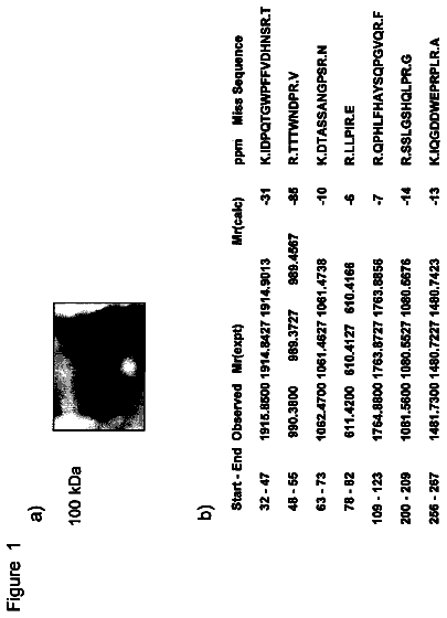
FIG. 2 includes MALDI-TOF MS and SDS PAGE analysis of a K.sub.7NH.sub.2Y--Z-domain.
FIG. 3 shows expression of Y.sub.731NH.sub.2Y-.alpha.2. Cells were grown in the presence or absence of IPTG and NH.sub.2Y/DTT as indicated and the level of expression assessed by SDS PAGE.
FIG. 4 provides a spectral trace showing reaction of Y.sub.730NH.sub.2Y-.alpha.2/ATP with wt .beta.2/CDP monitored by EPR spectroscopy.
FIG. 5 provides a graph showing microwave power dependence of Y.sub.122. and NH.sub.2Y.sub.730. signal intensities.
FIG. 6 Comparison of the NH.sub.2Y.sub.730. (dotted line, FIG. 4) and NH.sub.2Y.sub.731. (dashed line, FIG. 15).
FIG. 7 Point-by-point reconstruction of the UV-vis spectrum of NH.sub.2Y.sub.730. (circles) and NH.sub.2Y.sub.731. (squares).
FIG. 8 is a graph showing stopped flow kinetics of NH.sub.2Y.sub.730. formation.
FIG. 9 provides dual spectrums showing formation of N. from N.sub.3ADP upon incubation with Y.sub.730NH.sub.2Y-.alpha.2, .beta.2 and dGTP.
FIG. 10 Spectral comparison of N. (black), Y.sub.122. (dotted line), and NH.sub.2Y.sub.730. (dashed line).
FIG. 11 shows mechanistic options for oxidation of C439 by NH2Y730.
FIG. 12 provides a schematic graphic of the strategy used to determine the mechanistic roles of amino acids Y.sub.730 and Y.sub.731 in the .alpha.2 subunit of E. coli ribonucleotide reductase.
FIG. 13 Expression of Y.sub.730NH.sub.2Y-.alpha.2. Cells were grown in the presence or absence of IPTG and NH.sub.2Y/DTT and at 25.degree. or 37.degree. C., as indicated, and the level of expression assessed by SDS PAGE. The position of protein bands for full-length .alpha. and truncated cc are denoted by arrows.
FIG. 14 shows SDS PAGE analysis of purified Y.sub.730NH.sub.2Y-.alpha.2 (A) and Y.sub.731NH.sub.2Y-.beta.2 (B).
FIG. 15 depicts the results of a reaction of Y.sub.731NH.sub.2Y-.alpha.2/ATP with wt .beta.2/CDP monitored by EPR.
FIG. 16 depicts stopped flow kinetics of NH.sub.2Y.sub.731. formation.
FIG. 17 shows the results of an N.sub.3ADP assay for Y.sub.731NH.sub.2Y-.alpha.2.
FIG. 18 provides various nucleotide sequences finding use with the invention.
Detailed description of the invention
The present invention is directed to reductases, such as ribonucleotide reductases, that comprise 3-aminotyrosine (NH.sub.2Y) residues. The NH.sub.2Y residue can be incorporated into a reductase using a system of orthogonal elements that comprises an aminoacyl tRNA synthetase specific for 3-aminotyrosine (NH.sub.2Y), an O-tRNA, and, optionally, the unnatural amino acid 3-aminotyrosine. Methods and compositions for producing such recombinant reductases and compositions that comprise high yields of these recombinant reductases are described in further detail hereinbelow.
Once incorporated into a reductase, e.g., any one of the reductases described hereinbelow, the NH.sub.2Y can advantageously serve as a probe for analyzing the mechanistic role of the natural amino acid it replaced in radical propagation in the reductase enzyme (see, for example, FIG. 1 and corresponding description). In general, this can be accomplished by detecting the formation of the unnatural amino acid radical intermediate NH.sub.2Y., e.g., via EPR, rapid-freeze-quench EPR, and/or stopped flow spectrometry.
Systems of Orthogonal Elements for NH.sub.2Y Incorporation
Orthogonal components for the incorporation of, e.g., NH.sub.2Y, have been described previously; see WO 2006/110182 A2 by Schultz et al. ORTHOGONAL TRANSLATION COMPONENTS FOR THE VIVO INCORPORATION OF UNNATURAL AMINO ACIDS. In general, synthetases with the desired NH.sub.2Y specificity can be produced by randomly or selectively mutating the active site of an existing synthetase and selecting the resulting library of mutant synthetases to screen for desired NH.sub.2Y incorporation activity. Typically, libraries are positively screened for NH.sub.2Y incorporation and then negatively screened to eliminate members that aminoacylate a tRNA with natural amino acids. Iterative rounds of positive and negative selection can be performed to obtain the synthetase, e.g., a synthetase with a specificity for NH2Y. Additional details regarding screening and selection of O-RS to identify those that aminoacylate a cognate O-tRNA with NH.sub.2Y can be found in the examples below.
The reductase enzymes of the invention optionally include additional unnatural amino acids, in addition to the NH.sub.2Y unnatural amino acid. In general, using systems of orthogonal components, it is possible to put, e.g., 1, 2, 3, 4, 5, or more different unnatural amino acids into, e.g., 1, 2, 3, 4, 5, or more selected sites in proteins, e.g., by including a desired selector codon in a corresponding nucleic acid. A cell can include, e.g., 1, 2, 3, 4, 5, or more different sets of cognate orthogonal components that each recognize a different given selector codon (stop codons and four or more base codons can be used as selector codons).
Details regarding methods for producing and/or altering the specificity of tRNAs and/or O-RSs, unnatural amino acids, selector codons, and orthogonal translation systems that are suitable for making proteins that include one or more unnatural amino acids are generally described in, for example, International Publication Numbers WO 2002/086075, entitled "METHODS AND COMPOSITION FOR THE PRODUCTION OF ORTHOGONAL tRNA-AMINOACYL-tRNA SYNTHETASE PAIRS;" WO 2002/085923, entitled "IN VIVO INCORPORATION OF UNNATURAL AMINO ACIDS;" and WO 2004/094593, entitled "EXPANDING THE EUKARYOTIC GENETIC CODE;" WO 2005/019415, filed Jul. 7, 2004; WO 2005/007870, filed Jul. 7, 2004 and WO 2005/007624, filed Jul. 7, 2004. Each of these applications is incorporated herein by reference in its entirety. See also, Wang and Schultz "Expanding the Genetic Code," Angewandte Chemie Int. Ed., 44(1):34-66 (2005); Deiters et al, Bioorganic & Medicinal Chemistry Letters 15:1521-1524 (2005); Chin et al., J. Am. Chem. Soc. 2002, 124, 9026-9027; and International Publication No. WO2006/034332, filed on Sep. 20, 2005, the contents of each of which are incorporated by reference in their entirety. Additional details are found in U.S. Pat. No. 7,045,337; No. 7,083,970; No. 7,238,510; No. 7,129,333; No. 7,262,040; No. 7,183,082; No. 7,199,222; and No. 7,217,809.
Reductases
A reductase is an enzyme that catalyses a reduction reaction. However, most reductases can, under the proper conditions, behave as reductases or oxidases. Accordingly, the term oxidoreductase is also used to describe this broad family of enzymes, any of which can be modified according to the present invention to include, e.g., one or more NH.sub.2Y residue. In general, this is accomplished by incorporating an appropriate selector codon into a nucleic acid that encodes the reductase and expressing the reductase in a cell that includes an appropriate O-RS specific for NH.sub.2Y, a cognate O-tRNA that recognizes the selector codon, and NH.sub.2Y.
Examples of reductase enzymes that can be modified in this way to include an NH.sub.2Y residue include those with Enzyme Commission number (EC number) "EC1." This includes EC 1.1 (oxidoreductases that act on the CH--OH group of donors (e.g., alcohol oxidoreductases); EC 1.2 (oxidoreductases that act on the aldehyde or oxo group of donors); EC 1.3 oxidoreductases that act on the CH--CH group of donors (e.g., CH--CH oxidoreductases); EC 1.4 (oxidoreductases that act on the CH--NH.sub.2 group of donors (Amino acid oxidoreductases, Monoamine oxidase); EC 1.5 (oxidoreductases that act on CH--NH group of donors); EC 1.6 (oxidoreductases that act on NADH or NADPH); EC 1.7 (oxidoreductases that act on other nitrogenous compounds as donors) EC 1.8 (oxidoreductases that act on a sulfur group of donors); EC 1.9 (oxidoreductases that act on a heme group of donors), EC 1.10 (oxidoreductases that act on diphenols and related substances as donors) EC 1.11 (oxidoreductases that act on peroxide as an acceptor (e.g., peroxidases)); EC 1.12 (oxidoreductases that act on hydrogen as donors); EC 1.13 (oxidoreductases that act on single donors with incorporation of molecular oxygen (e.g., oxygenases)); EC 1.14 (oxidoreductases that act on paired donors with incorporation of molecular oxygen); EC 1.15 (oxidoreductases that act on superoxide radicals as acceptors); EC 1.16 (oxidoreductases that oxidize metal ions); EC 1.17 (oxidoreductases that act on CH or CH2 groups); EC 1.18 (oxidoreductases that act on iron-sulfur proteins as donors); EC 1.19 (oxidoreductases that act on reduced flavodoxin as a donor); EC 1.20 (oxidoreductases that act on phosphorus or arsenic in donors); EC 1.21 (oxidoreductases that act on X--H and Y--H to form an X--Y bond); EC 1.97 (other oxidoreductases); EC 1.98 (enzymes using H.sub.2 as a reductant); and EC 1.99 (enzymes using O.sub.2 as an oxidant).
One particularly preferred embodiment of the invention relates to the incorporation of NH.sub.2Y into the reductases of EC 1.17 (reductases acting on CH or CH2 groups), including xanthine oxidases and, especially, ribonucleotide reductases (RNRs). Ribonucleotide reductases (RNRs can catalyze the reduction of ribonucleotides, e.g., CDP, ADP, GDP, and UDP, to deoxyribonucleotides in all organisms. Because RNRs maintain the relative ratios of cellular dNTP levels, these enzymes play a central role in nucleic acid metabolism, DNA repair, genome maintenance, and cell proliferation (Sjoberg
"Ribonucleotide reductases--a group of enzymes with different metallosites and a similar reaction mechanism." Struct Bonding (Berlin) 88: 139-173; Reichard
"From RNA to DNA, why so many reductases?" Science 260: 1773-1777). (dTDP is produced by thymidilate synthase). The mechanism by which RNRs reduce NDPs involves complex and highly regulated radical-dependent redox chemistry (Stubbe
"Ribonucleotide reductases: amazing and confusing." J Biol Chem 265: 5329-5332; Jordan, et al.
"Ribonucleotide reductases." Annu Rev Biochem 67: 71-98). In general, RNRs, except those of herpesviruses, are allosterically regulated by deoxyribonucleoside triphosphates and ATP, such that DNA precursors are supplied in pools balanced according to the base composition of the different genomes (Hendricks, et al.
"Regulation of T4 phage aerobic ribonucleotide reductase. Simultaneous assay of the four activities." J Biol Chem 272: 2861-2865; Hendricks, et al.
"Allosteric regulation of vaccinia virus ribonucleotide reductase, analyzed by simultaneous monitoring of its four activities." J Biol Chem 273: 29512-29518). In addition to controlling RNR activity, the allosteric mechanism also regulates the substrate specificity (Jordan, et al.
"Ribonucleotide reductases." Annu Rev Biochem 67: 71-98).
RNRs are structurally diverse, and the metal cofactors, e.g., electron donors, they require are, likewise, structurally and chemically diverse. In fact, RNRs can be divided into four classes based on their metal cofactors. Class I rRNRs and generate a stable tyrosyl radical on the protein through activation of O.sub.2 by a diferric center. Class I reductases are further divided into class IA and class IB, based upon differences in enzyme regulation. Class IA reductases are distributed in eukaryotes, eubacteria, bacteriophages, and viruses. Class IB reductases are found in eubacteria and can produce radicals using manganese. Class II RNRs, which can function wither in the presence or absence of O.sub.2, generate a transient 5'-deoxyadenosyl radical through cleavage of the C--CO bond in adenosylcobalamin (AdoCbl). Class III RNRs are generally anaerobic and generate a stable glycyl radical on the protein by cleavage of S-adenosylmethionine. Class IV RNRs are proposed to comprise a manganese cofactor adjacent to a tyrosyl radical.
Class I RNRs comprise RNR1 and RNR2 subunits, which can associate to form active heterodimeric tetramers. A general mechanistic model for class I RNRs comprises three steps: 1) generation of the tyrosyl radical by the diiron center in subunit R2; 2) radical transfer to generate the proposed thiyl radical near the substrate bound in subunit R1; and 3) catalytic reduction of the bound ribonucleotide. Amino acid- or substrate-derived radicals are involved in all three major reactions. In preferred embodiments of the invention, tyrosine residues in class I RNRs can advantageously be replaced with the unnatural amino acid NH.sub.2Y, e.g., to elucidate the mechanistic function of each naturally occurring tyrosine residue in radical propagation. The tyrosine residues that can be replaced, e.g., in an E. coli Class I RNR, using the methods provided by the invention can include ribonucleotide reductase comprising an NH.sub.2Y mutation at one or more of Y.sub.730, Y.sub.731, Y.sub.122, and/or Y.sub.356 of the .beta.2 subunit.
Further details regarding the structure, mechanism, and/or regulation of ribonucleotide reductases can be found in, e.g., Stubbe & van der Donk
"Ribonucleotide reductases: radical enzymes with suicidal tendencies." Chem Biol 2: 793-801; Torrents et al.
"Ribonucleotide Reductases: Divergent Evolution of an Ancient Enzyme" Journal of Molecular Evolution 55: 138-152; Norlund, et al.
"Ribonucleotide reductases." Annu Rev Biochem 75: 681-706; Kolberg, et al.
"Structure, function, and mechanism of ribonucleotide reductases." Biochim Biophys Acta 1699: 1-34. Further details regarding the elucidation of the mechanism of radical propagation in a class I RNR are explained in the example below.
Expressing, Purifying and Isolating Reductases Comprising NH.sub.2Y Residues
The nucleic acids of the invention (e.g., nucleic acids derived from reductase nucleic acids that comprise selector codons and, e.g., encode one or more polypeptide chain of a reductase enzyme) can be produced according to standard cloning methods. Procedures for isolating, cloning, and amplifying nucleic acids; and for providing nucleic acid constructs to and expressing nucleic acid constructs in cells and cell free systems are replete in the literature and can be used in the present invention to provide and express a nucleic acid that comprises a selector codon, e.g., to produce a reductase protein, e.g., a class I or class IV ribonucleotide reductase, that comprises an NH.sub.2Y residue. The recombinant reductase enzyme can be derived from any of a variety of sources, including eukaryotes, e.g., humans, mice, yeast and others, prokaryotes, archea, and viruses, e.g., a herpes simplex virus. In preferred embodiments, a nucleic acid of the invention can encode a recombinant E. coli ribonucleotide reductase comprising an NH.sub.2Y mutation at a Y.sub.730, Y.sub.731, Y.sub.122, and/or Y.sub.356 in the .alpha.2 subunit. Alternatively, a nucleic acid of the invention can optionally encode any ribonucleotide reductase included in classes I-IV or any reductase with an EC number between 1.1 and 1.99, as described elsewhere herein.
Further details regarding nucleic acid cloning and expression techniques can be found in Berger and Kimmel, Guide to Molecular Cloning Techniques, Methods in Enzymology volume 152 Academic Press, Inc., San Diego, Calif. (Berger); Sambrook et al., Molecular Cloning--A Laboratory Manual (3rd Ed.), Vol. 1-3, Cold Spring Harbor Laboratory, Cold Spring Harbor, N.Y., 2000 ("Sambrook"); The Nucleic Acid Protocols Handbook Ralph Rapley (ed)
Cold Spring Harbor, Humana Press Inc (Rapley); Current Protocols in Molecular Biology, F. M. Ausubel et al., eds., Current Protocols, a joint venture between Greene Publishing Associates, Inc. and John Wiley & Sons, Inc., (supplemented through 2007) ("Ausubel")); PCR Protocols A Guide to Methods and Applications (Innis et al. eds) Academic Press Inc. San Diego, Calif.
(Innis); Chen et al. (ed) PCR Cloning Protocols, Second Edition (Methods in Molecular Biology, volume 192) Humana Press; in Viljoen et al.
Molecular Diagnostic PCR Handbook Springer; and Demidov and Broude (eds)
DNA Amplification: Current Technologies and Applications. Horizon Bioscience, Wymondham, UK. Other useful references, e.g., for cell isolation and culture (e.g., for subsequent nucleic acid isolation), include Freshney
Culture of Animal Cells, a Manual of Basic Technique, third edition, Wiley-Liss, New York and the references cited therein; Payne et al.
Plant Cell and Tissue Culture in Liquid Systems John Wiley & Sons, Inc. New York, N.Y.; Gamborg and Phillips (eds)
Plant Cell, Tissue and Organ Culture; Fundamental Methods Springer Lab Manual, Springer-Verlag (Berlin Heidelberg New York) and Atlas and Parks (eds) The Handbook of Microbiological Media
CRC Press, Boca Raton, Fla.
Assessing the mechanistic role of an amino acid, e.g., a tyrosine amino acid, in a reductase enzyme, e.g., an E. coli-derived ribonucleotide reductase, entails the purification of a recombinant reductase in which the natural amino acid of interest has been replaced with an NH.sub.2Y residue. High yields of recombinant reductase can be obtained from cells pastes or extracts, e.g., derived from cells in which a recombinant reductase of the invention has been expressed. A cell paste or extract can comprise, e.g., about 2 mg/g reductase, or, more preferably, 4-6 mg/g recombinant reductase. A variety of protein purification methods are well known in the art and can be applied to the purification and analysis of reductase variants that include at least one NH.sub.2Y residue. These techniques, and others that are necessary for the analysis of polypeptides, include those set forth in R. Scopes, Protein Purification, Springer-Verlag, N.Y. (1982); Deutscher, Methods in Enzymology Vol. 182: Guide to Protein Purification, Academic Press, Inc. N.Y. (1990); Sandana
Bioseparation of Proteins, Academic Press, Inc.; Bollag et al.
Protein Methods, 2nd Edition Wiley-Liss, NY; Walker
The Protein Protocols Handbook Humana Press, NJ; Harris and Angal
Protein Purification Applications: A Practical Approach IRL Press at Oxford, Oxford, England; Harris and Angal Protein Purification Methods: A Practical Approach IRL Press at Oxford, Oxford, England; Scopes
Protein Purification: Principles and Practice 3rd Edition Springer Verlag, NY; Janson and Ryden
Protein Purification: Principles, High Resolution Methods and Applications, Second Edition Wiley-VCH, NY; and Walker
Protein Protocols on CD-ROM Humana Press, NJ; and the references cited therein.
Further details regarding protocols for expression and isolation of reductase proteins such as E. coli RNR are described in the examples hereinbelow.
Methods of Detecting NH.sub.2Y in Reductase Enzymes
Example 1 below provides details regarding the detection of NH.sub.2Y residues in reductase enzymes, e.g., for detection of NH.sub.2Y residue incorporation and, e.g., as a molecular probe for detecting radical formation. Two common methods that can be used include EPR spectroscopy and spectral analysis, e.g., using UV-vis spectroscopy. These methods are commonly available, and one of skill is familiar with their use in the detection of amino acid residues, radicals, and other moieties of interest.
Electron paramagnetic resonance (EPR), which is also known as electron spin resonance (ESR) and electron magnetic resonance (EMR), is a form of spectroscopy that can be used to detect and identify free radicals and paramagnetic centers. EPR spectroscopy measures the absorption of microwave radiation by an unpaired electron when it is placed in a strong static magnetic field, B.sub.0, and exposed to a low-amplitude, high-frequency magnetic field, B.sub.1, that is perpendicular to B.sub.0. Highly unstable free radicals can, in many cases, be stabilized for EPR characterization by spin trapping. In fact, EPR spin-trapping can be advantageously applied to the detection and analysis of free radical intermediates in generated by metalloproteins, such as certain RNR reductases, as discussed herein.
Relatedly, rapid freeze quench (RFQ) EPR can be used to measure the kinetics of formation and decay of reaction intermediates that are EPR-active, e.g., free radicals. RFQ EPR entails arresting a reaction, e.g., catalyzed by an RNR, by rapid freezing and sustained low temperature after a reaction is allowed to proceed for a specified time. The trapped species are then analyzed by EPR. This method can be beneficially applied to the detection of, e.g., NH2Y radical formation, by a recombinant reductase of the invention. Briefly, in RFQ EPR, the reactants are mixed together rapidly and the reaction carried out for a pre-determined time period, e.g., on the millisecond to second timescale. The reaction is stopped or quenched by squirting the mixture rapidly into, e.g., liquid isopentane that is maintained at -140.degree. C. The crystals formed upon quenching of the reaction are packed into an EPR tube and the EPR spectrum is subsequently acquired to determine the kinetics and structure of the radical that is monitored.
Further details regarding EPR techniques can be found in Weil and Bolton
Electron Paramagnetic Resonance: Elementary Theory and Practical Applications Second Edition, Wiley; and in Graslund, et al.
"Electron Paramagnetic Resonance and Nuclear Magnetic Resonance Studies of Class I Ribonucleotide Reductase." Annu Rev Biophys Biomolec Struct 25: 259-286. Other resonance methods such as nuclear magnetic resonance (NMR) can also be used to analyze NH.sub.2Y residues in reductase proteins. For a general discussion of NMR techniques, see, e.g., Introduction to Solid-State NMR Spectroscopy
Melinda J. Duer (Editor), Wiley.
Other spectroscopic methods, including stopped flow spectroscopy, can also be used, e.g., to acquire kinetic data on the kinetics of free radical formation and/or propagation during, e.g., an RNR-catalyzed reaction. For a discussion of UV-vis spectroscopy and other spectroscopic techniques that can be used to analyze the mechanistic role of NH.sub.2Y residues in reductase proteins, and/or to monitor reductase reaction kinetics, see, e.g., Tong, et al.
"Characterization of Y122F R2 of Escherichia coli Ribonucleotide Reductase by Time-Resolved Physical Biochemical Methods and X-ray Crystallography." Biochem 37: 5840-5848; Lassman, et al.
"An advanced EPR stopped-flow apparatus based on a dielectric ring resonator." J Mag Res 172: 312-323; Pavia
Introduction to Spectroscopy: A Guide for Students of Organic Chemistry, Harcourt College Pub; Sorrel
Interpreting Spectra of Organic Molecules University Science Books; and Mohan
Molecular Spectroscopy: An Introduction ISBN: 978-81-7319-549-5.
Additional Details Regarding Term Definitions
A "reductase enzyme" is an enzyme that catalyses a reduction reaction. Because such enzymes catalyze reactions in either direction, most reductases can, under the proper conditions, behave as a reductase or an oxidase; accordingly, the term oxidoreductase is also used to describe this broad family of structurally diverse enzymes. Examples of reductase enzymes that can be used with the invention include those with Enzyme Commission number (EC number) "EC1". Further details regarding classes of reductases are elaborated elsewhere herein.
Derived from: As used herein, the term "derived from" refers to a component that is isolated from or made using a specified molecule or organism, or information from the specified molecule or organism. For example, a polypeptide that is derived from a second polypeptide can include an amino acid sequence that is identical or substantially similar to the amino acid sequence of the second polypeptide, e.g., other than the incorporation of unnatural amino acids into the second polypeptide. In the case of polypeptides, the derived species can be obtained by, for example, mutagenesis. The mutagenesis used to derive polypeptides can be intentionally directed or intentionally random, or a mixture of each. The mutagenesis of a polypeptide to create a different polypeptide derived from the first can be a random event (e.g., caused by polymerase infidelity) and the identification of the derived polypeptide can be made by appropriate screening methods, e.g., as discussed herein. Mutagenesis of a polypeptide typically entails manipulation of the polynucleotide that encodes the polypeptide.
Derivation of one protein or nucleic acid sequence from another can be identified by detection of homology between the molecules. Two molecules are homologous when they derive from a common ancestral molecule. Homology is ordinarily detected by detecting sequence identity or similarity. The precise cut-off for recognizing homology by assessing sequence identity or similarity varies, but it is common to identify homology when sequence similarity is as low as about 25%. Higher percentages of similarity, e.g., 35%, 45%, 55%, 65%, 75%, 85% 95%, 98% or higher are useful for identifying homology. Sequence similarity/identity can be identified using publicly available programs such as BLASTP (for proteins) and BLASTN (for nucleic acids), e.g., using default parameters (BLASTP and BLASTN are widely available, e.g., from the NCBI, e.g., on the world wide web at ncbi(dot)nlm(dot)nih(dot)gov/blast.
Orthogonal: As used herein, the term "orthogonal" refers to functional molecules, e.g., an orthogonal tRNA (O-tRNA) and/or an orthogonal aminoacyl-tRNA synthetase (O-RS), that function poorly or not at all with endogenous components of a cell, when compared to a corresponding molecule (tRNA or RS) that is endogenous to the cell or translation system. Orthogonal components are usefully provided as cognate components that function well with each other, e.g., an O-RS can be provided that can efficiently aminoacylates a cognate O-tRNA in a cell, even though the O-tRNA functions poorly or not at all as a substrate for the endogenous RS of the cell, and the O-RS functions poorly or not at all with endogenous tRNAs of the cell. Various comparative efficiencies of the orthogonal and endogenous components can be evaluated. For example, an O-tRNA will typically display poor or non-existent activity as a substrate, under typical physiological conditions, with endogenous RSs, e.g., the O-tRNA is less than 10% as efficient as a substrate as endogenous tRNAs for any endogenous RS, and will typically be less than 5%, and usually less than 1% as efficient a substrate. At the same time, the tRNA can be highly efficient as a substrate for the O-RS, e.g., at least 50%, and often 75%, 95%, or even 100% or more as efficient as an aminoacylation substrate as any endogenous tRNA is for its endogenous RS.
Orthogonal aminoacyl-tRNA synthetase: As used herein, an orthogonal aminoacyl-tRNA synthetase (O-RS) is an enzyme that preferentially aminoacylates an O-tRNA with an amino acid in a translation system of interest. The amino acid that the O-RS loads onto the O-tRNA in the present invention is a 3-aminotyrosine (NH.sub.2Y)
Orthogonal tRNA: As used herein, an orthogonal tRNA (O-tRNA) is a tRNA that is orthogonal to a translation system of interest. The O-tRNA can exist charged with, e.g., a 3-aminotyrosine, or can exist in an uncharged state. It is also to be understood that an O-tRNA is optionally charged (aminoacylated) by a cognate orthogonal aminoacyl-tRNA synthetase with a 3-aminotyrosine. Indeed, it will be appreciated that the O-tRNA described herein is used to insert a 3-aminotyrosine into a growing polypeptide, during translation, in response to a selector codon.
Cognate: The term "cognate" refers to components that function together, e.g., an orthogonal tRNA and an orthogonal aminoacyl-tRNA synthetase. The components can also be referred to as being complementary.
Preferentially aminoacylates: As used herein in reference to orthogonal translation systems, an O-RS "preferentially aminoacylates" a cognate O-tRNA when the O-RS charges the O-tRNA with 3-aminotyrosine more efficiently than it charges any endogenous tRNA in an expression system. That is, when the O-tRNA and any given endogenous tRNA are present in a translation system in approximately equal molar ratios, the O-RS will charge the O-tRNA more frequently than it will charge the endogenous tRNA. Preferably, the relative ratio of O-tRNA charged by the O-RS to endogenous tRNA charged by the O-RS is high, preferably resulting in the O-RS charging the O-tRNA exclusively, or nearly exclusively, when the O-tRNA and endogenous tRNA are present in equal molar concentrations in the translation system. The relative ratio between O-tRNA and endogenous tRNA that is charged by the O-RS, when the O-tRNA and O-RS are present at equal molar concentrations, is greater than 1:1, preferably at least about 2:1, more preferably 5:1, still more preferably 10:1, yet more preferably 20:1, still more preferably 50:1, yet more preferably 75:1, still more preferably 95:1, 98:1, 99:1, 100:1, 500:1, 1,000:1, 5,000:1 or higher. The O-RS "preferentially aminoacylates an O-tRNA with a 3-aminotyrosine" when (a) the O-RS preferentially aminoacylates the O-tRNA compared to an endogenous tRNA, and (b) where that aminoacylation is specific for the 3-aminotyrosine, as compared to aminoacylation of the O-tRNA by the O-RS with any natural amino acid. For example, when a 3-aminotyrosine and natural amino acids are present in equal molar amounts in a translation system comprising a relevant O-RS of the sequence listing herein and a relevant O-tRNA of the sequence listing herein, the O-RS will load the O-tRNA with 3-aminotyrosine more frequently than with any natural amino acid. Preferably, the relative ratio of O-tRNA charged with 3-aminotyrosine to O-tRNA charged with the natural amino acid is high. More preferably, O-RS charges the O-tRNA exclusively, or nearly exclusively, with the 3-aminotyrosine. The relative ratio between charging of the O-tRNA with the 3-aminotyrosine and charging of the O-tRNA with a natural amino acid, when both the natural amino acid and 3-aminotyrosine are present in the translation system in equal molar concentrations, is greater than 1:1, preferably at least about 2:1, more preferably 5:1, still more preferably 10:1, yet more preferably 20:1, still more preferably 50:1, yet more preferably 75:1, still more preferably 95:1, 98:1, 99:1, 100:1, 500:1, 1,000:1, 5,000:1 or higher.
The description continues in the full USPTO document.
 Lapsed, fee not paid10 drawings
