Cross reference to a related application
This application is a National Stage Application of International Application Number PCT/JP2009/058515, filed Apr. 30, 2009; which claims priority to Japanese Patent Application No. 2008-119348, filed Apr. 30, 2008; which are incorporated herein by reference in their entirety.
Technical field
The present invention relates to agents that mobilize bone marrow-derived pluripotent stem cells into peripheral circulation.
Background art
In recent years, it has been revealed that various stem cells contribute towards the repairing process of damaged tissues, and development of novel regenerative medicines that induce functional tissue regeneration by mobilizing a large number of stem cells to lesion sites is in progress. To bring these novel regenerative medicines to realization, (i) stem cells that are mobilizable to lesion sites must be present abundantly in vivo; and (ii) factors that mobilize stem cells to lesion sites must be isolated/identified.
Examples of stem cells that are mobilizable to lesion sites include tissue stem cells present in lesion areas or nearby tissues, and bone marrow-derived stem cells present in peripheral blood. In recent years, it has been reported that bone marrow-derived cells contribute to many types of damaged tissue regenerations, but the mechanism for mobilizing bone marrow-derived cells to lesion sites is unknown. Bone marrow-derived cells as used herein are distinguished from hematopoietic stem cells which have the potential to differentiate into blood cells (leukocytes and erythrocytes), and include stem cells represented by cells called bone marrow mesenchymal stem cells, or tissue progenitor cell groups present in the bone marrow. Bone marrow mesenchymal stem cells are undifferentiated stem cells with the potential to differentiate into osteoblasts, adipocytes, and chondrocytes, and can further differentiate into other mesenchymal cells such as fibroblasts, muscle cells, stromal cells, and tendon cells. Recently, it has been proved that bone marrow mesenchymal stem cells differentiate into nerve cells and further to epithelial cells (such as skin keratinocytes) and vascular endothelial cells (Non-patent Document 9). Tissue progenitor cells are defined as undifferentiated cells having a unidirectional potential to differentiate into specific tissues/cells other than those of the blood system, and include undifferentiated cells with the potential to differentiate into mesenchymal tissue, epithelial tissue, nerve tissue, parenchymatous organs, and vascular endothelium, as mentioned above.
HMGB1 (High Mobility Group Box 1: High mobility group 1 protein) is a protein with molecular weight of about 25,000 that exists in almost all types of cells in vivo. According to previous reports, the following functions are known: 1) HMGB1 regulates gene expression by intracellularly binding with DNA to control chromatin structure (Non-patent Document 1); 2) HMGB1 is secreted from monocytes or macrophages present in inflammatory tissues by the action of inflammatory cytokines TNF-.alpha., IL-1, and LPS, and extracellularly binds to RAGE (Receptor for Advanced Glycation End products) (Non-patent Document 2) to induce strong inflammatory reactions (Non-patent Document 3); 3) HMGB1 is released from hypoperfusion-induced necrosed cells into surrounding tissues (Non-patent Document 4); 4) HMGB1 is associated with the progression of inflammation in patients with septicemia, a severe infectious disease (Non-patent Document 5); 5) administration of HMGB1 to infarcted areas in a myocardial infarction model promotes the division/proliferation of stem cells present in the myocardium, and therefore the regeneration/functional recovery of the myocardium (Patent Document 1); 6) administration of HMGB1 to a model animal with hypoperfusive liver failure prior to the induction of hypoperfusive conditions alleviates the degree of hepatic impairment (Non-patent Document 6); 7) administration of HMGB1 to lesion sites in a muscle injury model directs simultaneously-administered vascular progenitor cells to lesion sites, and therefore promotes muscular tissue regeneration (Non-patent Document 7); and 8) HMGB1 induces neurite formation in nerve cells (Non-patent Document 8). However, no previous reports showed that bone marrow-derived stem cells, in particular those mesenchymal stem cells that can differentiate into osteoblasts, chondrocytes, adipocytes, and the like, are mobilized to damaged tissues.
Conventionally it was thought that central nerve cells in the brain and spinal cord cannot be regenerated once damaged. However, recently the existence of neural stem cells became known and induction of these cells was made possible. The neural stem cell niche within the nominal nerve system has also been identified. Therefore, recovery of damaged central neurons, which was long considered impossible, is now expected to be feasible. Currently, research related to neuronal regeneration for brain and spinal cord injury, degenerative diseases, and the like is being expanded.
The main causes of brain tissue (cells) injury are traumatic cerebral contusion and cerebral ischemic diseases. Other causes can be injury resulting from brain surgeries such as brain tumor removal. In particular, complete removal of neuroglioma that have developed from cerebral parenchymal cells is difficult, and there is no choice but to stop at partial removal to avoid damage to motor and language functions. Moreover, malignant neuroglioma has a worse prognosis, and none of the treatments of active research in recent years ranging from chemotherapy and radiotherapy to immunotherapy/gene therapy has achieved satisfactory effects. Accordingly, an ideal treatment would be one that can remove as many tumor cells as possible, and restore damage to cerebral functions that results from the removal.
Prior art documents
Patent Documents
[Patent Document 1] Japanese Patent Application Kohyo Publication No. (JP-A) 2005-537253 (unexamined Japanese national phase publication corresponding to a non-Japanese international publication)
Non-patent Documents
[Non-patent Document 1] Bustin et al., Mol Cell Biol, 19: 5237-5246, 1999
[Non-patent Document 2] Hori et al., J. Biol. Chem., 270, 25752-25761, 1995
[Non-patent Document 3] Wang et al., Science, 285: 248-251, 1999
[Non-patent Document 4] Muller et al., EMBO J, 20: 4337-4340, 2001
[Non-patent Document 5] Wang et al., Science, 285: 248-251, 1999
[Non-patent Document 6] Germani et al., J. Leukoc. Biol., Jan; 81(1): 41-5, 2007
[Non-patent Document 7] Palumbo et al., J. Cell Biol., 164: 441-449, 2004
[Non-patent Document 8] Merenmies et al., J. Biol. Chem., 266: 16722-16729, 1991
[Non-patent Document 9] Wu Y et al., Stem cells, 25:2648-2659, 2007
Disclosure of the invention
Problems to be Solved by the Invention
It is known that mesenchymal stem cells capable of differentiating into bone tissues, cartilage tissues, and adipose tissues exist among stem cells in bone marrow. In recent years, it has been revealed that pluripotent stem cells that differentiate into epithelial cells and neural cells exist.
Meanwhile, methods for treating intractable cutaneous ulcer include treatments by skin grafting. Studies of the present inventors have revealed that the skin is regenerated through reconstruction of epidermis, dermis, hair follicles (tissue constituting the hair), and such from bone marrow-derived cells in grafted skin after ulcer treatment. Thus, there is an expectation that a simple and efficient method for collecting such cell populations with tissue-repairing ability from the bone marrow may be established. To date, however, such method still remains to be developed.
Thus, an objective of the present invention is to provide methods for mobilizing a large number of bone marrow-derived pluripotent stem cells into peripheral blood.
Means for Solving the Problems
There is a possibility that during the survival process of a grafted skin on biological tissue, bone marrow-derived cells are mobilized to the skin graft from non-skin tissue and thus participate in skin tissue regeneration. This suggests a potential mechanism for mobilizing such pluripotent bone marrow-derived cells to peripheral blood. The present invention enables to mobilize a large number of bone marrow-derived pluripotent stem cells into peripheral blood by intravenously administering a skin tissue extract or bone marrow-derived pluripotent stem cell inducer. Specifically, the present invention revealed for the first time in the world that:
bone marrow-derived pluripotent tissue stem cells can be induced in peripheral blood by intravenously administering a tissue extract prepared from isolated skin pieces;
the substance in isolated skin pieces, which is responsible for mobilizing bone marrow-derived pluripotent tissue stem cells to peripheral blood, is HMGB1; and
HMGB1 that has the activity of mobilizing bone marrow-derived pluripotent stem cells to peripheral blood can be easily purified from cultured cells.
Based on the findings described above, the present invention provides the following inventions: [1] an agent for mobilizing a bone marrow cell to peripheral blood from bone marrow, which is administered to blood vessel or muscle, and which comprises the component of any one of: (a) an HMGB1 protein; (b) a cell that secretes an HMGB1 protein; (c) a vector inserted with a DNA encoding an HMGB1 protein; (d) an HMGB2 protein; (e) a cell that secretes an HMGB2 protein; (f) a vector inserted with a DNA encoding an HMGB2 protein; (g) an HMGB3 protein; (h) a cell that secretes an HMGB3 protein; and (i) a vector inserted with a DNA encoding an HMGB3 protein; [2] an agent for mobilizing a bone marrow cell to peripheral blood from bone marrow, which is produced by a method comprising the step of immersing a cell or tissue in a solvent, and which is administered to blood vessel or muscle; [3] an agent for mobilizing a bone marrow cell to peripheral blood from bone marrow, which is administered to blood vessel or muscle, and which comprises a heparin-binding fraction produced by a method comprising the steps of: (a) immersing a cell or tissue in a solvent; (b) contacting immobilized heparin with an extract prepared in step (a); and (c) eluting a heparin-binding fraction from the immobilized heparin; [4] a method for assessing whether or not a factor that mobilizes a bone marrow cell to peripheral blood from bone marrow is contained in an extract of a cell or tissue, and for determining that the factor is contained in the extract of cell or tissue when the activity of mobilizing a bone marrow cell to peripheral blood from bone marrow in step (b) is higher than that of the control, wherein the method comprises the steps below: (a) preparing a cell extract; and (b) measuring the activity of mobilizing a bone marrow cell to peripheral blood from bone marrow in the extract prepared in step (a); [5] a method of screening for an extract of a cell or tissue containing a factor that mobilizes a bone marrow cell to peripheral blood from bone marrow, which comprises the steps of: (a) assessing multiple extracts by the method of [4] on whether or not a factor that mobilizes a bone marrow cell to peripheral blood from bone marrow is contained in the extract; and (b) selecting an extract which is assessed to contain a factor that mobilizes a bone marrow cell to peripheral blood from bone marrow by step (a); [6] a method for identifying a factor that mobilizes a bone marrow cell to peripheral blood from bone marrow, which comprises the step of purifying a factor that mobilizes a bone marrow cell to peripheral blood from bone marrow from an extract that is determined to contain a factor that mobilizes a bone marrow cell to peripheral blood from bone marrow by the method of [4] or [5], using the activity of mobilizing a bone marrow cell to peripheral blood from bone marrow as an indicator; [7] a kit for mobilizing a bone marrow cell to peripheral blood from bone marrow, comprising a composition to be administered to blood vessel or muscle and which comprises the substance of any one of: (a) an HMGB1 protein; (b) a cell that secretes an HMGB1 protein; (c) a vector inserted with a DNA encoding an HMGB1 protein; (d) an HMGB2 protein; (e) a cell that secretes an HMGB2 protein; (f) a vector inserted with a DNA encoding an HMGB2 protein; (g) an HMGB3 protein; (h) a cell that secretes an HMGB3 protein; and (i) a vector inserted with a DNA encoding an HMGB3 protein; [8] a kit for mobilizing a bone marrow cell to peripheral blood from bone marrow, comprising a extract of a cell or tissue to be administered to blood vessel or muscle and which is produced by a method comprising the step of immersing a cell or tissue in a solvent; [9] a kit for mobilizing a bone marrow cell to peripheral blood from bone marrow, comprising a heparin-binding fraction to be administered to blood vessel or muscle and which is produced by a method comprising the steps of: (a) immersing a cell or tissue in a solvent; (c) eluting a heparin-binding fraction from the immobilized heparin; [10] a method for mobilizing a bone marrow cell to peripheral blood from bone marrow, comprising the step of administering to blood vessel or muscle the substance of any one of: (a) an HMGB1 protein; (b) a cell that secretes an HMGB1 protein; (c) a vector inserted with a DNA encoding an HMGB1 protein; (d) an HMGB2 protein; (e) a cell that secretes an HMGB2 protein; (f) a vector inserted with a DNA encoding HMGB2 protein; (g) an HMGB3 protein; (h) a cell that secretes an HMGB3 protein; and (i) a vector inserted with a DNA encoding an HMGB3 protein; [11] a method for mobilizing a bone marrow cell to peripheral blood from bone marrow, which comprises the step of administering to blood vessel or muscle an extract of a cell or tissue prepared by a method comprising the step of immersing a cell or tissue in a solvent; [12] a method for mobilizing a bone marrow cell to peripheral blood from bone marrow, comprising the step of administering to blood vessel or muscle a heparin-binding fraction prepared by a method comprising the steps of: (a) immersing a cell or tissue in a solvent; (b) contacting immobilized heparin with the extract prepared in step (a): and (c) eluting a heparin-binding fraction from the immobilized heparin; [13] use of any one of the following substances (a) to (i) in the production of and agent for mobilizing a bone marrow cell to peripheral blood from bone marrow, which is administered to blood vessel of muscle: (a) an HMGB1 protein; (b) a cell that secretes an HMGB1 protein; (c) a vector inserted with a DNA encoding an HMGB1 protein; (d) an HMGB2 protein; (e) a cell that secretes an HMGB2 protein; (f) a vector inserted with a DNA encoding an HMGB2 protein; (g) an HMGB3 protein; (h) a cell that secretes an HMGB3 protein; and (i) a vector inserted with a DNA encoding an HMGB3 protein; [14] use of an extract of a cell or tissue produced by a method comprising the step of immersing a cell or tissue in a solvent, in the preparation of an agent for mobilizing a bone marrow cell to peripheral blood from bone marrow, which is to be administered to blood vessel or muscle; [15] use of a heparin-binding fraction produced by a method comprising the steps of: (a) immersing a cell or tissue in a solvent; (b) contacting immobilized heparin with the extract prepared in step (a): and (c) eluting a heparin-binding fraction from the immobilized heparin; in the preparation of an agent for mobilizing a bone marrow cell to peripheral blood from bone marrow, which is to be administered to blood vessel or muscle; [16] the substance of any one of: (a) an HMGB1 protein; (b) a cell that secretes an HMGB1 protein; (c) a vector inserted with a DNA encoding an HMGB1 protein; (d) an HMGB2 protein; (e) a cell that secretes an HMGB2 protein; (f) a vector inserted with a DNA encoding an HMGB2 protein; (g) an HMGB3 protein; (h) a cell that secretes an HMGB3 protein; and (i) a vector inserted with a DNA encoding an HMGB3 protein; which is used in a method for mobilizing a bone marrow cell to peripheral blood from bone marrow, which is to be administered to blood vessel or muscle; [17] an extract of a cell or tissue produced by a method comprising the step of immersing a cell or tissue in a solvent, which is used in a method for mobilizing a bone marrow cell to peripheral blood from bone marrow, and which is to be administered to blood vessel or muscle; and [18] a heparin-binding fraction produced by a method comprising the steps of: (a) immersing a cell or tissue in a solvent; (b) contacting immobilized heparin with the extract prepared in step (a); and (c) eluting a heparin-binding fraction from the immobilized heparin; which is used in a method for mobilizing a bone marrow cell to peripheral blood from bone marrow, and which is to be administered into blood vessel or muscle.
Brief description of the drawings
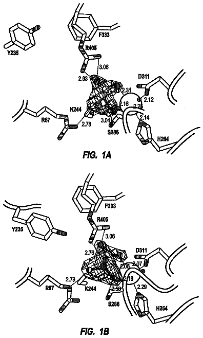
FIG. 1 presents a diagram showing an HMGB1 expression vector.
FIG. 2 presents a diagram showing administration of skin extract (SE) to a mouse via caudal vein, followed by collection of peripheral blood.
FIG. 3 shows in a diagram flow cytometric fractionation of a mouse peripheral blood mononuclear cell fraction fluorescently labeled with anti-mouse PDGFR.alpha. antibody and anti-mouse CD44 antibody 12 hours after administration of skin extract (SE). The upper three charts correspond to the PBS-administered group (n=3) as a negative control, while the lower three charts correspond to the skin extract (SE)-administered group (n=3). The vertical axis indicates the expression level of CD44, and the horizontal axis indicates the expression level of PDGFR.alpha.. The area boxed with blue line corresponds to a population of CD44 and PDGFR.alpha. double-positive cells. The population was increased in the skin extract-administered group (SE) as compared to the PBS-administered group.
FIG. 4 shows in a diagram administration of HMGB1 to a mouse via caudal vein, followed by collection of peripheral blood.
FIG. 5 shows in a diagram flow cytometric fractionation of mouse peripheral blood mononuclear cell fraction fluorescently labeled with anti-mouse PDGFR.alpha. antibody and anti-mouse CD44 antibody 12 hours after administration of HMGB1. The left chart corresponds to the PBS-administered mice as a negative control, while the right chart corresponds to the HMGB1-administered mice. The vertical axis indicates the expression level of CD44, and the horizontal axis indicates the expression level of PDGFR.alpha.. The area boxed with blue line corresponds to a population of CD44 and PDGFR.alpha. double-positive cells. The population was increased in the HMGB1-administered mice as compared to the PBS-administered mice.
FIG. 6 shows in a set of photographs Western blot detection of the HMGB family in neonatal mouse skin extract.
FIG. 7 shows in a set of photographs Western blot results for the purified recombinant Flag tag-HMGB family-fusion proteins expressed in HEK293 cells.
FIG. 8 presents a set of graphs showing the migration activity of bone marrow mesenchymal stem cells by recombinant HMGB1/HMGB2/HMGB3 using a Boyden chamber. All recombinant proteins showed higher migration activities as compared to the control groups.
FIG. 9 shows in a set of graphs the result of treatment in a mouse cutaneous ulcer treatment model using the HMGB family proteins. HMGB1, HMGB2, and HMGB3 all showed significant effects on reducing the ulcer area as compared to the control groups.
FIG. 10 shows in a photograph the activity of human HMGB1 and a human skin extract to induce the migration of human bone marrow-derived mesenchymal stem cells as confirmed using a Boyden chamber.
FIG. 11 shows in a set of photographs the activity of activators purified on a heparin column from mouse heart, brain, and skin extracts to induce bone marrow mesenchymal stem cells as confined using a Boyden chamber.
FIG. 12 shows in a set of photographs the activity of cultured cell line HEK293 extract and HeLa extract to induce the migration of human bone marrow mesenchymal stem cells as confirmed using a Boyden chamber. Both cultured cell lines showed chemotactic activity for human bone marrow mesenchymal stem cells.
FIG. 13A shows in a photograph a mouse fixed to a brain stereotaxic apparatus and subjected to a midline incision in the head with a scalpel, followed by trepanation using a drill. FIG. 13B is a photograph showing the brain to which a negative pressure is applied using a syringe to suck part of the brain tissue. FIG. 13C is a photograph of a mouse after injection of 5 .mu.l heparin-column purified fraction of a skin extract dissolved in fibrin adhesive formulation (fibrinogen), and a subsequent injection of 5 .mu.l of fibrin glue formulation (thrombin). FIGS. 13D and 13E are photographs of the brain injury model taken 2 weeks after the treatment. Higher accumulation of GFP-positive cells was observed in the treatment group using the heparin-column purified fraction of skin extract in E compared to the control in D. FIGS. 13F and 13G are photographs of the brain injury model taken 6 weeks after the treatment. Higher accumulation of GFP-positive cells was observed in the treatment group using the heparin-column purified fraction of skin extract in G compared to the control in F.
FIG. 14A shows in a diagram the flow cytometry result that shows the presence of cells having CD44 and PDGFR.alpha.. HMGB1 administration increased both populations of PDGFR.alpha. and CD44 double-positive cells, and PDGFR.alpha.-positive CD44-negative cells in peripheral blood. FIGS. 14B and 14C show results of comparison between the PBS- and HMGB1-administered groups on the presence of PDGFR.alpha. and CD44 double-positive cells, and PDGFR.alpha.-positive CD44-negative cells in peripheral blood, respectively. Both cell populations were statistically significantly increased in the HMGB1-administered group.
Mode for carrying out the invention
The present invention provides pharmaceutical agents for mobilizing bone marrow cells to peripheral blood from bone marrow, comprising any one of the following ingredients (a) to (i), which is to be administered to peripheral blood or muscle: (a) an HMGB1 protein; (b) a cell secreting an HMGB1 protein; (c) a vector inserted with a DNA which encodes an HMGB1 protein; (d) an HMGB2 protein; (e) a cell secreting an HMGB2 protein; (f) a vector inserted with a DNA which encodes an HMGB2 protein; (g) an HMGB3 protein; (h) a cell secreting an HMGB3 protein; and (i) a vector inserted with a DNA which encodes an HMGB3 protein.
Bone marrow tissue stem cells are mobilized into peripheral circulation by administering the above-described pharmaceutical agents to blood vessel or muscle, and thus regeneration of the damaged tissue can be promoted. Further, besides the use of an above pharmaceutical agent as an inducer/promoter of functional tissue regeneration, its use as a so-called preventive drug which prevents deteriorations in tissue/organ functions caused by the reduction of tissue stem cells, or as an anti-aging drug which delays the progress of age-related alterations is anticipated.
Alternatively, such treatment can be achieved by administering the above-described pharmaceutical agent, collecting and concentrating the pluripotent stem cells mobilized to peripheral blood outside the body, and administering the cells to lesion sites. Conventional therapy using bone marrow mesenchymal stem cells is invasive because cells are collected from the bone marrow located deeply inside the body. Meanwhile, by using the pharmaceutical agents of the present invention, bone marrow mesenchymal stem cells can be collected from peripheral blood in a less-invasive fashion and used for cell transplantation or such.
The present invention relates to pharmaceutical agents for mobilizing bone marrow cells to peripheral blood from bone marrow, which comprise extracts of cells or tissues to be administered to blood vessel or muscle and that are prepared by methods comprising the step of immersing cells or tissues in a solvent.
Cells or tissues to be immersed in a solvent are not specifically limited, and examples include tissue-derived cells and cell lines established from tissue-derived cells (such as HeLa and HEK293, but not limited thereto), isolated cells, non-isolated cells (such as cells existing in isolated tissues), cells introduced with a DNA encoding an HMGB1, HMGB2, or HMGB3 protein. Any tissue may be used as the tissue described as above. For example, such tissues include, but are not limited to, living skin tissues or tissues obtained from internal biopsies (operations) (such as brain, lung, heart, liver, stomach, small intestine, large intestine, pancreas, kidney, bladder, spleen, uterus, testis, and blood).
Examples of the above solvent include, but are not limited to, physiological saline, PBS (phosphate-buffered saline), and TBS (Tris-buffered saline). Moreover, the immersion time of cells or tissue in a solvent should be a duration necessary and sufficient for inducing cell necrosis, that is, 1 hour to 48 hours (such as 6 to 48 hours), and preferably 12 to 24 hours, but is not limited thereto. Therefore, the "step of immersing cells in a solvent" can be rephrased as "step of immersing cells in a solvent for a duration necessary and sufficient for inducing necrosis" or "step of necrosing cells". Moreover, examples of the temperature for immersing cells or tissue in a solvent include, but are not limited to, 4.degree. C. to 25.degree. C. (such as 4.degree. C. to 8.degree. C.), and preferably 4.degree. C. Further, examples of the pH for immersing cells or tissue in a solvent include, without limitation, pH 7 to 8, and preferably pH 7.5. Examples of the buffer include, without limitation, a phosphate buffer solution at a concentration of 10 mM to 50 mM, preferably 10 to 20 mM.
Moreover, in the present invention, cells or tissues can be removed from a solvent containing them after they are immersed in the solvent. The method for removing cells or tissues from a solvent is not particularly limited as long as the method is well known to those skilled in the art. For example, cells or tissues can be removed from a solvent by centrifuging at a gravity acceleration of 10 G to 100,000 G (for example, 440 G) at 4.degree. C. to 25.degree. C. (for example, 4.degree. C.), followed by separation of the supernatant, but the removal method is not limited thereto. The supernatant can be used as an extract of cells or tissues.
The extracts of cells or tissues of the present invention prepared by methods comprising the step of immersing cells or tissues in a solvent include, for example, skin extract and peripheral blood mononuclear cell extract (peripheral blood extract), but are not limited thereto.
The peripheral blood extract is prepared by the following method: after collecting blood with a syringe or the like, the cells are frozen in a freezer or liquid nitrogen, on dry ice, or such, and then thawed at a temperature of 0.degree. C. or higher. Then, to remove insoluble cellular components, the sample is centrifuged, for example, at a gravity of 10 to 100,000 G (for example, at 440 G) and 4.degree. C. to 25.degree. C. (for example, at 4.degree. C.), and the resulting supernatant is collected. The insoluble cellular components can be removed from the solvent by the method described above. However, methods for removing insoluble cellular components are not limited to the above example. The resulting supernatant can be used as an extract of cells or tissues. Alternatively, instead of centrifugation, insoluble cellular components can be removed by filtration through a nitrocellulose filter with micro pores of 0.45 .mu.m, or the like. Alternatively, collected peripheral blood may be allowed to stand for three to 48 hours at 4.degree. C. to induce cell necrosis. The intracellular components can be released from peripheral blood cells by this treatment. Then, to remove insoluble cellular components from the solvent, the sample is centrifuged at a gravity of 10 to 100,000 G (for example, at 440 G), and the resulting supernatant is collected. The insoluble cellular components can be removed from the solvent by the method described above, but are not limited thereto. The resulting supernatant can be used as an extract of cells or tissues. Alternatively, instead of centrifugation, insoluble cellular components can be removed by filtration through a nitrocellulose filter with micro pores of 0.45 .mu.m of the like.
Meanwhile, cell extracts are prepared from peripheral blood mononuclear cells by the following method: peripheral whole blood is collected using a syringe or the like, and then the whole sample is diluted up to 4 ml with PBS. After 3 ml of Ficoll-Paque Plus (GE) is placed into a centrifuge tube, the diluted blood is overlaid onto the Ficoll layer. The tube is centrifuged at 400 G and 18.degree. C. for 40 minutes. The resulting middle layer containing mononuclear cells is transferred into a fresh centrifuge tube, and 45 ml of PBS is added thereto. The sample is centrifuged at 800 G and 18.degree. C. for five minutes, and the resulting supernatant is removed. 45 ml of PBS is added to the cells again, and the sample is centrifuged at 800 G and 18.degree. C. for five minutes. The resulting supernatant is removed. 200 .mu.l of PBS is added to the precipitated cells and suspended. The cell suspension is allowed to stand at -80.degree. C. in a freezer for 30 minutes. The frozen suspension is taken out of the freezer and thawed on ice. The freeze-thaw treatment is repeated three times. Then, the sample is centrifuged at 800 G and 4.degree. C. for 15 minutes, and the supernatant is collected. Alternatively, instead of freezing the cells, the cell sample may be allowed to stand at 4.degree. C. for three to 48 hours to induce cell necrosis. The intracellular components can be released by this treatment. Alternatively, the cells may be disrupted by sonication with cooling on ice. The intracellular components can be released by this treatment. In either case, after releasing the intracellular components to the outside of the cells, the sample is centrifuged at a gravity of 440 to 1,000,000 preferably 20,000 to 100,000 G. The resulting supernatant is collected as a cell extract. Alternatively, instead of centrifugation, insoluble components can be removed by filtration through a nitrocellulose or cellulose acetate filter with micro pores of 0.45 .mu.m, or the like. The resulting filtrate is used as a cell extract.
Further, the present invention relates to pharmaceutical agents to be administered to blood vessel or muscle for use in mobilizing bone marrow cells to peripheral blood from bone marrow, comprising a heparin-binding fraction produced by a method that comprises the following steps: (a) immersing the cells or tissue in a solvent; (b) contacting an extract obtained by the step (a) with immobilized heparin; and (c) eluting a heparin-binding fraction (may also be expressed as heparin-purified fraction or heparin-column purified fraction) from the immobilized heparin.
"Immobilized heparin" refers to heparin covalently bound to an insoluble carrier. Examples of the insoluble carrier include, but are not limited to, Sepharose beads (such as Sepharose 4B, Sepharose 6B and such: GE Healthcare). In the present invention, a commercially available immobilized heparin (Hitrap Heparin HP column: GE Healthcare) may also be used.
Examples of conditions for contacting an extract of cells or tissues with immobilized heparin include, but are not limited to, about pH 7 to 8 (preferably pH 7.5), and a salt concentration of 0 to 200 mM, and preferably about 100 to 200 mM. The time the extract is in contact with immobilized heparin is not specifically limited, but the contact is preferably retained for 5 minutes or more in view of sufficient adsorption of the heparin-binding fraction onto immobilized heparin. Examples of the temperature include, but are not limited to, 4 to 8.degree. C., and preferably 4.degree. C. Further, examples of the elution condition of the heparin-binding fraction adsorbed onto the immobilized heparin include, but are not limited to, a pH of about 7 to 8 and a salt concentration of 200 to 1,000 mM (preferably about 1,000 mM).
When administered to blood vessel or muscle, pharmaceutical agents containing the extract or fraction described above mobilize bone marrow tissue stem cells into the peripheral blood circulation and can promote the regeneration of damaged tissues. Furthermore, the above-described pharmaceutical agents are expected to be used not only as an inducer/promoter for functional tissue regeneration, but also as a so-called preventive drug to prevent the functional impairment of tissues/organs caused by reduction in the number of tissue stem cells or as an anti-aging drug to delay the progress of age-related changes.
Alternatively, such treatment can be achieved by administering the agent described above; collecting and concentrating the pluripotent stem cells mobilized to peripheral blood outside the body, and then administering the cells to lesion sites. Conventional therapy using bone marrow mesenchymal stem cells is invasive because cells are collected from the bone marrow located deeply inside the body. Meanwhile, by using the agents of the present invention, bone marrow mesenchymal stem cells can be collected from peripheral blood in a less invasive manner and used for bone marrow mesenchymal stem cell transplantation or such.
The present invention also provides kits for mobilizing a bone marrow cell to peripheral blood from bone marrow, which consists of compositions to be administered to blood vessel or muscle and which comprise the materials of any one of: (a) an HMGB1 protein; (b) a cell that secretes an HMGB1 protein; (c) a vector inserted with a DNA encoding an HMGB1 protein; (d) an HMGB2 protein; (e) a cell that secretes an HMGB2 protein; (f) a vector inserted with a DNA encoding an HMGB2 protein; (g) an HMGB3 protein; (h) a cell that secretes an HMGB3 protein; and (i) a vector inserted with a DNA encoding an HMGB3 protein.
Furthermore, the present invention provides kits for mobilizing bone marrow cells to peripheral blood from bone marrow, which comprise extracts of cells or tissues produced by a method comprising the step of immersing cells or tissues in a solvent and which are to be administered to blood vessel or muscle.
The present invention also provides kits for mobilizing bone marrow cells to peripheral blood from bone marrow, which comprise a heparin-binding fraction to be administered to blood vessel or muscle and which is produced by a method comprising the steps of: (a) immersing a cell or tissue in a solvent; (b) contacting immobilized heparin with the extract prepared in step (a); and (c) eluting a heparin-binding fraction from the immobilized heparin.
The above-described kits for mobilizing bone marrow cells to peripheral blood are characterized in that administration to blood vessel or muscle mobilizes bone marrow tissue stem cells into peripheral blood circulation.
Examples of the above-described kits include: kits for promoting tissue regeneration comprising
the above-mentioned extract or the above-mentioned fraction or such dissolved in fibrinogen, and
thrombin; or alternatively, kits for promoting tissue regeneration comprising
the above-mentioned extract or the above-mentioned fraction or such,
fibrinogen, and
thrombin. In the present invention, commercially available fibrinogen and thrombin can be used. Examples include, but are not limited to, fibrinogen HT-Wf (Benesis-Mitsubishi Pharma), Beriplast (ZLB Behring), Tisseel (Baxter), Bolheal (Kaketsuken), and TachoComb (ZLB Behring).
Bone marrow-derived cells that are mobilized to the damaged tissue differentiate into various types of cells to contribute to functional regeneration of the damaged tissue and maintenance/enhancement of the functions. In the present invention, examples of damaged tissue include, but are not limited to, tissues damaged by various pathological conditions, trauma, burns, inflammation, autoimmunity, gene abnormalities, and the like causing ischemic/hypoperfusive/hypoxic conditions. Damaged tissue also includes necrosed tissues.
Tissues in the present invention are not particularly limited as long as bone marrow-derived cells can differentiate into the tissues. All types of tissues in the living body can be exemplified, such as skin tissue, bone tissue, cartilage tissue, muscle tissue, adipose tissue, cardiac muscle tissue, neurological tissue, pulmonary tissue, gastrointestinal tissues, hepatic/biliary/pancreatic tissues, and genitourinary organs. Moreover, with use of the above tissue regeneration promoters, treatments for inducing functional tissue regeneration becomes possible not only in cutaneous diseases such as intractable cutaneous ulcers, skin wounds, bullosis, and alopecia, but also in tissue damages such as cerebral infarction, myocardial infarction, bone fracture, pulmonary infarction, gastric ulcers, and enteritis. The types of animals to be administered with the above tissue regeneration promoters include human and non-human animals, which can be exemplified by, but are not limited to, humans, mice, rats, monkeys, pigs, dogs, rabbits, hamsters, and guinea pigs.
Bone marrow cells of the present invention are cells other than hematopoietic stem cells, or cells derived therefrom such as leukocytes, erythrocytes, and platelets, and include stem cells represented by cells which have been hitherto called bone marrow mesenchymal stem cells, bone marrow stromal pluripotent stem cells, or bone marrow pluripotent stem cells and tissue progenitor cell populations existing in the bone marrow. Bone marrow cells of the present invention can be isolated from bone-marrow extracts (bone marrow cell extracts) or peripheral blood collection. Hematopoietic stem cells are nonadherent, while bone marrow cells of the present invention are obtained as adherent cells by means of a cell culture of a mononuclear cell fraction of blood obtained from the bone marrow extracts (bone marrow cell extracts) or peripheral blood collection. Moreover, bone marrow cells of the present invention include mesenchymal stem cells, and have a potential to differentiate into, preferably, osteoblasts (the induction of differentiation can be identified by observing calcification), chondrocytes (which can be identified by alcian blue positive staining, safranin O positive staining, or the like), adipocytes (which can be identified by Sudan III positive staining), and other mesenchymal cells such as fibroblasts, smooth muscle cells, stromal cells, and tendon cells; and further nerve cells, epithelial cells (for example, epidermal keratinocytes and intestinal epithelial cells express cytokeratin family), and vascular endothelial cells. However, the cells to be differentiated into are not limited to the above cells, and the potential to differentiate into cells of parenchymatous organs such as liver, kidney, and pancreas are also included.
The description continues in the full USPTO document.
 Lapsed, fee not paid89 drawings