 Lapsed, fee not paid14 drawings
Biotech & Lab · US 8,636,956 B2
Sensor for measuring carbon dioxide in respiratory gas
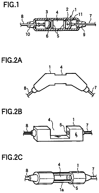
A sensor main unit 1 is axially split into three sub-segments.
US 8,637,016 B2 · Assignee: KaloBios Pharmaceuticals, Inc. · Inventors: Lackmann; Martin et al.
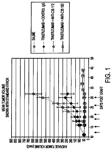
Sheet 1 of 6 from the published document. All sheets in the USPTO PDF
The invention provides methods and compositions comprising anti-EphA3 antibodies for the treatment of solid tumors.
Eph receptor tyrosine kinases (Ephs) belong to a large group of receptor tyrosine kinases (RTKs), kinases that phosphorylate proteins on tyrosine residues. Ephs and their membrane bound ephrin ligands (ephrins) control cell positioning and tissue organization (Poliakov, et al., Dev Cell 7:465-80, 2004). In contrast to other receptor tyrosine kinases, Eph receptor activation does not only require ligand binding and dimerization, but also involves preformed ligand oligomers. Thus, tyrosine phosphorylation of Eph receptors requires presentation of ephrin ligands in their clustered or membrane-attached forms (Davis et al., Science 266:816-819, 1994). Functional and biochemical Eph responses occur at higher ligand oligomerization states (Stein et al., Genes Dev 12:667-678, 1998). Among other patterning functions, various Ephs and ephrins have been shown to play a role in vascular development.
All 6 drawing sheets from the published document, cropped to the drawing.
What the patent claimed, word for word. All of it is now free to use.
Eph receptor tyrosine kinases (Ephs) belong to a large group of receptor tyrosine kinases (RTKs), kinases that phosphorylate proteins on tyrosine residues. Ephs and their membrane bound ephrin ligands (ephrins) control cell positioning and tissue organization (Poliakov, et al., Dev Cell 7:465-80, 2004). In contrast to other receptor tyrosine kinases, Eph receptor activation does not only require ligand binding and dimerization, but also involves preformed ligand oligomers. Thus, tyrosine phosphorylation of Eph receptors requires presentation of ephrin ligands in their clustered or membrane-attached forms (Davis et al., Science 266:816-819, 1994). Functional and biochemical Eph responses occur at higher ligand oligomerization states (Stein et al., Genes Dev 12:667-678, 1998).
Among other patterning functions, various Ephs and ephrins have been shown to play a role in vascular development. Knockout of EphB4 and ephrin-B2 results in a lack of the ability to remodel capillary beds into blood vessels (Poliakov, et al., supra) and embryonic lethality. Persistent expression of some Eph receptors and ephrins has also been observed in newly-formed, adult micro-vessels (Brantley-Sieders, et al., Curr Pharm Des 10:3431-42, 2004; Adams, J Anat 202:105-12, 2003).
The de-regulated re-emergence of some ephrins and their receptors in adults also has been observed to contribute to tumor invasion, metastasis and neo-angiogenesis (Nakamoto, et al., Microsc Res Tech 59:58-67, 2002; Brantley-Sieders, et al., surpa). Furthermore, some Eph family members have been found to be over-expressed on tumor cells from a variety of human tumors (Brantley-Sieders, D. et al., supra); Marme, Ann Hematol 81 Suppl 2:S66, 2002; Booth, et al., Nat Med 8:1360-1, 2002).
Dominant-negative, soluble EphA2 or A3 proteins exhibit effects on ephrin-induced endothelial cell functions in vitro, and tumor angiogenesis and progression in vivo (Brantley, et al. Oncogene 21:7011-26, 2002; Cheng, et al. Neoplasia 5:445-56, 2003; Dobrzanski, et al. Cancer Res 64:910-9, 2004). However, because of lack of specificity of ephrin-A family members for Eph A receptors, these studies do not indicate whether EphA3 itself plays a role in the vascular endothelium in either tumor or normal tissues.
In summary, prior to the current invention, there has been no evidence that EphA3 is expressed on endothelial cells present in the tumor vasculature. Indeed, no vasculature abnormalities have been reported in EphA3 knockout mice (see, e.g., Vaidya et al. Mol. Cell. Biol. 23:8092-8098, 2003). Thus, although certain Eph receptors and ephrins have been implicated as playing a role in angiogenesis and tumor formation and progression, there have been no specific therapies that target EphA3 expression on tumor endothelial cells. This invention therefore provides new therapeutic targets and methods of treating tumors.
The invention is based on the discovery that EphA3 is expressed on the vasculature of solid tumors. Thus, in one aspect, the invention provides a method of inhibiting growth of a solid tumor that does not express EphA3 on tumor cells, the method comprising administering an anti EphA3 antibody. In some embodiments, the anti EphA3 antibody clusters EphA3, e.g., through Fc receptor binding, on the surface of cells that express it, particularly endothelial cells of the vasculature of a tumor. In some embodiments, the EphA3 antibody activates EphA3, even when a natural ligand is bound to EphA3.
The invention also provides a method of inhibiting tumor growth, comprising administering to a patient that has a solid tumor: a) an anti-EphA3 antibody that clusters EphA3, e.g., through Fc receptor binding, and b) a cancer therapeutic agent. In some embodiments, the cancer therapeutic agent disrupts tubulin assembly. The therapeutic agent can be administered concurrently with the anti EphA3 antibody, or following treatment with the anti-EphA3 antibody. In some embodiments, the therapeutic agent is covalently linked to the anti-EphA3 antibody. In other embodiments, the therapeutic agent is a separate molecule that is not linked to the EphA3 antibody. In some embodiments, the anti EphA3 antibody competes for EphA3 binding with monoclonal antibody IIIA4 (mAb IIIA4) and clusters EphA3. In some embodiments, the antibody activates EphA3.
In another aspect, the invention provides a composition comprising an anti EphA3 antibody having an active human isotype, where the antibody clusters EphA3, e.g., through Fc receptor binding. In one embodiment, the anti EphA3 antibody competes with mAb IIIA4 for binding to EphA3. In some embodiments, the antibody competes with mAb IIIA4 for binding to EphA3 and does not block binding of an ephrin, e.g., ephrin-A5, to EphA3. In another embodiment, the antibody binds to EphA3 and clusters EphA3, but does not compete with mAb IIIA4 for binding to EphA3. In some embodiments, the antibody activates EphA3. A composition of the invention can also include another agent that inhibits tumor growth, e.g., an agent that inhibits tubulin assembly.
An anti-EphA3 antibody for use in the methods and/or compositions of the invention can be a recombinant or chimeric antibody. In another embodiment, the antibody is a human antibody, e.g., a humaneered antibody or a humanized antibody. In a further embodiment, the antibody is a polyclonal antibody. Alternatively, the antibody can be a monoclonal antibody. In an additional embodiment, the antibody is a multivalent antibody that comprises an antibody fragment that is a Fab, a Fab', or an Fv. In another embodiment, the antibody has an active human isotype, e.g., IgG1, IgG3, IgM, IgA, or IgE, that binds to Fc receptors on immune effector cells. Thus, in some embodiments the antibody comprises a human heavy chain constant region, e.g., an IgG1 or IgG3 gamma region. In some embodiments, the antibody may be chemically cross-linked IgG.
In some embodiments, an antibody for use in the methods and/or compositions of the invention comprises the V.sub.H and V.sub.L regions of mAb IIIA4. In other embodiments, the antibody comprises the V.sub.H and V.sub.L region CDR1, CDR2 and CDR3 of mAb IIIA4. In further embodiments, the antibody comprises the V.sub.H region CDR3 and V.sub.L region CDR3 of mAb IIIA4. In some embodiments, the antibody comprises a heavy chain CDR1, CDR2, and CDR3 from Table 1 and a light chain CDR1, CDR2, and CDR3 from Table 1. In additional embodiments, the antibody comprises a heavy chain CDR3 from Table 1 and a light chain CDR3 from Table 1.
The invention additionally provide a method of inhibiting the growth of solid tumors (whether or not the tumor cells express EpA3) by administering a monomeric, non-aggregated antibody preparation, where the antibody can cluster EphA3. Such antibodies, e.g., have an active isotype, e.g., have a human IgG1 or IgG3 gamma region. In some embodiments, the antibody activates EphA3. The antibody can be a recombinant or chimeric antibody. In another embodiment, the antibody is a human antibody, e.g., a humaneered antibody or a humanized antibody. In some embodiments, the antibody is a Fab, a Fab', or an Fv that is in a multivalent form, e.g., a tri-Fab. In some embodiments, an antibody for use in the methods and/or compositions of the invention comprises the V.sub.H and V.sub.L regions of mAb IIIA4. In other embodiments, the antibody comprises the V.sub.H and V.sub.L region CDR1, CDR2 and CDR3 of mAb IIIA4; or a heavy chain CDR1, CDR2 and CDR3 from Table 1 and a light chain CDR1, CDR2, and CDR3 from Table 1. In further embodiments, the antibody comprises the V.sub.H region CDR3 and V.sub.L region CDR3 of mAb IIIA4. In additional embodiments, the antibody comprises a heavy chain CDR3 from Table 1 and a light chain CDR3 from Table 1.
In some embodiments, the invention provides a method of inhibiting the growth of solid tumors by administering an antibody to EphA3 that clusters EphA3, e.g., through Fc receptor binding, present on tumor vascular endothelial cells with the proviso that antibody is not conjugated to a therapeutic agent such as a radiometal or toxin. In some embodiments, the antibody activates EphA3.
In some embodiments, the invention provides a method of inhibiting the growth of solid tumors by administering an antibody to EphA3 that is chemically cross-linked.
In another aspect, the invention provides a method of treating solid tumors that comprises administering a smaller dose of antibody compared to treatment regimens that target proteins on the surface of tumor cells. The method targets the EphA3 receptors present on tumor vasculature endothelial cells. Accordingly, an antibody can be administered at a dose of less than about 1.0 mg/kg, preferably less than about 0.5 mg/kg, or less than 0.1 mg/kg, to inhibit growth of the tumor. Such an antibody can be any antibody of the invention as described herein that binds to and clusters EphA3. In some embodiments, the antibody activates EphA3.
The invention also provides a method of inhibiting tumor growth by administering an EphA3 binding agent, e.g., a multivalent form of a scaffolded protein, or an antibody, that specifically binds to EphA3 and clusters, the EphA3 receptor. In typical embodiments, clustering induced by an EphA3 binding agent such as an antibody can take place even when natural ligand, for example and ephrin such as ephrin-A5, is bound to EphA3. The binding agent can be a multivalent form of scaffolded proteins that bind to EphA3. In some embodiments, the EphA3 binding agent competes with mAb IIIA4 for binding to EphA3. Administration of anti-EphA3 binding agents to tumors is exemplified by the use of anti-EphA3 antibodies. However, the methods described herein can also be used for other anti-EphA3 binding agents.
The EphA3 binding agents of the invention, e.g., an EphA3 antibody that clusters EphA3 receptors, can also be used for the treatment of other diseases that involve neovascularization. For example, an EphA3 antibody related as described herein can be used for the treatment of retinal vascular diseases, such as age-related macular degeneration or other intraocular neovascular syndromes. Thus, other non-neoplastic conditions that can be treated with the EphA3 agents described herein include rheumatoid arthritis, psoriasis, atherosclerosis, diabetic and other proliferative retinopathies including retinopathy of prematurity, retrolental fibroplasia, neovascular glaucoma, age-related macular degeneration, thyroid hyperplasias (including Grave's disease), hemangiomas, corneal and other tissue transplantation, preeclampsia, and chronic inflammation.
FIGS. 1a-c. Detection of EphA3 using the EphA3 monoclonal antibody IIIA4 (mAb IIIA4) in (a) human malignant melanoma sections or (b), by analysis of various endothelial cell lines using flow cytometry, and (c) by IP/Western Blot analysis of parental or EphA3-overexpressing HEK293T cells, SK-MeI melanoma cells, TEC-28 kidney tumor endothelial cells, brain microvascular endothelial cells (b-MVEC) or myometrial MVECS (m-MVEC).
FIGS. 2a-b. Expression of EphA3 on endometrial endothelial cells is lost during extended tissue culture. (a) The expression of the various cell surface markers as well as EphA3, detected by immunocytochemical analysis in FIG. 4, was examined by flow cytometry. The EphA3 expression profile of EphA3/HEK-293T cells is shown for comparison. (b) IP/Western blot analysis of EphA3 expression in successive passages of endometrium-derived MVECS as indicated (P4-P9). EphA3 was immunoprecipitated from whole-cell lysates with IIIA4-Sepharose and Western blots probed with anti-EphA3 polyclonal antibodies.
FIG. 3. Estimation of EphA3 mRNA expression levels by quantitative real-time PCR. Total mRNA was extracted from mMVECS isolated from various endometrial tissue samples. The levels of .beta.-actin were determined in parallel as internal reference, while mRNA from HEK293T cells served as a positive control for EphA3 expression, expressed as ratio between .beta.-actin and EphA3 mRNA levels. Mean and SD from three independent samples are illustrated.
FIGS. 4a-c. Expression of EphA3 in human 22RV1 prostate carcinoma cells. (a) EphA3 expression was examined by flow cytometry, using mAb IIIA4 and fluorescine-conjugated anti-mouse antibody for detection. (b) The levels of EphA3 expression was estimated and compared to the expression in EphA3/HEK293T cells by IP/Western Blot analysis of whole cell lysates. In parallel samples the EphA3 tyrosine phosphorylation following stimulation of cells with pre-clustered ephrin-A5 Fc or ch-IIIA4 was assessed using an anti-PY EphA3 polyclonal antibody for Western blot analysis. (c) 22RV1 cells were cultured on fibronectin-coated glass slides and incubated with Alexa .sup.546IIIA4 in the presence or absence of ephrin-A5 Fc, as indicated. The actin cytoskeleton of fixed and permeabilised cells was stained with Alexa.sup.488Phalloidin.
FIGS. 5a-b. Anti-EphA3 antibody inhibits the growth of EphA3 antigen-negative tumor xenografts in vivo. Nude mice bearing human DU-145 prostate cancer cells were treated twice weekly for 6 weeks with chimeric IIIA4 antibody (10 mg/kg; i.p.) or vehicle control. a) mean tumor volumes (determined using vernier calipers) up to 50 days after the end of treatment. b) tumor weights (mean.+-.standard deviation) at necropsy 50 days after the end of treatment.
FIGS. 6a-b. Anti-EphA3 antibody inhibits the growth of EphA3 antigen-positive tumor xenografts in vivo. Nude mice bearing human LNCaP prostate cancer cells were treated twice weekly for 6 weeks with chimeric IIIA4 antibody (10 mg/kg; i.p.) or vehicle control. a) mean tumor volumes (determined using vernier calipers) up to 22 days after the end of treatment. b) tumor weights (mean.+-.standard deviation) at necropsy 25 days after the end of treatment.
Definitions
As used herein "solid tumor" refers to an abnormal mass of tissue. Solid tumors may be benign or malignant. Solid tumors that can be treated using the methods and compositions of the invention are characterized by neovascularization. The tumor vasculature (also referred to as microvasculature) is characterized by rapid proliferation of the endothelial cells, poor wall structure, increased permeability to plasma proteins, and a limited ability to increase blood flow in response to demand. The tumor vasculature allows the tumor cells of the tumor mass to acquire a growth advantage compared to the normal cells. Solid tumors are named for the type of cells that form them. Examples of solid tumors are sarcomas, carcinomas (epithelial tumors), melanomas, and glioblastomas.
"Inhibiting growth of a tumor" in the context of the invention refers to slowing tumor growth and/or reducing tumor size. "Inhibiting growth of a tumor" thus includes killing tumor cells as well as slowing or arresting tumor cell growth.
The term "tumor cell" as used herein refers to a neoplastic cell. The term includes cancer cells that are benign as well as malignant. Neoplastic transformation is associated with phenotypic changes of the tumor cell relative to the cell type from which it is derived. The changes can include loss of contact inhibition, morphological changes, and aberrant growth. (see, Freshney, Culture of Animal Cells a Manual of Basic Technique (3.sup.rd edition, 1994). In the context of the current invention, a "tumor cell" does not refer to the cells of the vasculature of the tumor.
As used herein, "tumor vasculature endothelial cells" are endothelial cells that are present in the vasculature of a tumor.
As used herein "EphA3" refers to the Eph receptor A3. This receptor has also been referred to as "Human embryo kinase", "hek", "eph-like tyrosine kinase 1", "etk1" or "tyro4". EphA3 belongs to the ephrin receptor subfamily of the protein-tyrosine kinase family. EPH and EPH-related receptors have been implicated in mediating developmental events. Receptors in the EPH subfamily typically have a single kinase domain and an extracellular region containing a Cys-rich domain and 2 fibronectin type III repeats. The ephrin receptors are divided into 2 groups based on the similarity of their extracellular domain sequences and their affinities for binding ephrin-A and ephrin-B ligands. EphA3 binds ephrin-A ligands. EphA3 nucleic acid and protein sequences are known. An exemplary human EphA3 amino acid sequence is available under accession number (EAW68857).
In the present invention, "activation" of EphA3 causes phosphorylation of EphA3 and typically, rounding of the cell.
As used herein, "clustering" or "cross-linking" of EphA3 refers to cross-linking of EphA3 molecules on the surface of a cell. Clustering generally forms an active signaling complex that causes phosphorylation of EphA3. "Clustering" is typically a hallmark of EphA3 activation.
The term "non-aggregated" as used herein with reference to a preparation of an antibody that has an active isotype refers to a preparation that has less than about 5%, and in some embodiments less than about 2%, or less than about 1%, of the antibody in an aggregated form, i.e., that is in a form that is more than monomeric.
A "monomeric" antibody as used herein refers to a divalent antibody that has two antigen binding sites.
A "multivalent" antibody or "multivalent" binding agent as used herein refers to an antibody or protein that has more than two antigen binding sites.
A "solid tumor that does not express EphA3 on tumor cells" as used herein refers to a solid tumor that has fewer than about 25% of cells that express EphA3 on the tumor cell. In some embodiments, the solid tumor has fewer than about 15%, or fewer than 10%, or fewer than 5% of cells that express EphA3 on the tumor cell. In further embodiments, a tumor cell that does not express EphA3 refers to a tumor cell that has little or no detectable EphA3 expression, e.g., as detected by immunohistochemistry. "Little detectable EphA3 expression" refers an amount of expression that is less than 2 times the background from a control cell that does not express EphA3.
In the present invention, "EphA3 antibody" or "anti EphA3 antibody" are used interchangeably to refer to an antibody that binds to EphA3. In some embodiments, the antibody clusters EphA3, e.g., through Fc receptor binding. The term encompasses antibodies that bind to EpbA3 in the presence of ephrin ligand (e.g., ephrin-A5) binding, as well as antibodies that bind to the ligand binding site.
An "EphA3 antibody that binds to EphA3 in the presence of binding of an ephrin ligand" refers to an antibody that does not significantly prevent binding of an ephrin ligand, such as ephrin-A5, to EphA3. The presence of such an antibody in a binding reaction comprising EphA3 and an ephrin ligand, e.g., ephrin-A5, reduces ephrin ligand binding to EphA3 by less than about 30%, typically less than 20% or 10%.
The term "mAb IIIA4" refers to monoclonal antibody IIIA4 that was originally raised against LK63 human acute pre-B leukemia cells to affinity isolate EphA3 (Boyd, et al. J Biol Chem 267:3262-3267, 1992). mAb IIIA4 binds to the native EphA3 globular ephrin-binding domain (e.g., Smith, et al., J. Biol. Chem 279:9522-9531, 2004). It is deposited in the European Collection of Animal Cell Cultures under accession no. 91061920 (see, e.g., EP patent no. EP0590030).
An "antibody having an active isotype" as used herein refers to an antibody that has a human Fc region that binds to an Fc receptor present on immune effector cells. "Active isotypes" include IgG1, IgG3, IgM, IgA, and IgE. The term encompasses antibodies that have a human Fc region that comprises modifications, such as mutations or changes to the sugar composition and/or level of glycosylation, that modulate Fc effector function.
An "Fc region" refers to the constant region of an antibody excluding the first constant region immunoglobulin domain. Thus, Fc refers to the last two constant region immunoglobulin domains of IgA, IgD, and IgG, and the last three constant region immunoglobulin domains of IgE and IgM, and the flexible hinge N-terminal to these domains. For IgA and IgM Fc may include the J chain. For IgG, Fc comprises immunoglobulin domains C.gamma.2 and C.gamma.3 and the hinge between C.gamma.1 and C.gamma.. It is understood in the art that the boundaries of the Fc region may vary, however, the human IgG heavy chain Fc region is usually defined to comprise residues C226 or P230 to its carboxyl-terminus, using the numbering is according to the EU index as in Kabat et al. (1991, NIH Publication 91-3242, National Technical Information Service, Springfield, Va.). The term "Fc region" may refer to this region in isolation or this region in the context of an antibody or antibody fragment. "Fc region" includes naturally occurring allelic variants of the Fc region as well as modifications that modulate effector function. Fc regions also include variants that don't result in alterations to biological function. For example, one or more amino acids can be deleted from the N-terminus or C-terminus of the Fc region of an immunoglobulin without substantial loss of biological function. Such variants can be selected according to general rules known in the art so as to have minimal effect on activity (see, e.g., Bowie, et al., Science 247:306-1310, 1990).
As used herein, an "antibody" refers to a protein functionally defined as a binding protein and structurally defined as comprising an amino acid sequence that is recognized by one of skill as being derived from the framework region of an immunoglobulin encoding gene of an animal producing antibodies. An antibody can consist of one or more polypeptides substantially encoded by immunoglobulin genes or fragments of immunoglobulin genes. The recognized immunoglobulin genes include the kappa, lambda, alpha, gamma, delta, epsilon and mu constant region genes, as well as myriad immunoglobulin variable region genes. Light chains are classified as either kappa or lambda. Heavy chains are classified as gamma, mu, alpha, delta, or epsilon, which in turn define the immunoglobulin classes, IgG, IgM, IgA, IgD and IgE, respectively.
A typical immunoglobulin (antibody) structural unit is known to comprise a tetramer. Each tetramer is composed of two identical pairs of polypeptide chains, each pair having one "light" (about 25 kD) and one "heavy" chain (about 50-70 kD). The N-terminus of each chain defines a variable region of about 100 to 110 or more amino acids primarily responsible for antigen recognition. The terms variable light chain (V.sub.L) and variable heavy chain (V.sub.H) refer to these light and heavy chains respectively.
The term "antibody" as used herein includes antibody fragments that retain binding specificity. For example, there are a number of well characterized antibody fragments. Thus, for example, pepsin digests an antibody below the disulfide linkages in the hinge region to produce F(ab)'.sub.2, a dimer of Fab which itself is a light chain joined to VH--CH1 by a disulfide bond. The F(ab)'.sub.2 may be reduced under mild conditions to break the disulfide linkage in the hinge region thereby converting the (Fab').sub.2 dimer into an Fab' monomer. The Fab' monomer is essentially an Fab with part of the hinge region (see, Fundamental Immunology, W. E. Paul, ed., Raven Press, N.Y. (1993), for a more detailed description of other antibody fragments). While various antibody fragments are defined in terms of the digestion of an intact antibody, one of skill will appreciate that fragments can be synthesized de novo either chemically or by utilizing recombinant DNA methodology. Thus, the term antibody, as used herein also includes antibody fragments either produced by the modification of whole antibodies or synthesized using recombinant DNA methodologies.
Antibodies include V.sub.H-V.sub.L dimers, including single chain antibodies (antibodies that exist as a single polypeptide chain), such as single chain Fv antibodies (sFv or scFv) in which a variable heavy and a variable light region are joined together (directly or through a peptide linker) to form a continuous polypeptide. The single chain Fv antibody is a covalently linked V.sub.H-V.sub.L which may be expressed from a nucleic acid including V.sub.H- and V.sub.L-encoding sequences either joined directly or joined by a peptide-encoding linker (e.g., Huston, et al. Proc. Nat. Acad. Sci. USA, 85:5879-5883, 1988). While the V.sub.H and V.sub.L are connected to each as a single polypeptide chain, the V.sub.H and V.sub.L domains associate non-covalently. Alternatively, the antibody can be another fragment. Other fragments can also be generated, e.g., using recombinant techniques, as soluble proteins or as fragments obtained from display methods. Antibodies can also include diantibodies and miniantibodies. Antibodies of the invention also include heavy chain dimers, such as antibodies from camelids. For the purposes of this invention, antibodies are employed in a form that can cluster EphA3 present on the surface of endothelial cells. Thus, in some embodiments an antibody is in a monomeric form that has an active isotype. In other embodiments the antibody is in a multivalent form, e.g., a trivalent or tetravalent form, that can cross-link EphA3.
As used herein, "V-region" refers to an antibody variable region domain comprising the segments of Framework 1, CDR1, Framework 2, CDR2, and Framework3, including CDR3 and Framework 4, which segments are added to the V-segment as a consequence of rearrangement of the heavy chain and light chain V-region genes during B-cell differentiation.
As used herein, "complementarity-determining region (CDR)" refers to the three hypervariable regions in each chain that interrupt the four "framework" regions established by the light and heavy chain variable regions. The CDRs are primarily responsible for binding to an epitope of an antigen. The CDRs of each chain are typically referred to as CDR1, CDR2, and CDR3, numbered sequentially starting from the N-terminus, and are also typically identified by the chain in which the particular CDR is located. Thus, a V.sub.H CDR3 is located in the variable domain of the heavy chain of the antibody in which it is found, whereas a V.sub.L CDR1 is the CDR1 from the variable domain of the light chain of the antibody in which it is found.
The sequences of the framework regions of different light or heavy chains are relatively conserved within a species. The framework region of an antibody, that is the combined framework regions of the constituent light and heavy chains, serves to position and align the CDRs in three dimensional space.
The amino acid sequences of the CDRs and framework regions can be determined using various well known definitions in the art, e.g., Kabat, Chothia, international ImMunoGeneTics database (IMGT), and AbM (see, e.g., Johnson et al., supra; Chothia & Lesk, 1987, Canonical structures for the hypervariable regions of immunoglobulins. J. Mol. Biol. 196, 901-917; Chothia C. et al., 1989, Conformations of immunoglobulin hypervariable regions. Nature 342, 877-883; Chothia C. et al., 1992, structural repertoire of the human V.sub.H segments J. Mol. Biol. 227, 799-817; Al-Lazikani et al., J. Mol. Biol. 1997, 273(4)). Definitions of antigen combining sites are also described in the following: Ruiz et al., IMGT, the international ImMunoGeneTics database. Nucleic Acids Res., 28, 219-221 (2000); and Lefranc, M.-P. IMGT, the international ImMunoGeneTics database. Nucleic Acids Res. January 1; 29(1):207-9 (2001); MacCallum et al, Antibody-antigen interactions: Contact analysis and binding site topography, J. Mol. Biol., 262 (5), 732-745 (1996); and Martin et al, Proc. Natl. Acad. Sci. USA, 86, 9268-9272 (1989); Martin, et al, Methods Enzymol., 203, 121-153, (1991); Pedersen et al, Immunomethods, 1, 126, (1992); and Rees et al, In Sternberg M. J. E. (ed.), Protein Structure Prediction. Oxford University Press, Oxford, 141-172 1996).
"Epitope" or "antigenic determinant" refers to a site on an antigen to which an antibody binds. Epitopes can be formed both from contiguous amino acids or noncontiguous amino acids juxtaposed by tertiary folding of a protein. Epitopes formed from contiguous amino acids are typically retained on exposure to denaturing solvents whereas epitopes formed by tertiary folding are typically lost on treatment with denaturing solvents. An epitope typically includes at least 3, and more usually, at least 5 or 8-10 amino acids in a unique spatial conformation. Methods of determining spatial conformation of epitopes include, for example, x-ray crystallography and 2-dimensional nuclear magnetic resonance. See, e.g., Epitope Mapping Protocols in Methods in Molecular Biology, Vol. 66, Glenn E. Morris, Ed (1996).
As used herein, "chimeric antibody" refers to an immunoglobulin molecule in which (a) the constant region, or a portion thereof, is altered, replaced or exchanged so that the antigen binding site (variable region) is linked to a constant region of a different or altered class, effector function and/or species, or an entirely different molecule which confers new properties to the chimeric antibody, e.g., an enzyme, toxin, hormone, growth factor, drug, etc.; or (b) the variable region, or a portion thereof, is altered, replaced or exchanged with a variable region, or portion thereof, having a different or altered antigen specificity; or with corresponding sequences from another species or from another antibody class or subclass.
As used herein, "humanized antibody" refers to an immunoglobulin molecule in which the CDRs of a recipient human antibody are replaced by CDRs from a donor non-human antibody. Humanized antibodies may also comprise residues of donor origin in the framework sequences. The humanized antibody can also comprise at least a portion of a human immunoglobulin constant region. Humanized antibodies may also comprise residues which are found neither in the recipient antibody nor in the imported CDR or framework sequences. Humanization can be performed using methods known in the art (e.g., Jones et al., Nature 321:522-525; 1986; Riechmann et al., Nature 332:323-327, 1988; Verhoeyen et al., Science 239:1534-1536, 1988); Presta, Curr. Op. Struct. Biol. 2:593-596, 1992; U.S. Pat. No. 4,816,567), including techniques such as "superhumanizing" antibodies (Tan et al., J. Immunol. 169: 1119, 2002) and "resurfacing" (e.g., Staelens et al., Mol. Immunol. 43: 1243, 2006; and Roguska et al., Proc. Natl. Acad. Sci. USA 91: 969, 1994).
A "humaneered" antibody in the context of this invention refers to is an engineered human antibody having a binding specificity of a reference antibody. The term refers to an immunoglobulin molecule that contains minimal sequence derived from the reference antibody. Typically, an antibody is "humaneered" by joining a DNA sequence encoding a binding specificity determinant (BSD) from the CDR3 region of the heavy chain of the reference antibody to human V.sub.H segment sequence and a light chain CDR3 BSD from the reference antibody to a human V.sub.L segment sequence. Methods for humaneering are provided in US patent application publication no. 20050255552 and US patent application publication no. 20060134098.
A "human" antibody as used herein encompasses humanized and humaneered antibodies, as well as human monoclonal antibodies that are obtained using known techniques.
The term "heterologous" when used with reference to portions of a nucleic acid indicates that the nucleic acid comprises two or more subsequences that are not normally found in the same relationship to each other in nature. For instance, the nucleic acid is typically recombinantly produced, having two or more sequences, e.g., from unrelated genes arranged to make a new functional nucleic acid. Similarly, a heterologous protein refers to two or more subsequences that are not found in the same relationship to each other in nature.
The term "recombinant" when used with reference, e.g., to a cell, or nucleic acid, protein, or vector, indicates that the cell, nucleic acid, protein or vector, has been modified by the introduction of a heterologous nucleic acid or protein or the alteration of a native nucleic acid or protein, or that the cell is derived from a cell so modified. Thus, e.g., recombinant cells express genes that are not found within the native (non-recombinant) form of the cell or express native genes that are otherwise abnormally expressed, under expressed or not expressed at all. By the term "recombinant nucleic acid" herein is meant nucleic acid, originally formed in vitro, in general, by the manipulation of nucleic acid, e.g., using polymerases and endonucleases, in a form not normally found in nature. In this manner, operable linkage of different sequences is achieved. Thus, an isolated nucleic acid, in a linear form, or an expression vector formed in vitro by ligating DNA molecules that are not normally joined, are both considered recombinant for the purposes of this invention. It is understood that once a recombinant nucleic acid is made and reintroduced into a host cell or organism, it will replicate non-recombinantly, i.e., using the in vivo cellular machinery of the host cell rather than in vitro manipulations; however, such nucleic acids, once produced recombinantly, although subsequently replicated non-recombinantly, are still considered recombinant for the purposes of the invention. Similarly, a "recombinant protein" is a protein made using recombinant techniques, i.e., through the expression of a recombinant nucleic acid as depicted above.
The phrase "specifically (or selectively) binds" to an antibody or "specifically (or selectively) immunoreactive with," when referring to a protein or peptide, refers to a binding reaction that is determinative of the presence of the protein in a heterogeneous population of proteins, such as a cell extract. Thus, under designated immunoassay conditions, the specified antibodies bind to a particular protein sequence at least two times the background and more typically more than 10 to 100 times background.
As used herein, "cancer therapeutic agent" refers to an agent that when administered to a patient suffering from cancer, in a therapeutically effective dose, will cure, or at least partially arrest the symptoms of the disease and complications associated with the disease.
The terms "identical" or percent "identity," in the context of two or more polypeptide (or nucleic acid) sequences, refer to two or more sequences or subsequences, e.g., an antibody sequence, that are the same or have a specified percentage of amino acid residues (or nucleotides) that are the same (i.e., about 60% identity, preferably 70%, 75%, 80%, 85%, 90%, 91%, 92%, 93%, 94%, 95%, 96%, 97%, 98%, 99%, or higher identity over a specified region, when compared and aligned for maximum correspondence over a comparison window or designated region) as measured using a BLAST or BLAST 2.0 sequence comparison algorithms with default parameters described below, or by manual alignment and visual inspection (see, e.g., NCBI web site). Such sequences are then said to be "substantially identical." "Substantially identical" sequences also includes sequences that have deletions and/or additions, as well as those that have substitutions, as well as naturally occurring, e.g., polymorphic or allelic variants, and man-made variants. As described below, the preferred algorithms can account for gaps and the like. Preferably, protein sequence identity exists over a region that is at least about 25 amino acids in length, or more preferably over a region that is 50-100 amino acids=in length, or over the length of a protein.
A "comparison window", as used herein, includes reference to a segment of one of the number of contiguous positions selected from the group consisting typically of from 20 to 600, usually about 50 to about 200, more usually about 100 to about 150 in which a sequence may be compared to a reference sequence of the same number of contiguous positions after the two sequences are optimally aligned. Methods of alignment of sequences for comparison are well-known in the art. Optimal alignment of sequences for comparison can be conducted, e.g., by the local homology algorithm of Smith & Waterman, Adv. Appl. Math. 2:482 (1981), by the homology alignment algorithm of Needleman & Wunsch, J. Mol. Biol. 48:443 (1970), by the search for similarity method of Pearson & Lipman, Proc. Nat'l. Acad. Sci. USA 85:2444 (1988), by computerized implementations of these algorithms (GAP, BESTFIT, FASTA, and TFASTA in the Wisconsin Genetics Software Package, Genetics Computer Group, 575 Science Dr., Madison, Wis.), or by manual alignment and visual inspection (see, e.g., Current Protocols in Molecular Biology (Ausubel et al., eds. 1995 supplement)).
Preferred examples of algorithms that are suitable for determining percent sequence identity and sequence similarity include the BLAST and BLAST 2.0 algorithms, which are described in Altschul et al., Nuc. Acids Res. 25:3389-3402
and Altschul et al., J. Mol. Biol. 215:403-410 (1990). BLAST and BLAST 2.0 are used, with the parameters described herein, to determine percent sequence identity for the nucleic acids and proteins of the invention. The BLASTN program (for nucleotide sequences) uses as defaults a wordlength (W) of 11, an expectation (E) of 10, M=5, N=-4 and a comparison of both strands. For amino acid sequences, the BLASTP program uses as defaults a wordlength of 3, and expectation (E) of 10, and the BLOSUM62 scoring matrix (see Henikoff & Henikoff, Proc. Natl. Acad. Sci. USA 89:10915 (1989)) alignments (B) of 50, expectation (E) of 10, M=5, N=-4, and a comparison of both strands.
An indication that two polypeptides are substantially identical is that the first polypeptide is immunologically cross reactive with the antibodies raised against the second polypeptide. Thus, a polypeptide is typically substantially identical to a second polypeptide, e.g., where the two peptides differ only by conservative substitutions.
The terms "isolated," "purified," or "biologically pure" refer to material that is substantially or essentially free from components that normally accompany it as found in its native state. Purity and homogeneity are typically determined using analytical chemistry techniques such as polyacrylamide gel electrophoresis or high performance liquid chromatography. A protein that is the predominant species present in a preparation is substantially purified. The term "purified" in some embodiments denotes that a protein gives rise to essentially one band in an electrophoretic gel. Preferably, it means that the protein is at least 85% pure, more preferably at least 95% pure, and most preferably at least 99% pure.
The terms "polypeptide," "peptide" and "protein" are used interchangeably herein to refer to a polymer of amino acid residues. The terms apply to amino acid polymers in which one or more amino acid residue is an artificial chemical mimetic of a corresponding naturally occurring amino acid, as well as to naturally occurring amino acid polymers, those containing modified residues, and non-naturally occurring amino acid polymer.
The description continues in the full USPTO document.
About 6,074 words. The USPTO PDF has it with every drawing.
Fees are due 3.5, 7.5 and 11.5 years after grant. This patent expired on January 28, 2026, so the fee marked "not paid" was the one that went unpaid.
EphA3 Antibodies for the Treatment of Solid Tumors
Filed Mar 2008 · published Nov 2008EphA3 antibodies for the treatment of solid tumors
Filed Mar 2008 · granted Jan 2014Earlier publications, parents and continuations. None of them can still be enforced, or this patent would not be listed.
Prior art cited by the examiner or applicant. Useful when you check your own idea for novelty.
Everything on this page comes from the documents linked above.