The present disclosure relates to methods, compositions and articles of manufacture useful for the treatment of Bacillus anthracis and certain related bacteria, spores thereof, and related conditions. The disclosure further relates to methods and compositions for the identification of a phage associated lytic enzyme to rapidly and specifically detect and kill Bacillus anthracis and certain related bacteria.
Background
A major problem in medicine has been the development of drug resistant bacteria as more antibiotics are used for a wide variety of illnesses and other conditions. The use of more antibiotics and the number of bacteria showing resistance has prompted longer treatment times. Furthermore, broad, non-specific antibiotics, some of which have detrimental effects on the patient, are now being used more frequently. A related problem with this increased use is that many antibiotics do not penetrate mucus linings easily. Additionally, the number of people allergic to antibiotics appears to be increasing. Accordingly, there is a commercial need for new antibiotics, especially those that operate in new modalities or provide new means to kill pathogenic bacteria.
Attempts have been made to treat bacterial diseases through the use of bacteriophages. However, the direct introduction of bacteriophages into an animal to prevent or fight diseases has certain drawbacks. Specifically, both the bacteria and the phage have to be in the correct and synchronized growth cycles for the phage to attach. Additionally, there must be the right number of phages to attach to the bacteria; if there are too many or too few phages, there will be either no attachment or no production of the lysing enzyme. The phage must also be active enough. The phages are also inhibited by many things including bacterial debris from the organism it is going to attack. Further complicating the direct use of a bacteriophage to treat bacterial infections is the possibility of immunological reactions, rendering the phage non-functional.
Consequently, others have explored the use of safer and more effective means to treat and prevent bacterial infections. In particular, the use of phage associated lytic enzymes has been explored.
Bacteriophage lysins are a class of bacteriolytic agents recently proposed for eradicating the nasopharyngeal carriage of pathogenic streptococci. (Loeffler, J. M., Nelson, D. & Fischetti, V. A. Rapid killing of Streptococcus pneumoniae with a bacteriophage cell wall hydrolase. Science 294, 2170-2 (2001); Nelson, D., Loomis, L. & Fischetti, V. A. Prevention and elimination of upper respiratory colonization of mice by group A streptococci by using a bacteriophage lytic enzyme. Proc Natl Acad Sci USA 98, 4107-12 (2001)). Lysins are part of the lytic mechanism used by double stranded DNA (dsDNA) phage to coordinate host lysis with completion of viral assembly. Wang, I. N., Smith, D. L. & Young, R. Holins: the protein clocks of bacteriophage infections. Annu Rev Microbiol 54, 799-825 (2000). Late in infection, lysin translocates into the cell wall matrix where it rapidly hydrolyzes covalent bonds essential for peptidoglycan integrity, causing bacterial lysis and concomitant progeny phage release. Lysin family members exhibit a modular design in which a usually well conserved catalytic domain is fused to a more divergent specificity or binding domain. See, Lopez, R., Garcia, E., Garcia, P. & Garcia, J. L. The pneumococcal cell wall degrading enzymes: a modular design to create new lysins? Microb Drug Resist 3, 199-211 (1997).
Summary
The dormant and durable spore form of Bacillus anthracis has been reported to be an ideal biological weapon. (Mock, M. & Fouet, A. Anthrax. Annu Rev Microbiol 55, 647-71 (2001), Inglesby, T. V. et al. Anthrax as a biological weapon, 2002: updated recommendations for management. Jama 287, 2236-52 (2002)). Bacillus anthracis spores are extremely virulent in humans and can remain viable in the environment almost indefinitely after release. Once inhaled, spores are transported by alveolar macrophages to mediastinal and peribronchial lymph nodes where they germinate; subsequent vegetative clonal expansion causes an overwhelming bacteremia and toxemia. Mortality rates associated with untreated inhalational anthrax can reach 99%, with antibiotic treatment being largely unsuccessful if initiated after the onset of non-specific febrile symptoms. The potential for naturally occurring and genetically engineered antibiotic resistance amplifies the threat of weaponized spores and accentuate needs for improved treatments and methods for spore detection following an intentional release.
An embodiment of the disclosure provides for the extraction and use of a variety of bacterial phage associated holin proteins, chimeric lytic enzymes, and shuffled lytic enzymes, in addition to lytic enzymes, for the treatment of Bacillus anthracis. More specifically, the present disclosure provides for a pharmaceutical composition comprising at least one bacteria-associated phage enzyme that is isolated from one or more bacterial species and includes at least one phage lytic enzyme and/or holin protein that may be used for the prophylactic and therapeutic treatment of Bacillus anthracis.
Furthermore, the embodiment of the disclosure concerns the extraction and use of a bacterial phage associated lytic enzymes for the treatment and prevention of Bacillus anthracis, also referred to simply as anthrax. In one such embodiment, the bacterial phage associated lytic enzyme is prepared by growing gamma phage in an infected bacterium and harvesting the enzyme. In another such embodiment, the bacterial phage associated lytic enzyme is prepared recombinantly by growing a transgenic bacterium that makes the enzyme and then extracting the enzyme from the bacterium.
Experiments were performed that demonstrate the inherent specificity of the enzymes for the rapid detection and killing of Bacillus anthracis. The PlyG lysin, isolated and purified from the highly specific gamma phage for Bacillus anthracis, quickly and specifically killed Bacillus anthracis and members of the anthracis "group" of bacilli.
In one embodiment of the disclosure, the PlyG lysin is sequenced.
In another embodiment, the PlyG lysin is used to prophylactically and therapeutically treat Bacillus anthracis.
In yet another embodiment of the disclosure, PlyG lysin is used to detect and identify Bacillus anthracis.
In yet another embodiment of the disclosure, shuffled lytic enzymes are used to prophylactically and therapeutically treat bacterial infections caused by Bacillus anthracis.
In yet another embodiment of the disclosure, holin proteins are used in conjunction with phage associated lytic enzymes to prophylactically and therapeutically treat bacterial diseases caused by Bacillus anthracis. In another embodiment of the disclosure, holin proteins alone are used to prophylactically and therapeutically treat bacterial infections caused by Bacillus anthracis. The holin proteins may be shuffled holin proteins or chimeric holin proteins, in either combination with or independent of the lytic enzymes caused by Bacillus anthracis.
In yet another embodiment of the disclosure, a chimeric and/or shuffled lytic enzyme is administered parenterally, wherein the phage associated lytic enzyme is administered intramuscularly, intrathecally, subdermally, subcutaneously, or intravenously to treat infections by Bacillus anthracis enzyme(s).
It is another object of the disclosure to apply a phage associated shuffled and/or chimeric lytic enzyme intravenously, to treat septicemia and general infections of Bacillus anthracis.
In yet another embodiment, chimeric lytic enzymes, shuffled lytic enzymes, "unaltered" versions of the PlyG lysin, holin proteins, and combinations thereof are used to prophylactically and therapeutically treat exposure to Bacillus anthracis. In another embodiment, chimeric lytic enzymes, shuffled lytic enzymes, "unaltered" versions of the PlyG lysin, holin proteins, and combinations thereof are used to detect and identify Bacillus anthracis. In one embodiment, the phage associated lytic enzyme specific for Bacillus anthracis may be used to identify Bacillus anthracis in its vegetative state.
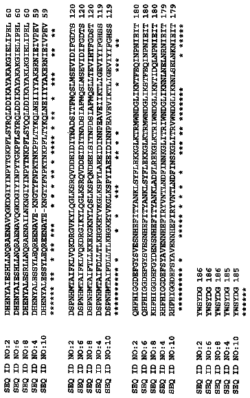
While the sequence of PlyG lysin has been isolated from the gamma phage as shown in FIG. 1, other lytic enzymes from bacteriophage specific for Bacillus anthracis may be used in place of PlyG. In one embodiment, the DNA encoding the lytic enzyme or holin protein, including their isozymes, analogs, or variants, has been genetically altered. In another embodiment, the lytic enzyme or holin protein, including their isozymes, analogs, or variants, has been chemically altered. In yet another embodiment, the lytic enzyme or holin protein, including their isozymes, analogs, or variants, are used in a combination of natural and modified (genetically or chemically altered) forms. The modified or altered forms of lytic enzymes and holin proteins are made synthetically by chemical synthesis and/or DNA recombinant techniques. The enzymes are made synthetically by chimerization and/or shuffling.
It should be understood that bacteriophage lytic enzyme are enzymes that specifically cleave bonds that are present in the peptidoglycan of bacterial cells. Since the bacterial cell wall peptidoglycan is highly conserved among all bacteria, there are only a few bonds to be cleaved to disrupt the cell wall. Enzymes that cleave these bonds are muramidases, glucosaminidases, endopeptidases, or N-acetyl-muramoyl-L-alanine amidases (hereinafter referred to as amidases). The majority of reported phage enzymes are either muramidases or amidases, and there have been no reports of bacteriophage glucosaminidases. Fischetti et al
reported that the C1 streptococcal phage lysin enzyme was an amidase. Garcia et al (1987, 1990) reported that the Cp1 lysin from a S. pneumoniae from a Cp-1 phage was a lysozyme. Caldentey and Bamford
reported that a lytic enzyme from the phi 6 Pseudomonas phage was an endopeptidase, splitting the peptide bridge formed by melodiaminopimilic acid and D-alanine. The E. coli T1 and T6 phage lytic enzymes are amidases as is the lytic enzyme from Listeria phage (ply) (Loessner et al, 1996). There are also other enzymes which cleave the cell wall.
The present disclosure uses such a lytic enzyme genetically coded for by a particular bacteriophage as either a prophylactic treatment for preventing those who have possibly been exposed to Bacillus anthracis, or as a therapeutic treatment for those who have already become ill from the infection. The phage associated lytic enzymes specific for Bacillus anthracis and genetically coded by a specific phage can effectively and efficiently break down the cell wall of the Bacillus anthracis. It is noted that the semipurified enzyme may lack proteolytic enzymatic activity and therefore may be nondestructive to mammalian proteins and tissues when present during the digestion of the bacterial cell wall.
Another embodiment also provides for chimeric proteins or peptides fragments which include fusion proteins for the aforesaid uses.
A definition of terms used and their applicability to the disclosure are provided as follows:
In this context of the embodiments, the term "lytic enzyme genetically coded for by a bacteriophage" means a polypeptide having at least some lytic activity against the host bacteria. The polypeptide has a sequence that encompasses a native sequence of a lytic enzyme and variants thereof. The polypeptide may be isolated from a variety of sources, such as from phage, or prepared by recombinant or synthetic methods, such as those by Garcia et al. Every polypeptide has two domains. One domain is a choline binding portion at the carboxyl terminal side and the other domain is an amidase activity that acts upon amide bonds in the peptidoglycan at the amino terminal side. Generally speaking, a lytic enzyme according to the disclosure is between 25,000 and 35,000 daltons in molecular weight and comprises a single polypeptide chain; however, this can vary depending on the enzyme chain. The molecular weight most conveniently is determined by assay on denaturing sodium dodecyl sulfate gel electrophoresis and comparison with molecular weight markers.
The term "isolated" means at least partially purified from a starting material. The term "purified" means that the biological material has been measurably increased in concentration by any purification process, including by not limited to, column chromatography, HPLC, precipitation, electrophoresis, etc., thereby partially, substantially or completely removing impurities such as precursors or other chemicals involved in preparing the material. Hence, material that is homogenous or substantially homogenous (e.g., yields a single protein signal in a separation procedure such as electrophoresis or chromatography) is included within the meanings of isolated and purified. Skilled artisans will appreciated that the amount of purification necessary will depend upon the use of the material. For example, compositions intended for administration to humans ordinarily must be highly purified in accordance with regulatory standards.
"A native sequence phage associated lytic enzyme" is a polypeptide having the same amino acid sequence as an enzyme derived from nature. Such native sequence enzyme can be isolated from nature or can be produced by recombinant or synthetic means. The term "native sequence enzyme" specifically encompasses naturally occurring forms (e.g., alternatively spliced or modified forms) and naturally-occurring variants of the enzyme. In one embodiment of the disclosure, the native sequence enzyme is a mature or full-length polypeptide that is genetically coded for by a gene from a bacteriophage specific for Bacillus anthracis. Of course, a number of variants are possible and known, as acknowledged in publications such as Lopez et al., Microbial Drug Resistance 3: 199-211 (1997); Garcia et al., Gene 86: 81-88 (1990); Garcia et al., Proc. Natl. Acad. Sci. USA 85: 914-918 (1988); Garcia et al., Proc. Natl. Acad. Sci. USA 85: 914-918 (1988); Garcia et al., Streptococcal Genetics (J. J. Ferretti and Curtis eds., 1987); Lopez et al., FEMS Microbiol. Lett. 100: 439-448 (1992); Romero et al., J. Bacteriol. 172: 5064-5070 (1990); Ronda et al., Eur. J. Biochem. 164: 621-624
and Sanchez et al., Gene 61: 13-19 (1987). The contents of each of these references, particularly the sequence listings and associated text that compares the sequences, including statements about sequence homologies, are specifically incorporated by reference in their entireties.
"A variant sequence phage associated lytic enzyme" means a functionally active lytic enzyme genetically coded for by a bacteriophage specific for Bacillus anthracis, as defined below, having at least 50%, 55%, 60%, 65%, 70%, 75%, 80%, 85%, 90%, 95%, 97%, 98%, 99%, or even at least 99.5% amino acid sequence identity with the sequence shown as SEQ ID No. 1. Of course a skilled artisan readily will recognize portions of this sequence that are associated with functionalities such as binding, and catalyzing a reaction. Accordingly, polypeptide sequences and nucleic acids that encode these sequences are contemplated that comprise at least 75%, 80%, 85%, 90%, 95%, 97%, 98%, 99% or more of each functional domain of SEQ ID No. 1. Such portions of the total sequence are very useful for diagnostics as well as therapeutics/prophylaxis. In fact, sequences as short as 5 amino acids long have utility as epitopic markers for the phage. More desirably, larger fragments or regions of protein having a size of at least 8, 9, 10, 12, 15 or 20 amino acids, and homologous sequences to these, have epitopic features and may be used either as small peptides or as sections of larger proteins according to embodiments. Nucleic acids corresponding to these sequences also are contemplated.
Such phage associated lytic enzyme variants include, for instance, lytic enzyme polypeptides wherein one or more amino acid residues are added, or deleted at the N or C terminus of the sequence of SEQ ID No. 1. In an embodiment one or more amino acids are substituted, deleted, and/or added to any position(s) in the sequence, or sequence portion. Ordinarily, a phage associated lytic enzyme will have at least about (e.g. exactly) 50%, 55%, 60%, 65%, 70%, 75%, amino acid sequence identity with native phage associated lytic enzyme sequences, more preferably at least about (e.g. exactly) 80%, 85%, 90%, 95%, 97%, 98%, 99% or 99.5% amino acid sequence identity. In other embodiments a phage associated lytic enzyme variant will have at least about 50% (e.g. exactly 50%), 55%, 60%, 65%, 70%, 75%, 80%, 85%, 90%, 95%, 97%, 98%, 99%, or even at least 99.5% amino acid sequence identity with the sequence shown as SEQ ID No. 1.
"Percent amino acid sequence identity" with respect to the phage associated lytic enzyme sequences identified herein is defined as the percentage of amino acid residues in a candidate sequence that are identical with the amino acid residues in the phage associated lytic enzyme sequence, after aligning the sequences in the same reading frame and introducing gaps, if necessary, to achieve the maximum percent sequence identity, and not considering any conservative substitutions as part of the sequence identity. Alignment for purposes of determining percent amino acid sequence identity can be achieved in various ways that are within the skill in the art, such as using publicly available computer software such as blast software. Those skilled in the art can determine appropriate parameters for measuring alignment, including any algorithms needed to achieve maximal alignment over the whole length of the sequences being compared.
In each case, of course conservative amino acid substitutions also may be made simultaneously in determining percent amino acid sequence identity. For example, a 15 amino acid long region of protein may have 50%, 55%, 60%, 65%, 70%, 75%, 80%, 85%, 90%, 95%, 97%, 98%, or 99% sequence homology with a region of SEQ ID No. 1. At the same time, the 15 amino acid long region of the protein may also have up to 0.5%, 1%, 2%, 5%, 10%, 15%, 20%, 30%, 40%, 50%, 65%, 75%, or more amino acids replaced with conservative substitutions. Preferably the region will have fewer than 30%, 20%, 10% or even less conservative substitutions. The "percent amino acid sequence identity" calculation in such cases will be higher than the actual percent sequence identity when conservative amino acid substitutions have been made.
"Percent nucleic acid sequence identity" with respect to the phage associated lytic enzyme sequences identified herein is defined as the percentage of nucleotides in a candidate sequence that are identical with the nucleotides in the phage associated lytic enzyme sequence, after aligning the sequences and introducing gaps, if necessary, to achieve the maximum percent sequence identity. Alignment for purposes of determining percent nucleic acid sequence identity can be achieved in various ways that are within the scope of those skilled in the art, including but not limited to the use of publicly available computer software. Those skilled in the art can determine appropriate parameters for measuring alignment, including any algorithms needed to achieve maximal alignment over the full length of the sequences being compared.
"Polypeptide" refers to a molecule comprised of amino acids which correspond to those encoded by a polynucleotide sequence which is naturally occurring. The polypeptide may include conservative substitutions wherein the naturally occurring amino acid is replaced by one having similar properties, where such conservative substitutions do not alter the function of the polypeptide (see, for example, Lewin "Genes V" Oxford University Press Chapter 1, pp. 9-13 1994).
A "chimeric protein" or "fusion protein" comprises all or (preferably a biologically active) part of a polypeptide of the disclosure operably linked to a heterologous polypeptide. Chimeric proteins or peptides are produced, for example, by combining two or more proteins having two or more active sites. Chimeric protein and peptides can act independently on the same or different molecules, and hence have a potential to treat two or more different bacterial infections at the same time. Chimeric proteins and peptides also are used to treat a bacterial infection by cleaving the cell wall in more than one location.
The term "operably linked" means that the polypeptide of the disclosure and the heterologous polypeptide are fused in-frame. The heterologous polypeptide can be fused to the N-terminus or C-terminus of the polypeptide of the disclosure. Chimeric proteins are produced enzymatically by chemical synthesis, or by recombinant DNA technology. A number of chimeric lytic enzymes have been produced and studied. Gene E-L, a chimeric lysis constructed from bacteriophages phi X174 and MS2 lysis proteins E and L, respectively, was subjected to internal deletions to create a series of new E-L clones with altered lysis or killing properties. The lytic activities of the parental genes E, L, E-L, and the internal truncated forms of E-L were investigated in this study to characterize the different lysis mechanism, based on differences in the architecture of the different membranes spanning domains. Electron microscopy and release of marker enzymes for the cytoplasmic and periplasmic spaces revealed that two different lysis mechanisms can be distinguished depending on penetration of the proteins of either the inner membrane or the inner and outer membranes of the E. coli. FEMS Microbiol. Lett. 1998 Jul. 1, 164(1); 159-67 (incorporated herein by reference).
In another experiment, an active chimeric cell wall lytic enzyme (TSL) was constructed by fusing the region coding for the N-terminal half of the lactococcal phage Tuc2009 lysin and the region coding for the C-terminal domain of the major pneumococcal autolysin. The chimeric enzyme exhibited a glycosidase activity capable of hydrolysing choline-containing pneumococcal cell walls. One example of a useful fusion protein is a GST fusion protein in which the polypeptide of the disclosure is fused to the C-terminus of a GST sequence. Such a chimeric protein can facilitate the purification of a recombinant polypeptide of the disclosure.
In another embodiment, the chimeric protein or peptide contains a heterologous signal sequence at its N-terminus. For example, the native signal sequence of a polypeptide of the disclosure can be removed and replaced with a signal sequence from another protein. For example, the gp67 secretory sequence of the baculovirus envelope protein can be used as a heterologous signal sequence (Current Protocols in Molecular Biology, Ausubel et al., eds., John Wiley & Sons, 1992, incorporated herein by reference). Other examples of eukaryotic heterologous signal sequences include the secretory sequences of melittin and human placental alkaline phosphatase (Stratagene; La Jolla, Calif.). In yet another example, useful prokaryotic heterologous signal sequences include the phoA secretory signal (Sambrook et al., supra) and the protein A secretory signal (Pharmacia Biotech; Piscataway, N.J.).
Another embodiment discloses an immunoglobulin fusion protein in which all or part of a polypeptide of the disclosure is fused to sequences derived from a member of the immunoglobulin protein family. An immunoglobulin fusion protein can be incorporated into a pharmaceutical composition and administered to a subject to inhibit an interaction between a ligand (soluble or membrane-bound) and a protein on the surface of a cell (receptor), to thereby suppress signal transduction in vivo. The immunoglobulin fusion protein can alter bioavailability of a cognate ligand of a polypeptide of the disclosure. Inhibition of ligand/receptor interaction may be useful therapeutically, both for treating bacterial-associated diseases and disorders for modulating (i.e. promoting or inhibiting) cell survival. Moreover, an immunoglobulin fusion protein of the disclosure can be used as an immunogen to produce antibodies directed against a polypeptide of the disclosure in a subject, to purify ligands and in screening assays to identify molecules which inhibit the interaction of receptors with ligands. Chimeric and fusion proteins and peptides of the disclosure can be produced by standard recombinant DNA techniques.
In another embodiment, the fusion gene can be synthesized by conventional techniques, including automated DNA synthesizers. Alternatively, PCR amplification of gene fragments can be carried out using anchor primers which give rise to complementary overhangs between two consecutive gene fragments which subsequently can be annealed and reamplified to generate a chimeric gene sequence (see, i.e., Ausubel et al., supra). Moreover, many expression vectors are commercially available that already encode a fusion moiety (i.e., a GST polypeptide). A nucleic acid encoding a polypeptide of the disclosure can be cloned into such an expression vector such that the fusion moiety is linked in-frame to the polypeptide of the disclosure.
As used herein, shuffled proteins or peptides, gene products, or peptides for more than one related phage protein or protein peptide fragments have been randomly cleaved and reassembled into a more active or specific protein. Shuffled oligonucleotides, peptides or peptide fragment molecules are selected or screened to identify a molecule having a desired functional property. This method is described, for example, in Stemmer, U.S. Pat. No. 6,132,970. (Method of shuffling polynucleotides); Kauffman, U.S. Pat. No. 5,976,862 (Evolution via Condon-based Synthesis) and Huse, U.S. Pat. No. 5,808,022 (Direct Codon Synthesis). The contents of these patents are incorporated herein by reference. Shuffling is used to create a protein that is 10 to 100 fold more active than the template protein. The template protein is selected among different varieties of lysin or holin proteins. The shuffled protein or peptides constitute, for example, one or more binding domains and one or more catalytic domains. Each binding or catalytic domain is derived from the same or a different phage or phage protein. The shuffled domains are either oligonucleotide based molecules, as gene or gene products, that either alone or in combination with other genes or gene products are translatable into a peptide fragment, or they are peptide based molecules. Gene fragments include any molecules of DNA, RNA, DNA-RNA hybrid, antisense RNA, Ribozymes, ESTs, SNIPs and other oligonucleotide-based molecules that either alone or in combination with other molecules produce an oligonucleotide molecule capable or incapable of translation into a peptide.
As noted above, the present disclosure discusses the use of holin proteins. Holin proteins produce holes in the cell membrane. More specifically, holins form lethal membrane lesions. Like the lytic proteins, holin proteins are coded for and carried by a phage. In fact, it is quite common for the genetic code of the holin protein to be next to or even within the code for the phage lytic protein. Most holin protein sequences are short, and overall, hydrophobic in nature, with a highly hydrophilic carboxy-terminal domain. In many cases, the putative holin protein is encoded on a different reading frame within the enzymatically active domain of the phage. In other cases, holin protein is encoded on the DNA next or close to the DNA coding for the cell wall lytic protein. Holin proteins are frequently synthesized during the late stage of phage infection and found in the cytoplasmic membrane where they cause membrane lesions.
Holins can be grouped into two general classes based on primary structure analysis. Class I holins are usually 95 residues or longer and may have three potential transmembrane domains. Class II holins are usually smaller, at approximately 65-95 residues, with the distribution of charged and hydrophobic residues indicating two TM domains (Young, et al. Trends in Microbiology v. 8, No. 4, March 2000). At least for the phages of gram-positive hosts, however, the dual-component lysis system may not be universal. Although the presence of holins has been shown or suggested for several phages, no genes have yet been found encoding putative holins for all phages. Holins have been shown to be present in several bacteria, including, for example, lactococcal bacteriophage Tuc2009, lactococcal NLC3, pneumococcal bacteriophage EJ-1, Lactobacillus gasseri bacteriophage Nadh, Staphylococcus aureus bacteriophage Twort, Listeria monocytogenes bacteriophages, pneumococcal phage Cp-1, Bacillus subtillis phage M29, Lactobacillus delbrueckki bacteriophage LL-H lysin, and bacteriophage N11 of Staphyloccous aureus. (Loessner, et al., Journal of Bacteriology, August 1999, p. 4452-4460).
The modified or altered form of the protein or peptides and peptide fragments, as disclosed herein, includes protein or peptides and peptide fragments that are chemically synthesized or prepared by recombinant DNA techniques, or both. These techniques include, for example, chimerization and shuffling. When the protein or peptide is produced by chemical synthesis, it is preferably substantially free of chemical precursors or other chemicals, i.e., it is separated from chemical precursors or other chemicals which are involved in the synthesis of the protein. Accordingly such preparations of the protein have less than about 30%, 20%, 10%, 5% (by dry weight) of chemical precursors or compounds other than the polypeptide of interest.
In one embodiment of the disclosure, a signal sequence of a polypeptide of can facilitate transmembrane movement of the protein and peptides and peptide fragments of the disclosure to and from mucous membranes, as well as by facilitating secretion and isolation of the secreted protein or other proteins of interest. Signal sequences are typically characterized by a core of hydrophobic amino acids which are generally cleaved from the mature protein during secretion in one or more cleavage events. Such signal peptides contain processing sites that allow cleavage of the signal sequence from the mature proteins as they pass through the secretory pathway. Thus, the disclosure can pertain to the described polypeptides having a signal sequence, as well as to the signal sequence itself and to the polypeptide in the absence of the signal sequence (i.e., the cleavage products). In one embodiment, a nucleic acid sequence encoding a signal sequence of the disclosure can be operably linked in an expression vector to a protein of interest, such as a protein which is ordinarily not secreted or is otherwise difficult to isolate. The signal sequence directs secretion of the protein, such as from an eukaryotic host into which the expression vector is transformed, and the signal sequence is subsequently or concurrently cleaved. The protein can then be readily purified from the extracellular medium by art recognized methods. Alternatively, the signal sequence can be linked to a protein of interest using a sequence which facilitates purification, such as with a GST domain.
In another embodiment, a signal sequence can be used to identify regulatory sequences, i.e., promoters, enhancers, repressors. Since signal sequences are the most amino-terminal sequences of a peptide, it is expected that the nucleic acids which flank the signal sequence on its amino-terminal side will be regulatory sequences that affect transcription. Thus, a nucleotide sequence which encodes all or a portion of a signal sequence can be used as a probe to identify and isolate the signal sequence and its flanking region, and this flanking region can be studied to identify regulatory elements therein. The present disclosure also pertains to other variants of the polypeptides of the disclosure. Such variants have an altered amino acid sequence which can function as either agonists (mimetics) or as antagonists. Variants can be generated by mutagenesis, i.e., discrete point mutation or truncation. An agonist can retain substantially the same, or a subset, of the biological activities of the naturally occurring form of the protein. An antagonist of a protein can inhibit one or more of the activities of the naturally occurring form of the protein by, for example, competitively binding to a downstream or upstream member of a cellular signaling cascade which includes the protein of interest. Thus, specific biological effects can be elicited by treatment with a variant of limited function. Treatment of a subject with a variant having a subset of the biological activities of the naturally occurring form of the protein can have fewer side effects in a subject relative to treatment with the naturally occurring form of the protein. Variants of a protein of the disclosure which function as either agonists (mimetics) or as antagonists can be identified by screening combinatorial libraries of mutants, i.e., truncation mutants, of the protein of the disclosure for agonist or antagonist activity. In one embodiment, a variegated library of variants is generated by combinatorial mutagenesis at the nucleic acid level and is encoded by a variegated gene library. A variegated library of variants can be produced by, for example, enzymatically ligating a mixture of synthetic oligonucleotides into gene sequences such that a degenerate set of potential protein sequences is expressible as individual polypeptides, or alternatively, as a set of larger fusion proteins (i.e., for phage display). There are a variety of methods which can be used to produce libraries of potential variants of the polypeptides of the disclosure from a degenerate oligonucleotide sequence. Methods for synthesizing degenerate oligonucleotides are known in the art (see, i.e., Narang
Tetrahedron 39:3; Itakura et al.
Annu. Rev. Biochem. 53:323; Itakura et al.
Science 198:1056; Ike et al.
Nucleic Acid Res. 11:477, all herein incorporated by reference).
In addition, libraries of fragments of the coding sequence of a polypeptide of the disclosure can be used to generate a variegated population of polypeptides for screening and subsequent selection of variants. For example, a library of coding sequence fragments can be generated by treating a double stranded PCR fragment of the coding sequence of interest with a nuclease under conditions wherein nicking occurs only about once per molecule, denaturing the double stranded DNA, renaturing the DNA to form double stranded DNA which can include sense/antisense pairs from different nicked products, removing single stranded portions from reformed duplexes by treatment with S1 nuclease, and ligating the resulting fragment library into an expression vector. By this method, an expression library can be derived which encodes N-terminal and internal fragments of various sizes of the protein of interest. Several techniques are known in the art for screening gene products of combinatorial libraries made by point mutations or truncation, and for screening cDNA libraries for gene products having a selected property. The most widely used techniques, which are amenable to high through-put analysis, for screening large gene libraries typically include cloning the gene library into replicable expression vectors, transforming appropriate cells with the resulting library of vectors, and expressing the combinatorial genes under conditions in which detection of a desired activity facilitates isolation of the vector encoding the gene whose product was detected. Recursive ensemble mutagenesis (REM), a technique which enhances the frequency of functional mutants in the libraries, can be used in combination with the screening assays to identify variants of a protein of the disclosure (Arkin and Yourvan
Proc. Natl. Acad. Sci. USA 89:7811-7815; Delgrave et al.
Protein Engineering 6(3):327-331).
Immunologically active portions of a protein or peptide fragment include regions that bind to antibodies that recognize the phage enzyme. In this context, the smallest portion of a protein (or nucleic acid that encodes the protein) according to embodiments is an epitope that is recognizable as specific for the phage that makes the lysin protein. Accordingly, the smallest polypeptide (and associated nucleic acid that encodes the polypeptide) that can be expected to bind antibody and is useful for some embodiments may be 8, 9, 10, 11, 12, 13, 15, 20, 25, 30, 35, 40, 45, 50, 55, 60, 65, 75, 85, or 100 amino acids long. Although small sequences as short as 8, 9, 10, 11, 12 or 15 amino acids long reliably comprise enough structure to act as epitopes, shorter sequences of 5, 6, or 7 amino acids long can exhibit epitopic structure in some conditions and have value in an embodiment. Thus, the smallest portion of the protein described by SEQ ID No. 1 includes polypeptides as small as 5, 6, 7, 8, 9, or 10 amino acids long.
Homologous proteins and nucleic acids can be prepared that share functionality with such small proteins and/or nucleic acids (or protein and/or nucleic acid regions of larger molecules) as will be appreciated by a skilled artisan. Such small molecules and short regions of larger molecules, that may be homologous specifically are intended as embodiments. Preferably the homology of such valuable regions is at least 50%, 65%, 75%, 85%, and more preferably at least 90%, 95%, 97%, 98%, or at least 99% compared to SEQ ID No. 1. These percent homology values do not include alterations due to conservative amino acid substitutions.
Of course, an epitope as described herein may be used to generate an antibody and also can be used to detect binding to molecules that recognize the lysin protein. Another embodiment is a molecule such as an antibody or other specific binder that may be created through use of an epitope such as by regular immunization or by a phase display approach where an epitope can be used to screen a library if potential binders. Such molecules recognize one or more epitopes of lysin protein or a nucleic acid that encodes lysin protein. An antibody that recognizes an epitope may be a monoclonal antibody, a humanized antibody, or a portion of an antibody protein. Desirably the molecule that recognizes an epitope has a specific binding for that epitope which is at least 10 times as strong as the molecule has for serum albumin. Specific binding can be measured as affinity (Km). More desirably the specific binding is at least 10.sup.2, 10.sup.3, 10.sup.4, 10.sup.5, 10.sup.6, 10.sup.7, 10.sup.8, or even higher than that for serum albumin under the same conditions.
In a desirable embodiment the antibody or antibody fragment is in a form useful for detecting the presence of the lysin protein. A variety of forms and methods for their synthesis are known as will be appreciated by a skilled artisan. The antibody may be conjugated (covalently complexed) with a reporter molecule or atom such as a fluor, an enzyme that creates an optical signal, a chemilumiphore, a microparticle, or a radioactive atom. The antibody or antibody fragment may be synthesized in vivo, after immunization of an animal, for example, The antibody or antibody fragment may be synthesized via cell culture after genetic recombination. The antibody or antibody fragment may be prepared by a combination of cell synthesis and chemical modification.
The description continues in the full USPTO document.
 Lapsed, fee not paid11 drawings